3.1: Horizontal Gene Transfer in Bacteria
- Page ID
- 3150
After completing this section you should be able to perform the following objectives.
- Compare and contrast mutation and horizontal gene transfer as methods of enabling bacteria to respond to selective pressures and adapt to new environments.
- Define horizontal gene transfer and state the most common form of horizontal gene transfer in bacteria.
- Briefly describe the mechanisms for transformation in bacteria.
- Briefly describe the following mechanisms of horizontal gene transfer in bacteria:
- generalized transduction
- specialized transduction
- Briefly describe the following mechanisms of horizontal gene transfer in bacteria:
- Transfer of conjugative plasmids, conjugative transposons, and mobilizable plasmids in Gram-negative bacteria
- F+ conjugation
- Hfr conjugation
- Describe R-plasmids and the significance of R-plasmids to medical microbiology.
Bacteria are able to respond to selective pressures and adapt to new environments by acquiring new genetic traits as a result of mutation, a modification of gene function within a bacterium, and as a result of horizontal gene transfer, the acquisition of new genes from other bacteria. Mutation occurs relatively slowly. The normal mutation rate in nature is in the range of 10-6 to 10-9 per nucleotide per bacterial generation, although when bacterial populations are under stress, they can greatly increase their mutation rate. Furthermore, most mutations are harmful to the bacterium. Horizontal gene transfer, on the other hand, enables bacteria to respond and adapt to their environment much more rapidly by acquiring large DNA sequences from another bacterium in a single transfer.
Horizontal gene transfer, also known as lateral gene transfer, is a process in which an organism transfers genetic material to another organism that is not its offspring. The ability of Bacteria and Archaea to adapt to new environments as a part of bacterial evolution most frequently results from the acquisition of new genes through horizontal gene transfer rather than by the alteration of gene functions through mutations. (It is estimated that as much as 20% of the genome of Escherichia coli originated from horizontal gene transfer.)
Horizontal gene transfer is able to cause rather large-scale changes in a bacterial genome. For example, certain bacteria contain multiple virulence genes called pathogenicity islands that are located on large, unstable regions of the bacterial genome. These pathogenicity islands can be transmitted to other bacteria by horizontal gene transfer. However, if these transferred genes provide no selective advantage to the bacteria that acquire them, they are usually lost by deletion. In this way the size of the bacterium's genome can remain approximately the same size over time.
There are three mechanisms of horizontal gene transfer in bacteria: transformation, transduction, and conjugation. The most common mechanism for horizontal gene transmission among bacteria, especially from a donor bacterial species to different recipient species, is conjugation. Although bacteria can acquire new genes through transformation and transduction, this is usually a more rare transfer among bacteria of the same species or closely related species.
Transformation
Transformation is a form of genetic recombination in which a DNA fragment from a dead, degraded bacterium enters a competent recipient bacterium and is exchanged for a piece of DNA of the recipient. Transformation usually involves only homologous recombination, a recombination of homologous DNA regions having nearly the same nucleotide sequences. Typically this involves similar bacterial strains or strains of the same bacterial species.
A few bacteria, such as Neisseria gonorrhoeae, Neisseria meningitidis, Hemophilus influenzae, Legionella pneomophila, Streptococcus pneumoniae, and Helicobacter pylori tend to be naturally competent and transformable. Competent bacteria are able to bind much more DNA than noncompetent bacteria. Some of these genera also undergo autolysis that then provides DNA for homologous recombination. In addition, some competent bacteria kill noncompetent cells to release DNA for transformation.
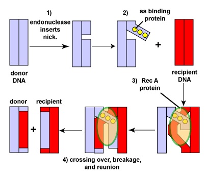
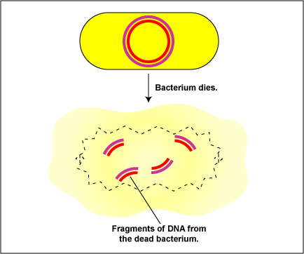
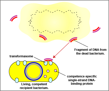
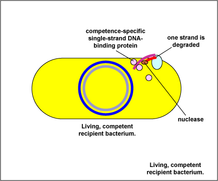
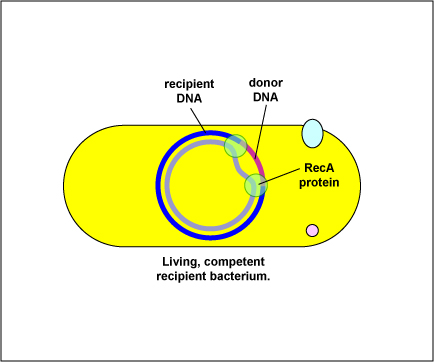
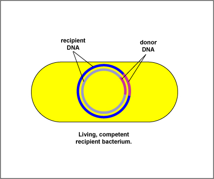
During transformation, DNA fragments (usually about 10 genes long) are released from a dead degraded bacterium and bind to DNA binding proteins on the surface of a competent living recipient bacterium. Depending on the bacterium, either both strands of DNA penetrate the recipient, or a nuclease degrades one strand of the fragment and the remaining DNA strand enters the recipient. This DNA fragment from the donor is then exchanged for a piece of the recipient's DNA by means of RecA proteins and other molecules and involves breakage and reunion of the paired DNA segments as seen in (Figure \(\PageIndex{1}\)). Transformation is summarized in Figure \(\PageIndex{2}\).
Transduction
Transduction involves the transfer of a DNA fragment from one bacterium to another by a bacteriophage. There are two forms of transduction: generalized transduction and specialized transduction.
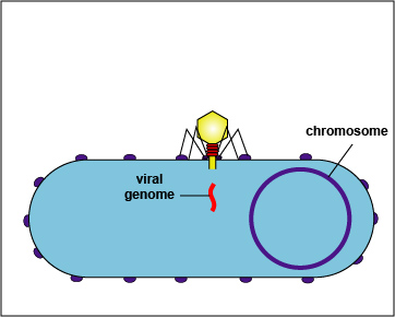
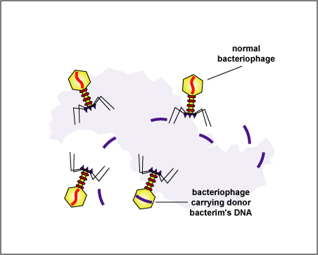
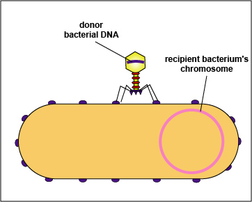
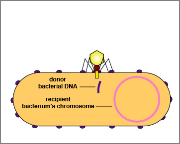
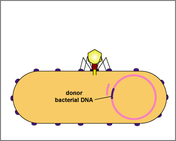
During the replication of lytic bacteriophages and temperate bacteriophages, occasionally the phage capsid accidently assembles around a small fragment of bacterial DNA. When this bacteriophage, called a transducing particle, infects another bacterium, it injects the fragment of donor bacterial DNA it is carrying into the recipient where it can subsequently be exchanged for a piece of the recipient's DNA by homologous recombination. Generalized transduction is summarized in Figure \(\PageIndex{3}\).
- Step 1: A bacteriophage adsorbs to a susceptible bacterium.
- Step 2: The bacteriophage genome enters the bacterium. The genome directs the bacterium's metabolic machinery to manufacture bacteriophage components and enzymes. Bacteriophage-coded enzymes will also breakup the bacterial chromosome.
- Step 3: Occasionally, a bacteriophage capsid mistakenly assembles around either a fragment of the donor bacterium's chromosome or around a plasmid instead of around a phage genome.
- Step 4: The bacteriophages are released as the bacterium is lysed. Note that one bacteriophage is carrying a fragment of the donor bacterium's DNA rather than a bacteriophage genome.
- Step 5: The bacteriophage carrying the donor bacterium's DNA adsorbs to a recipient bacterium.
- Step 6: The bacteriophage inserts the donor bacterium's DNA it is carrying into the recipient bacterium.
- Step 7: Homologous recombination occurs and the donor bacterium's DNA is exchanged for some of the recipient's DNA. (Figure \(\PageIndex{1}\) shows the functions of the RecA proteins involved in homologous recombination.)
Generalized transduction occurs in a variety of bacteria, including Staphylococcus, Escherichia, Salmonella, and Pseudomonas.

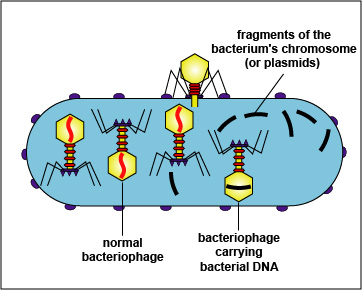
Plasmids, such as the penicillinase plasmid of Staphylococcus aureus, may also be carried from one bacterium to another by generalized transduction.
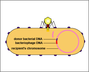
Specialized transduction: This may occur occasionally during the lysogenic life cycle of a temperate bacteriophage. During spontaneous induction, a small piece of bacterial DNA may sometimes be exchanged for a piece of the bacteriophage genome, which remains in the bacterial nucleoid. This piece of bacterial DNA replicates as a part of the bacteriophage genome and is put into each phage capsid. The bacteriophages are released, adsorb to recipient bacteria, and inject the donor bacterium DNA/phage DNA complex into the recipient bacterium where it inserts into the bacterial chromosome (Figure \(\PageIndex{4}\)).
Conjugation
Genetic recombination in which there is a transfer of DNA from a living donor bacterium to a living recipient bacterium by cell-to-cell contact. In Gram-negative bacteria it typically involves a conjugation or sex pilus.
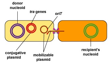
Conjugation is encoded by plasmids or transposons. It involves a donor bacterium that contains a conjugative plasmid and a recipient cell that does not. A conjugative plasmid is self-transmissible, in that it possesses all the necessary genes for that plasmid to transmit itself to another bacterium by conjugation. Conjugation genes known as tra genes enable the bacterium to form a mating pair with another organism, while oriT (origin of transfer) sequences determine where on the plasmid DNA transfer is initiated by serving as the replication start site where DNA replication enzymes will nick the DNA to initiate DNA replication and transfer. In addition, mobilizable plasmids that lack the tra genes for self-transmissibility but possess the oriT sequences for initiation of DNA transfer may also be transferred by conjugation if the bacterium containing them also possesses a conjugative plasmid. The tra genes of the conjugative plasmid enable a mating pair to form, while the oriT of the mobilizable plasmid enable the DNA to moves through the conjugative bridge (Figure \(\PageIndex{5}\)).
Transposons ("jumping genes") are small pieces of DNA that encode enzymes that enable the transposon to move from one DNA location to another, either on the same molecule of DNA or on a different molecule. Transposons may be found as part of a bacterium's chromosome (conjugative transposons) or in plasmids and are usually between one and twelve genes long. A transposon contains a number of genes, such as those coding for antibiotic resistance or other traits, flanked at both ends by insertion sequences coding for an enzyme called transpoase. Transpoase is the enzyme that catalyzes the cutting and resealing of the DNA during transposition.
Conjugative transposons, like conjugative plasmids, carry the genes that enable mating pairs to form for conjugation. Therefore, conjugative transposons also enable mobilizable plasmids and nonconjugative transposons to be transferred to a recipient bacterium during conjugation.
Many conjugative plasmids and conjugative transposons possess rather promiscuous transfer systems that enables them to transfer DNA not only to like species, but also to unrelated species. The ability of bacteria to adapt to new environments as a part of bacterial evolution most frequently results from the acquisition of large DNA sequences from another bacterium by conjugation.
a. General mechanism of transfer of conjugative plasmids by conjugation in Gram-negative bacteria
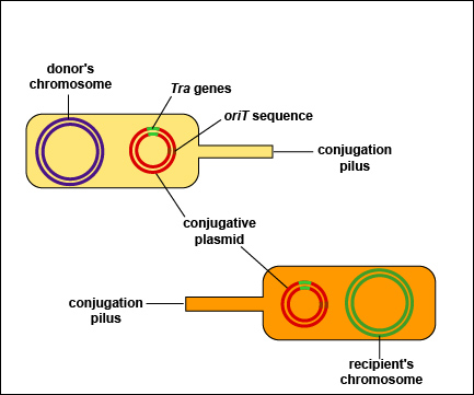
In Gram-negative bacteria, the first step in conjugation involves a conjugation pilus (sex pilus or F pilus) on the donor bacterium binding to a recipient bacterium lacking a conjugation pilus. Typically the conjugation pilus retracts or depolymerizes pulling the two bacteria together. A series of membrane proteins coded for by the conjugative plasmid then forms a bridge and an opening between the two bacteria, now called a mating pair.
Using the rolling circle model of DNA replication, a nuclease breaks one strand of the plasmid DNA at the origin of transfer site (oriT) of the plasmid and that nicked strand enters the recipient bacterium. The other strand remains behind in the donor cell. Both the donor and the recipient plasmid strands then make a complementary copy of themselves. Both bacteria now possess the conjugative plasmid. This process is summarized in Figure \(\PageIndex{6}\)).
This is the mechanism by which resistance plasmids (R-plasmids), coding for multiple antibiotic resistance and conjugation pilus formation, are transferred from a donor bacterium to a recipient. This is a big problem in treating opportunistic Gram-negative infections such as urinary tract infections, wound infections, pneumonia, and septicemia by such organisms as E. coli, Proteus, Klebsiella, Enterobacter, Serratia, and Pseudomonas, as well as with intestinal infections by organisms like Salmonella and Shigella.
There is also evidence that the conjugation pilus may also serve as a direct channel through which single-stranded DNA may be transferred during conjugation.
b. F+ conjugation
This results in the transfer of an F+ plasmid possessing tra genes coding only for a conjugation pilus and mating pair formation from a donor bacterium to a recipient bacterium. One strand of the F+ plasmid is broken with a nuclease at the origin of transfer (oriT) sequence that determines where on the plasmid DNA transfer is initiated by serving as the replication start site where DNA replication enzymes will nick the DNA to initiate DNA replication and transfer. The nicked strand enters the recipient bacterium while the other plasmid strand remains in the donor. Each strand then makes a complementary copy. The recipient then becomes an F+ male and can make a sex pilus (see 7A through 7D).
In addition, mobilizable plasmids that lack the tra genes for self-transmissibility but possess the oriT sequences for initiation of DNA transfer, may also be transferred by conjugation. The tra genes of the F+ plasmid enable a mating pair to form and the oriT sequences of the mobilizable plasmid enable the DNA to moves through the conjugative bridge (Figure \(\PageIndex{5}\)).
c. Hfr (high frequency recombinant) conjugation
Hfr conjugation begins when an F+ plasmid with tra genes coding for mating pair formation inserts or integrates into the chromosome to form an Hfr bacterium. (A plasmid that is able to integrate into the host nucleoid is called an episome.) A nuclease then breaks one strand of the donor's DNA at the origin of transfer (oriT) location of the inserted F+ plasmid and the nicked strand of the donor DNA begins to enter the recipient bacterium. The remaining non-nicked DNA strand remains in the donor and makes a complementary copy of itself.
The bacterial connection usually breaks before the transfer of the entire chromosome is completed so the remainder of the F+ plasmid seldom enters the recipient. As a result, there is a transfer of some chromosomal DNA, which may be exchanged for a piece of the recipient's DNA through homologous recombination, but not the ability to form a conjugation pilus and mating pairs (see Figure \(\PageIndex{8}\)A through 8E).
- A strain of living Streptococcus pneumoniae that cannot make a capsule is injected into mice and has no adverse effect. This strain is then mixed with a culture of heat-killed Streptococcus pneumoniae that when alive was able to make a capsule and kill mice. After a period of time, this mixture is injected into mice and kills them. In terms of horizontal gene transfer, describe what might account for this.
- A gram-negative bacterium that was susceptible to most common antibiotics suddenly becomes resistant to several of them. It also appears to be spreading this resistance to others of its kind. Describe the mechanism that most likely accounts for this.
Summary
- Mutation is a modification of gene function within a bacterium and while it enables bacteria to adapt to new environments, it occurs relatively slowly.
- Horizontal gene transfer enables bacteria to respond and adapt to their environment much more rapidly by acquiring large DNA sequences from another bacterium in a single transfer.
- Horizontal gene transfer is a process in which an organism transfers genetic material to another organism that is not its offspring.
- Mechanisms of bacterial horizontal gene transfer include transformation, transduction, and conjugation.
- During transformation, a DNA fragment from a dead, degraded bacterium enters a competent recipient bacterium and is exchanged for a piece of DNA of the recipient. Typically this involves similar bacterial strains or strains of the same bacterial species.
- Transduction involves the transfer of either a chromosomal DNA fragment or a plasmid from one bacterium to another by a bacteriophage.
- Conjugation is a transfer of DNA from a living donor bacterium to a living recipient bacterium by cell-to-cell contact. In Gram-negative bacteria it involves a conjugation pilus.
- A conjugative plasmid is self-transmissible, that is, it possesses conjugation genes known as tra genes enable the bacterium to form a mating pair with another organism, and oriT (origin of transfer) sequences that determine where on the plasmid DNA transfer is initiated.
- Mobilizable plasmids that lack the tra genes for self-transmissibility can be co-transfered in a bacterium possessing a conjugative plasmid.
- Transposons ("jumping genes") are small pieces of DNA that encode enzymes that enable the transposon to move from one DNA location to another, either on the same molecule of DNA or on a different molecule.
- Conjugative transposons carry the genes that enable mating pairs to form for conjugation.
- F+ conjugation is the transfer of an F+ plasmid possessing tra genes coding only for a conjugation pilus and mating pair formation from a donor bacterium to a recipient bacterium. Mobilizable plasmids may be co-transfered during F+ conjugation.
- During Hfr conjugation, an F+ plasmid with tra genes coding for mating pair formation inserts into the bacterial chromosome to form an Hfr bacterium. This results in a transfer of some chromosomal DNA from the donor to the recipient which may be exchanged for a piece of the recipient's DNA through homologous recombination.


