
1.1: Mitosis and Meiosis
- Page ID
- 73666

\( \newcommand{\vecs}[1]{\overset { \scriptstyle \rightharpoonup} {\mathbf{#1}} } \)
\( \newcommand{\vecd}[1]{\overset{-\!-\!\rightharpoonup}{\vphantom{a}\smash {#1}}} \)
\( \newcommand{\dsum}{\displaystyle\sum\limits} \)
\( \newcommand{\dint}{\displaystyle\int\limits} \)
\( \newcommand{\dlim}{\displaystyle\lim\limits} \)
\( \newcommand{\id}{\mathrm{id}}\) \( \newcommand{\Span}{\mathrm{span}}\)
( \newcommand{\kernel}{\mathrm{null}\,}\) \( \newcommand{\range}{\mathrm{range}\,}\)
\( \newcommand{\RealPart}{\mathrm{Re}}\) \( \newcommand{\ImaginaryPart}{\mathrm{Im}}\)
\( \newcommand{\Argument}{\mathrm{Arg}}\) \( \newcommand{\norm}[1]{\| #1 \|}\)
\( \newcommand{\inner}[2]{\langle #1, #2 \rangle}\)
\( \newcommand{\Span}{\mathrm{span}}\)
\( \newcommand{\id}{\mathrm{id}}\)
\( \newcommand{\Span}{\mathrm{span}}\)
\( \newcommand{\kernel}{\mathrm{null}\,}\)
\( \newcommand{\range}{\mathrm{range}\,}\)
\( \newcommand{\RealPart}{\mathrm{Re}}\)
\( \newcommand{\ImaginaryPart}{\mathrm{Im}}\)
\( \newcommand{\Argument}{\mathrm{Arg}}\)
\( \newcommand{\norm}[1]{\| #1 \|}\)
\( \newcommand{\inner}[2]{\langle #1, #2 \rangle}\)
\( \newcommand{\Span}{\mathrm{span}}\) \( \newcommand{\AA}{\unicode[.8,0]{x212B}}\)
\( \newcommand{\vectorA}[1]{\vec{#1}} % arrow\)
\( \newcommand{\vectorAt}[1]{\vec{\text{#1}}} % arrow\)
\( \newcommand{\vectorB}[1]{\overset { \scriptstyle \rightharpoonup} {\mathbf{#1}} } \)
\( \newcommand{\vectorC}[1]{\textbf{#1}} \)
\( \newcommand{\vectorD}[1]{\overrightarrow{#1}} \)
\( \newcommand{\vectorDt}[1]{\overrightarrow{\text{#1}}} \)
\( \newcommand{\vectE}[1]{\overset{-\!-\!\rightharpoonup}{\vphantom{a}\smash{\mathbf {#1}}}} \)
\( \newcommand{\vecs}[1]{\overset { \scriptstyle \rightharpoonup} {\mathbf{#1}} } \)
\(\newcommand{\longvect}{\overrightarrow}\)
\( \newcommand{\vecd}[1]{\overset{-\!-\!\rightharpoonup}{\vphantom{a}\smash {#1}}} \)
\(\newcommand{\avec}{\mathbf a}\) \(\newcommand{\bvec}{\mathbf b}\) \(\newcommand{\cvec}{\mathbf c}\) \(\newcommand{\dvec}{\mathbf d}\) \(\newcommand{\dtil}{\widetilde{\mathbf d}}\) \(\newcommand{\evec}{\mathbf e}\) \(\newcommand{\fvec}{\mathbf f}\) \(\newcommand{\nvec}{\mathbf n}\) \(\newcommand{\pvec}{\mathbf p}\) \(\newcommand{\qvec}{\mathbf q}\) \(\newcommand{\svec}{\mathbf s}\) \(\newcommand{\tvec}{\mathbf t}\) \(\newcommand{\uvec}{\mathbf u}\) \(\newcommand{\vvec}{\mathbf v}\) \(\newcommand{\wvec}{\mathbf w}\) \(\newcommand{\xvec}{\mathbf x}\) \(\newcommand{\yvec}{\mathbf y}\) \(\newcommand{\zvec}{\mathbf z}\) \(\newcommand{\rvec}{\mathbf r}\) \(\newcommand{\mvec}{\mathbf m}\) \(\newcommand{\zerovec}{\mathbf 0}\) \(\newcommand{\onevec}{\mathbf 1}\) \(\newcommand{\real}{\mathbb R}\) \(\newcommand{\twovec}[2]{\left[\begin{array}{r}#1 \\ #2 \end{array}\right]}\) \(\newcommand{\ctwovec}[2]{\left[\begin{array}{c}#1 \\ #2 \end{array}\right]}\) \(\newcommand{\threevec}[3]{\left[\begin{array}{r}#1 \\ #2 \\ #3 \end{array}\right]}\) \(\newcommand{\cthreevec}[3]{\left[\begin{array}{c}#1 \\ #2 \\ #3 \end{array}\right]}\) \(\newcommand{\fourvec}[4]{\left[\begin{array}{r}#1 \\ #2 \\ #3 \\ #4 \end{array}\right]}\) \(\newcommand{\cfourvec}[4]{\left[\begin{array}{c}#1 \\ #2 \\ #3 \\ #4 \end{array}\right]}\) \(\newcommand{\fivevec}[5]{\left[\begin{array}{r}#1 \\ #2 \\ #3 \\ #4 \\ #5 \\ \end{array}\right]}\) \(\newcommand{\cfivevec}[5]{\left[\begin{array}{c}#1 \\ #2 \\ #3 \\ #4 \\ #5 \\ \end{array}\right]}\) \(\newcommand{\mattwo}[4]{\left[\begin{array}{rr}#1 \amp #2 \\ #3 \amp #4 \\ \end{array}\right]}\) \(\newcommand{\laspan}[1]{\text{Span}\{#1\}}\) \(\newcommand{\bcal}{\cal B}\) \(\newcommand{\ccal}{\cal C}\) \(\newcommand{\scal}{\cal S}\) \(\newcommand{\wcal}{\cal W}\) \(\newcommand{\ecal}{\cal E}\) \(\newcommand{\coords}[2]{\left\{#1\right\}_{#2}}\) \(\newcommand{\gray}[1]{\color{gray}{#1}}\) \(\newcommand{\lgray}[1]{\color{lightgray}{#1}}\) \(\newcommand{\rank}{\operatorname{rank}}\) \(\newcommand{\row}{\text{Row}}\) \(\newcommand{\col}{\text{Col}}\) \(\renewcommand{\row}{\text{Row}}\) \(\newcommand{\nul}{\text{Nul}}\) \(\newcommand{\var}{\text{Var}}\) \(\newcommand{\corr}{\text{corr}}\) \(\newcommand{\len}[1]{\left|#1\right|}\) \(\newcommand{\bbar}{\overline{\bvec}}\) \(\newcommand{\bhat}{\widehat{\bvec}}\) \(\newcommand{\bperp}{\bvec^\perp}\) \(\newcommand{\xhat}{\widehat{\xvec}}\) \(\newcommand{\vhat}{\widehat{\vvec}}\) \(\newcommand{\uhat}{\widehat{\uvec}}\) \(\newcommand{\what}{\widehat{\wvec}}\) \(\newcommand{\Sighat}{\widehat{\Sigma}}\) \(\newcommand{\lt}{<}\) \(\newcommand{\gt}{>}\) \(\newcommand{\amp}{&}\) \(\definecolor{fillinmathshade}{gray}{0.9}\)- Describe the cell cycle.
- Explain the events of all stages of mitosis.
- Track chromosome and chromatid number through all stages of mitosis.
- Explain the events of all stages of meiosis.
- Track chromosome and chromatid number through all stages of meiosis.
- Describe the role of meiosis in gamete formation and how it relates to inheritance.
Introduction
Multicellular organisms such as plants and animals are composed of millions to trillions (1,000,000,000) of cells that work together. The cells that make up different tissues have different shapes and perform different functions for the plant or animal. Even though they have diverse functions, each somatic cell in the organism normally has the same chromosomes and therefore the same genetic makeup. Furthermore, the millions of cells that makeup a mature organism originated from a single cell formed when the male and female gametes from the parents of the organism fused. This single cell established the life of the organism. Understanding multicellular organisms requires an understanding of the lifecycle of the cells that make up the organism.
The Cell Cycle
Let us think about the cell cycle from a personal point of view. Your age plus about nine months ago you were a zygote, a single cell formed when the sperm and egg from your biological parents fused in a fallopian tube (or possibly in a test tube if in vitro fertilization factored into your birth). You have come a long way since then, progressing one cell cycle at a time. The cell cycle is the life cycle of a single cell. We depict the cell cycle in a circular diagram although the cells in your body do not actually go around in circles. The main idea is that when new cells are made from existing cells, the new cells start their lifecycle, and the old cells end theirs.

The first part of the cell cycle is the G1 phase. Cells can go through growth and development in this phase. Some cells differentiate into specialized cells and then never leave this phase. However, a zygote does not grow in size, but instead continues the cell cycle so it can quickly give rise to more cells.
The second part of the cell cycle is the S phase (synthesis phase). Here, the cell replicates its chromosomes so that it has a copy of each chromosome to pass on to daughter cells. The third part of the cell cycle is the G2 phase where the cell prepares for division. The G1, S and G2 phases together are called interphase. The M phase completes the cell cycle. ’M’ could be mitosis or meiosis depending on the type of cell. For the zygote, the goal is to make more somatic cells. Therefore, it goes through mitosis and gives rise to two daughter cells. This completes the life cycle of the zygote and starts the lifecycle of the new cells. Rounds of the cell cycle continued over and over to form the body you have today. As long as you live, some of your cells must be able to complete the cell cycle.
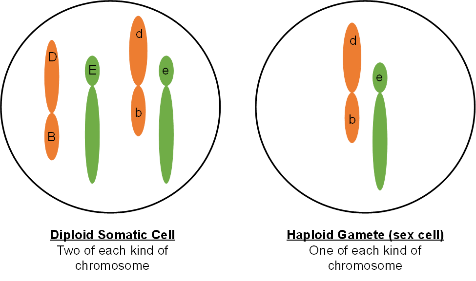
From a cytogenetics point of view, you have two types of cells in your body. You have cells with 46 chromosomes (somatic or body cells) and cells with 23 chromosomes (gamete or sex cells). Since you started as a single cell with 46 chromosomes, there must be two types of cell division taking place in your body to accommodate both somatic and gamete cells. Mitosis and meiosis are the two types of cell division.
Mitosis: Somatic cell division
The objective of mitosis is to make two genetically identical cells from a single cell. In the cells of our body, we start with 46 chromosomes in a single cell and end up with 46 chromosomes in two cells. Obviously, replicating the chromosomes is a prerequisite to mitosis. Remember, replication takes place during interphase when the chromosomes are dispersed structures in the nucleus. Mitosis is an organized procession of activity in the cell that allows the replicated chromosomes to be properly divided into two identical cells. Chromosomes are important because they contain genes. Therefore, we will include genes on our chromosome diagrams and slide show. These pictures depict the four stages of mitosis. We will describe the events at each stage that are important in understanding the distribution of genes during cell division.
Mitosis: Prophase
Prophase is the beginning of mitosis (Figure 3).
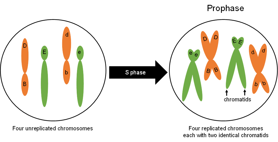
During interphase, the chromosomes look like a plate of spaghetti in the nucleus. It is difficult to pick out an individual chromosome because they are each so spread out. The chromosomes in the nucleus change from being loosely dispersed to becoming more condensed. This change in chromosome structure makes them easier to move around the cell, an important issue for what is about to happen. As the chromosomes condense, they get shorter and thicker and can be seen through the microscope as individual structures (Figure 4). The chromosomes at prophase will consist of two identical parts called sister chromatids that stay connected at the centromere. It is now clear that the chromosomes have been replicated. Chromosome replication occurs during the S-phase of interphase (Figure 1). Next, the nucleus is dissolved. At the end of prophase the replicated chromosomes are moved by the spindle apparatus to the center of the cell.
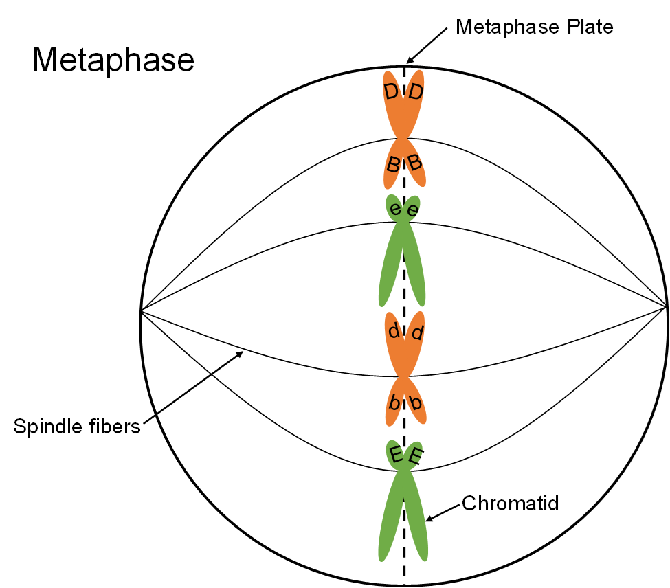
Mitosis: Metaphase
When the chromosomes are maneuvered to the center of the cell, metaphase begins (Figure 5). The spindle fiber network connects the centromere of the replicated chromosome to the outer part of each cell. The chromosomes now resemble the line of scrimmage of a football team, poised and waiting for some cellular signal to begin the next phase.
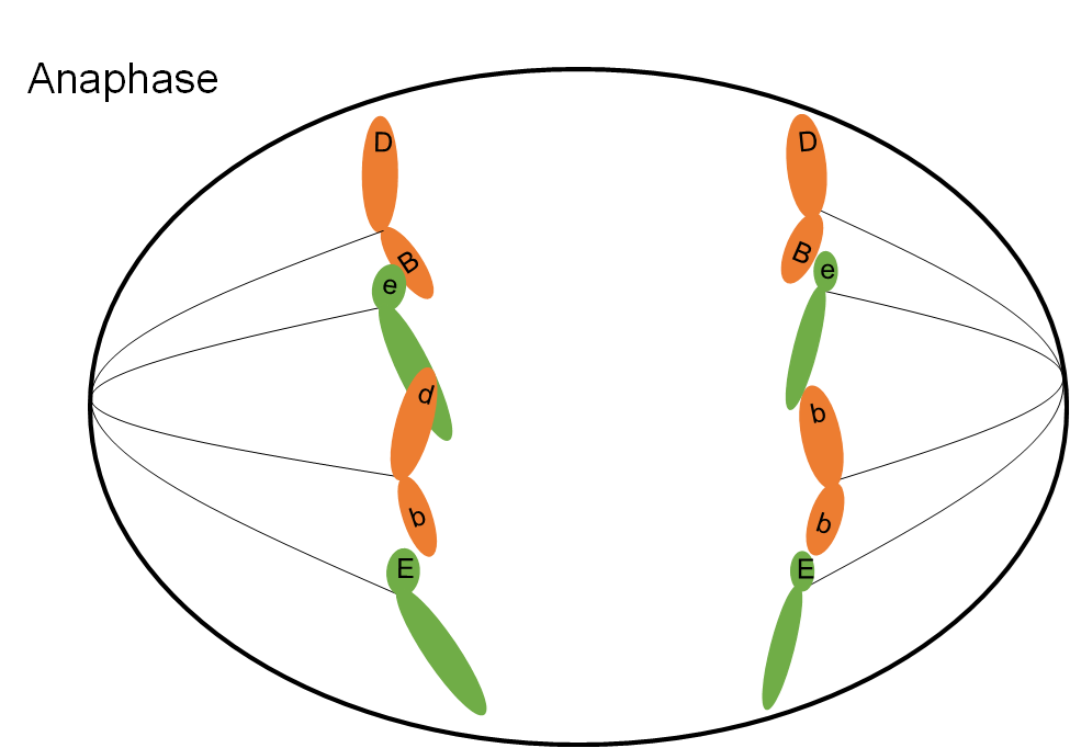
Mitosis: Anaphase
At anaphase (Figure 6) the chromatids making up each chromosome are pulled apart and begin to move away from each other under the control of the spindle fibers. Once they are pulled apart, the chromatids are now considered individual chromosomes. As anaphase progresses, the chromosomes on each end of the cell are pulled into a bundle. When they stop moving, telophase begins.
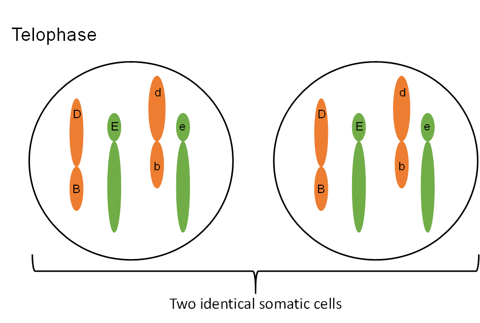
Mitosis: Telophase
The final stage of mitosis is telophase (Figure 7). A nuclear envelope will form around each bundle of chromosomes. The cell will undergo cytokinesis and the cytoplasm is split between the two identical daughter cells. Cell membranes (and cell walls in plants) form between the two cells. The chromosomes begin to decondense. They loosen up and spread around the nucleus because they no longer will need to move around. The new cells have now begun their lifecycle.
Meiosis: Gamete formation
The objective of meiosis is to make four cells from a single somatic cell. The four cells each have half the chromosome number found in the somatic cell. In our human bodies, the four gametes will each have 23 chromosomes which means the 46 chromosomes in the somatic cell must replicate during interphase prior to meiosis just as they would before mitosis. Meiosis occurs in specialized cells of the body called germline cells.
To appreciate meiosis and gamete formation it is important to first understand two ideas, chromosome sets and homologous chromosomes.
Chromosome sets: The 46 chromosomes you have consist of two sets. You are a diploid organism (‘di’ means two and ‘ploid’ means sets). One set of chromosomes came from each parent when their gametes fused. Therefore, human gametes are haploid (one set).
Homologous chromosomes: The 46 chromosomes in a somatic cell can be arranged into 23 homologous or similar pairs. One chromosome from each pair came from the male parent, the other from the female. Homologous chromosomes have the same genes although in heterozygous people the genes would be different alleles (A,a). The exception to this would be the sex (X and Y) chromosomes. Passing on a complete set of human genes requires one chromosome from each pair to end up in each gamete.
There are several key differences between meiosis and mitosis that are summarized in the following table:
|
Mitosis |
Meiosis
Chromosome number stays the same
Chromosome number is halved
One division occurs to make two cells. Four stages of this division.
Two divisions occur to make four cells. Eight stages in these divisions.
Similar or homologous chromosomes do not pair.
Homologous chromosomes pair during prophase l. Pairing is called synapsis.
Crossover exchanges between homologous chromosomes is rare.
Synapsis allows crossing over between homologous chromosomes.
Two cells made are genetically identical.
Four cells made are genetically different.
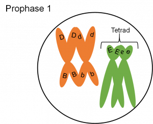
Meiosis: Prophase I
This stage starts meiosis and is the same as prophase of mitosis with one important change. As the chromosomes condense, they form groups of four chromatids called tetrads or bivalents. Close inspection reveals that each chromosome is replicated and consists of two sister chromatids. The two chromosomes in each cell that are homologous and have the same genes (but perhaps different alleles if the organism is heterozygous) will pair closely. This close association, or synapsis, allows the homologous chromosomes to crossover and exchange identical parts. The impact of crossing over is that genes that are on the same chromosome (Figure 8) can be recombined so that they are not always inherited together. The tetrad or bivalent formed during synapsis remains assembled as prophase progresses. The tetrad therefore moves as a unit to the center of the cell.
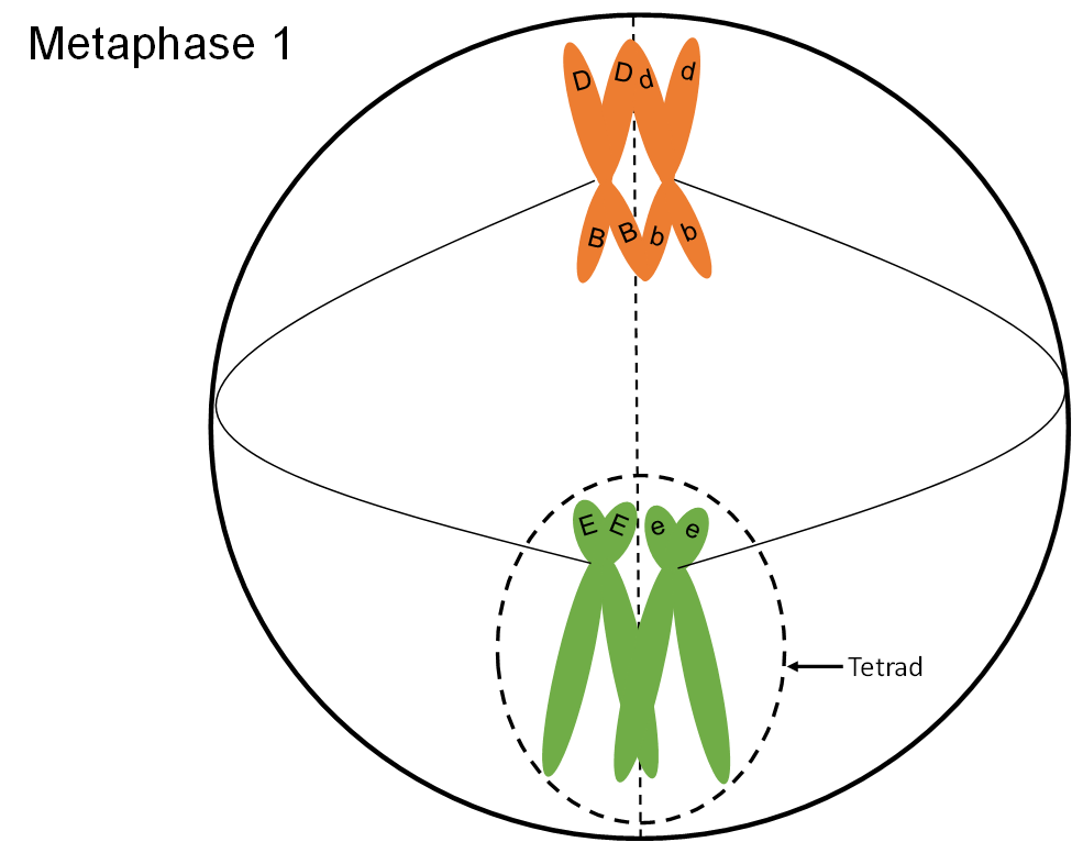
Meiosis: Metaphase I
Metaphase I starts when the tetrads are at the center of the cell (Figure 9). The tetrads have stayed together which insures that during the first division, each cell will get one chromosome from each homologous pair. The chromosomes remain at the center of the cell until the homologous pairs are ready to move away from each other.
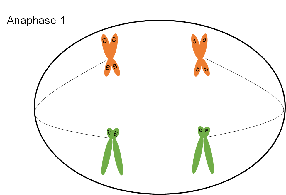
Meiosis: Anaphase I
The chromosomes that make up each tetrad separate during anaphase I (Figure 10). However, the sister chromatids will stay connected at the centromere. Anaphase I proceeds until the chromosomes are pulled into a bundle at opposite ends of the cell.
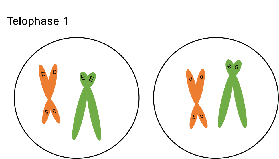
Meiosis: Telophase l
The cell divides into two cells during telophase I (Figure 11). The bundle of chromosomes may have a nuclear envelope develop around them. The germline cells in some organisms such as human females, go through the first four stages of meiosis prior to birth. The germline cells remain at telophase I for some time. The second round of division occurs when the gamete is needed for reproduction. In other situations, telophase I is an abbreviated stage, and the second round of division proceeds without delay.
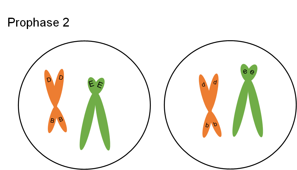
Meiosis: Prophase II
If the chromosomes became dispersed in telophase I, they will condense again at prophase II. The spindle apparatus moves the chromosomes to the middle of the cell. Look! The centromeres are still holding the sister chromatids together (Figure 12).
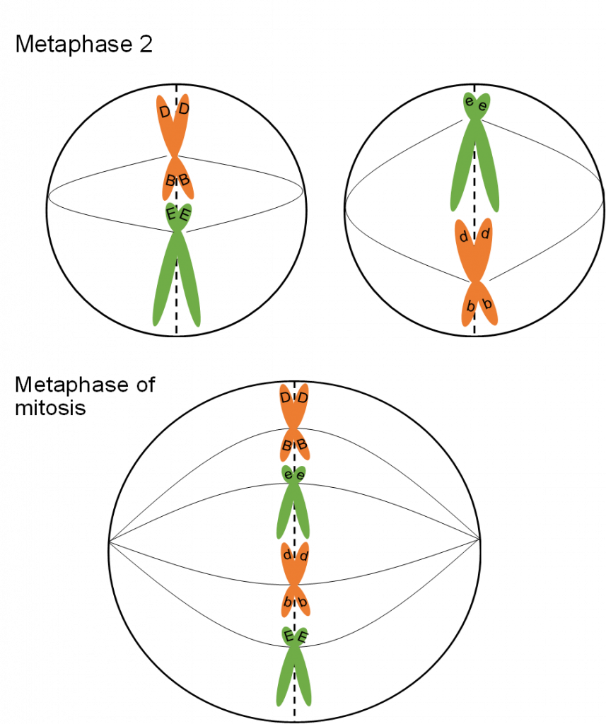
Meiosis: Metaphase II
In metaphase II the chromosomes are aligned at the center of the cell (Figure 13). This time there are not homologous chromosomes to be paired with. This metaphase looks similar to metaphase of mitosis but there is a key difference. What is the difference? (Compare Figures 5 and 13).
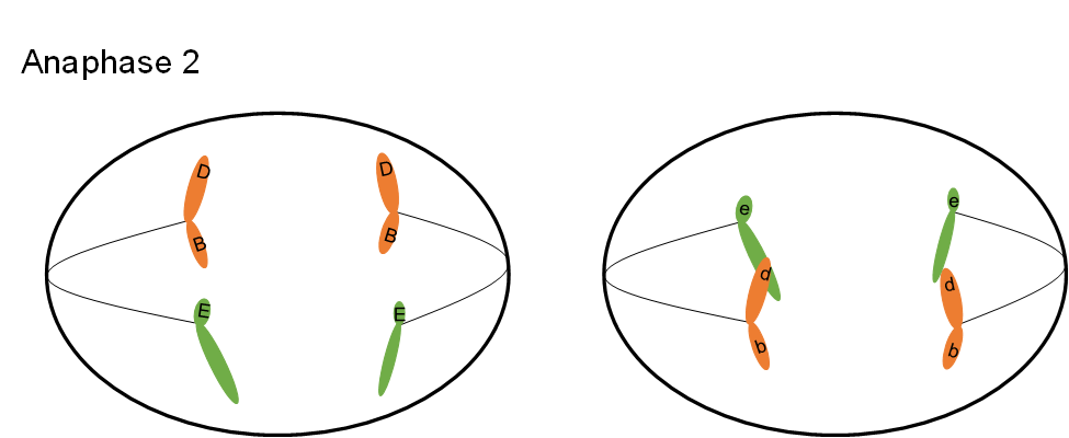
Meiosis: Anaphase II
During anaphase II, the chromatids are pulled apart by the spindle fibers. Now they are classified as chromosomes, not chromatids. The chromosomes move apart to opposite ends of the cell (Figure 14).
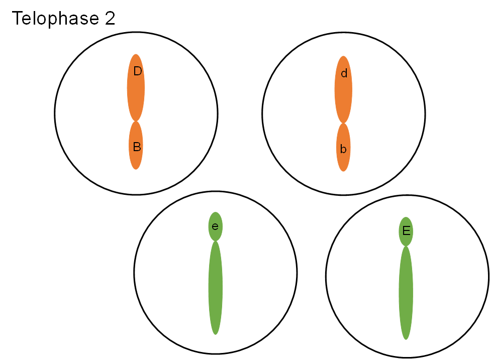
Meiosis: Telophase II
In the final stage of meiosis, telophase II, the nucleus forms around the bundle of chromosomes (Figure 15). The cell divides. Now four cells exist that originated from one germline cell. Each cell is a gamete with half the number of chromosomes and genes as a somatic cell.
Parallel Behavior and the Chromosome Theory
The successful completion of mitosis or meiosis requires the cell to move large objects with precision and control many detailed events. The process surpasses any engineering accomplishments of NASA. The importance of mitosis and meiosis to an organism is obvious when we consider that genes are a part of the chromosome and the genes must be copied and distributed properly to produce viable daughter cells. The mechanisms of these events are far from being completely understood. From our current understanding, we can appreciate how the principles of segregation and independent assortment are controlled by the mechanics of meiosis.
When cell division was first observed and described by cytogeneticists, biologists were just beginning to accept the idea that genes were tiny objects that controlled traits and existed in the cells of living things. Two biologists, a German named Boveri and an American graduate student named Sutton, recognized that chromosome behavior during meiosis matched Mendel’s principles of gene behavior. Both scientists proposed the idea that while genes had not yet been directly observed, they must be a part of the chromosome. Sutton and Boveri are both given credit for proposing this chromosome theory; genes are a part of chromosomes.
Segregation predicts gene behavior that matches the chromosome behavior observed by cytogeneticists. Genes are in pairs because chromosomes are in pairs. The gene pairs associate during gamete formation when the homologous chromosomes pair in prophase I. The gene pairs separate when the homologous pairs separate in anaphase I followed by chromatid separation in anaphase II. Thus, chromosome behavior dictates the gene’s segregation behavior.
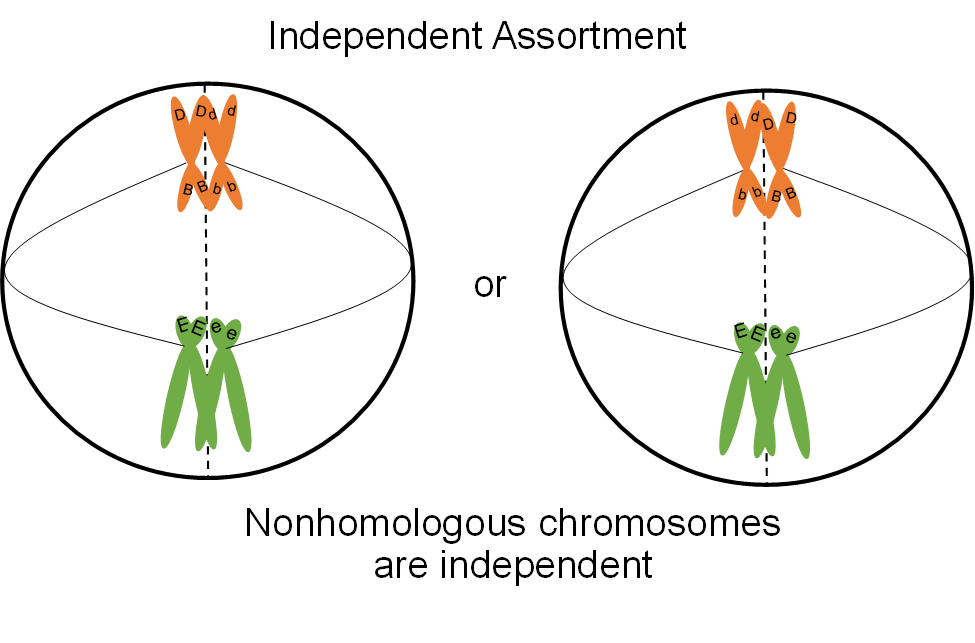
Independent assortment of gene pairs also correlates with chromosome behavior. Let us consider a dihybrid individual with the genotype BbEe (Figure 16). How many kinds of gametes can it make? The four possible combinations (BE, bE, Be and be) are made at equal frequencies. We can understand why this occurs if we think about what happens to chromosomes at metaphase I of meiosis. The chromosome pair that carries the ‘E’ and ‘e’ genes will be moved independently from the chromosome pair with the ‘B’ and ‘b’ chromosomes. When the chromosomes arrive at the center of the cell at metaphase I, the two tetrads may be aligned so that the ‘E’ and ‘B’ genes move to one cell and the ‘e’ and ‘b’ genes move to the other during the first division. In the other half of the cells that go through meiosis, the tetrads will line up so that the ‘E’ genes will be passed on with the ‘b’ genes and the ‘e’ genes will go with the ‘B’ genes. Keep in mind that organisms that make gametes make thousands of them. The chance alignment of the tetrads at metaphase I therefore dictates the overall frequency of gametes with different combinations of genes when the genes are on separate chromosomes.
When more gene pairs are considered, the same scenarios described above will be true as long as the genes are on separate chromosomes. When genes are on the same chromosome, the role of crossing over on gene inheritance needs to be considered in more detail. We will cover the inheritance of genes on the same chromosome in a later lesson.
- Watch this video about Mitosis from Daily Med Ed
- Watch this video about Meiosis from Daily Med Ed