
19: Acid-Fast Stain
- Page ID
- 3626

\( \newcommand{\vecs}[1]{\overset { \scriptstyle \rightharpoonup} {\mathbf{#1}} } \)
\( \newcommand{\vecd}[1]{\overset{-\!-\!\rightharpoonup}{\vphantom{a}\smash {#1}}} \)
\( \newcommand{\dsum}{\displaystyle\sum\limits} \)
\( \newcommand{\dint}{\displaystyle\int\limits} \)
\( \newcommand{\dlim}{\displaystyle\lim\limits} \)
\( \newcommand{\id}{\mathrm{id}}\) \( \newcommand{\Span}{\mathrm{span}}\)
( \newcommand{\kernel}{\mathrm{null}\,}\) \( \newcommand{\range}{\mathrm{range}\,}\)
\( \newcommand{\RealPart}{\mathrm{Re}}\) \( \newcommand{\ImaginaryPart}{\mathrm{Im}}\)
\( \newcommand{\Argument}{\mathrm{Arg}}\) \( \newcommand{\norm}[1]{\| #1 \|}\)
\( \newcommand{\inner}[2]{\langle #1, #2 \rangle}\)
\( \newcommand{\Span}{\mathrm{span}}\)
\( \newcommand{\id}{\mathrm{id}}\)
\( \newcommand{\Span}{\mathrm{span}}\)
\( \newcommand{\kernel}{\mathrm{null}\,}\)
\( \newcommand{\range}{\mathrm{range}\,}\)
\( \newcommand{\RealPart}{\mathrm{Re}}\)
\( \newcommand{\ImaginaryPart}{\mathrm{Im}}\)
\( \newcommand{\Argument}{\mathrm{Arg}}\)
\( \newcommand{\norm}[1]{\| #1 \|}\)
\( \newcommand{\inner}[2]{\langle #1, #2 \rangle}\)
\( \newcommand{\Span}{\mathrm{span}}\) \( \newcommand{\AA}{\unicode[.8,0]{x212B}}\)
\( \newcommand{\vectorA}[1]{\vec{#1}} % arrow\)
\( \newcommand{\vectorAt}[1]{\vec{\text{#1}}} % arrow\)
\( \newcommand{\vectorB}[1]{\overset { \scriptstyle \rightharpoonup} {\mathbf{#1}} } \)
\( \newcommand{\vectorC}[1]{\textbf{#1}} \)
\( \newcommand{\vectorD}[1]{\overrightarrow{#1}} \)
\( \newcommand{\vectorDt}[1]{\overrightarrow{\text{#1}}} \)
\( \newcommand{\vectE}[1]{\overset{-\!-\!\rightharpoonup}{\vphantom{a}\smash{\mathbf {#1}}}} \)
\( \newcommand{\vecs}[1]{\overset { \scriptstyle \rightharpoonup} {\mathbf{#1}} } \)
\(\newcommand{\longvect}{\overrightarrow}\)
\( \newcommand{\vecd}[1]{\overset{-\!-\!\rightharpoonup}{\vphantom{a}\smash {#1}}} \)
\(\newcommand{\avec}{\mathbf a}\) \(\newcommand{\bvec}{\mathbf b}\) \(\newcommand{\cvec}{\mathbf c}\) \(\newcommand{\dvec}{\mathbf d}\) \(\newcommand{\dtil}{\widetilde{\mathbf d}}\) \(\newcommand{\evec}{\mathbf e}\) \(\newcommand{\fvec}{\mathbf f}\) \(\newcommand{\nvec}{\mathbf n}\) \(\newcommand{\pvec}{\mathbf p}\) \(\newcommand{\qvec}{\mathbf q}\) \(\newcommand{\svec}{\mathbf s}\) \(\newcommand{\tvec}{\mathbf t}\) \(\newcommand{\uvec}{\mathbf u}\) \(\newcommand{\vvec}{\mathbf v}\) \(\newcommand{\wvec}{\mathbf w}\) \(\newcommand{\xvec}{\mathbf x}\) \(\newcommand{\yvec}{\mathbf y}\) \(\newcommand{\zvec}{\mathbf z}\) \(\newcommand{\rvec}{\mathbf r}\) \(\newcommand{\mvec}{\mathbf m}\) \(\newcommand{\zerovec}{\mathbf 0}\) \(\newcommand{\onevec}{\mathbf 1}\) \(\newcommand{\real}{\mathbb R}\) \(\newcommand{\twovec}[2]{\left[\begin{array}{r}#1 \\ #2 \end{array}\right]}\) \(\newcommand{\ctwovec}[2]{\left[\begin{array}{c}#1 \\ #2 \end{array}\right]}\) \(\newcommand{\threevec}[3]{\left[\begin{array}{r}#1 \\ #2 \\ #3 \end{array}\right]}\) \(\newcommand{\cthreevec}[3]{\left[\begin{array}{c}#1 \\ #2 \\ #3 \end{array}\right]}\) \(\newcommand{\fourvec}[4]{\left[\begin{array}{r}#1 \\ #2 \\ #3 \\ #4 \end{array}\right]}\) \(\newcommand{\cfourvec}[4]{\left[\begin{array}{c}#1 \\ #2 \\ #3 \\ #4 \end{array}\right]}\) \(\newcommand{\fivevec}[5]{\left[\begin{array}{r}#1 \\ #2 \\ #3 \\ #4 \\ #5 \\ \end{array}\right]}\) \(\newcommand{\cfivevec}[5]{\left[\begin{array}{c}#1 \\ #2 \\ #3 \\ #4 \\ #5 \\ \end{array}\right]}\) \(\newcommand{\mattwo}[4]{\left[\begin{array}{rr}#1 \amp #2 \\ #3 \amp #4 \\ \end{array}\right]}\) \(\newcommand{\laspan}[1]{\text{Span}\{#1\}}\) \(\newcommand{\bcal}{\cal B}\) \(\newcommand{\ccal}{\cal C}\) \(\newcommand{\scal}{\cal S}\) \(\newcommand{\wcal}{\cal W}\) \(\newcommand{\ecal}{\cal E}\) \(\newcommand{\coords}[2]{\left\{#1\right\}_{#2}}\) \(\newcommand{\gray}[1]{\color{gray}{#1}}\) \(\newcommand{\lgray}[1]{\color{lightgray}{#1}}\) \(\newcommand{\rank}{\operatorname{rank}}\) \(\newcommand{\row}{\text{Row}}\) \(\newcommand{\col}{\text{Col}}\) \(\renewcommand{\row}{\text{Row}}\) \(\newcommand{\nul}{\text{Nul}}\) \(\newcommand{\var}{\text{Var}}\) \(\newcommand{\corr}{\text{corr}}\) \(\newcommand{\len}[1]{\left|#1\right|}\) \(\newcommand{\bbar}{\overline{\bvec}}\) \(\newcommand{\bhat}{\widehat{\bvec}}\) \(\newcommand{\bperp}{\bvec^\perp}\) \(\newcommand{\xhat}{\widehat{\xvec}}\) \(\newcommand{\vhat}{\widehat{\vvec}}\) \(\newcommand{\uhat}{\widehat{\uvec}}\) \(\newcommand{\what}{\widehat{\wvec}}\) \(\newcommand{\Sighat}{\widehat{\Sigma}}\) \(\newcommand{\lt}{<}\) \(\newcommand{\gt}{>}\) \(\newcommand{\amp}{&}\) \(\definecolor{fillinmathshade}{gray}{0.9}\)Objectives
- Learn to perform the acid-fast stain.
- Differentiate between acid-fast and nonacid-fast bacteria.
Robert Koch was the first person to isolate and identify Mycobacterium tuberculosis from a patient with tuberculosis. He developed a stain for the bacterium, although it was not very effective for visualizing this slender bacillus. Paul Ehrlich is the first to describe the acid-fast properties of the bacterium. In the 1890s, Friedrich Neelsen and Franz Ziehl modified the stain by adding phenol (carbolic acid) and basic fuschin. The name of the dye carbol fuschin comes from the phenol and basic fuschin ingredients of the stain.
The ability of the bacteria to resist decolorization with ACID alcohol confers acid fastness to the bacterium. Acid-fast bacteria, of which there are very few---the major genus Mycobacterium, have a high concentration of mycolic acid, a lipid, in their walls. Although difficult to stain, once the stain goes into the wall, the cell will not de-stain or decolorize easily. The ability of the bacteria to resist decolorization with acid (1%) alcohol confers acid -fastness to the bacterium. It is thought that the phenol in the carbon fuschin facilitates the dye going into the waxy wall of the bacterium. Although gram positive, acid-fast bacteria do not take the crystal violet into the wall well, appearing very light purple rather than the deep purple of normal gram positive bacteria (the waxy lipid in the acid-fast wall repels the aqueous crystal violet stain).
As in the spore stain, steam is used as a gimmick to get the carbol fuschin primary dye to go into the wall. Once in, it will not come out: But the acid alcohol decolorizer WILL take it out of the nonacid-fast wall since the primary dye does not bind strongly to the cell wall. Nonacid-fast bacteria will also take up the carbol fuschin, but the acid alcohol decolorizer will remove it from wall since the primary dye does not bind strongly to the cell wall.
Acid-fastness is an uncommon characteristic shared by the genera Mycobacterium and Nocardia (weakly acid-fast). Because of this feature, this stain is extremely helpful in identification in diseases caused by acid-fast bacteria, particularly tuberculosis and leprosy. In addition, the stain is used to determine the presence of AF bacteria from lung tissue in patients undergoing antibiotic therapy.


Acid-fast bacteria Nonacid-fast bacteria
MATERIALS NEEDED
- dye kit
- stain rack
- hot plate
- beaker
- paper towel (cut the size of the slide)
- cultures: Mycobacterium and E. coli
THE PROCEDURE (Ziehl-Neelsen method)
- Prepare the bacterial smears---air-dry, and heat-fix. (or the Mycobacterium smear may be given to you already air-dried on a slide).
- Put a beaker of water on the hot plate and boil until steam is coming up from the water. Then turn the hot plate down so that the water is barely boiling.
- Place the wire stain rack over the beaker which now has steam coming up from the boiled water.
- Cut a small piece of paper towel and place it on top of the smear on the slide. The towel will keep the dye from evaporating too quickly, thereby giving more contact time between the dye and the bacterial walls.
- Flood the smear with the primary dye, carbol fuschin, and leave for 5 minutes. Keep the paper towel moist with the carbol fuschin. DO NOT let the dye dry on the towel.
- Remove the piece of paper towel and discard in the garbage. Wash the slide really WELL with water.
- Add the decolorizer acid alcohol (1% HCl + ethanol) and decolorize for 15-20 seconds. (The slide can be held, titled at an angle, and decolorizer dripped down the slide).
- Wash WELL with water.
- Flood the smear with the counterstain dye, methylene blue, and leave for 1 minute.
- Rinse well with water. Blot dry with bibulous paper.
Note: Alternate cold method acid fast stain (Kinyoun)
The only difference is that without heat you have to stain the smear with carbol fuschin for 10 minutes.
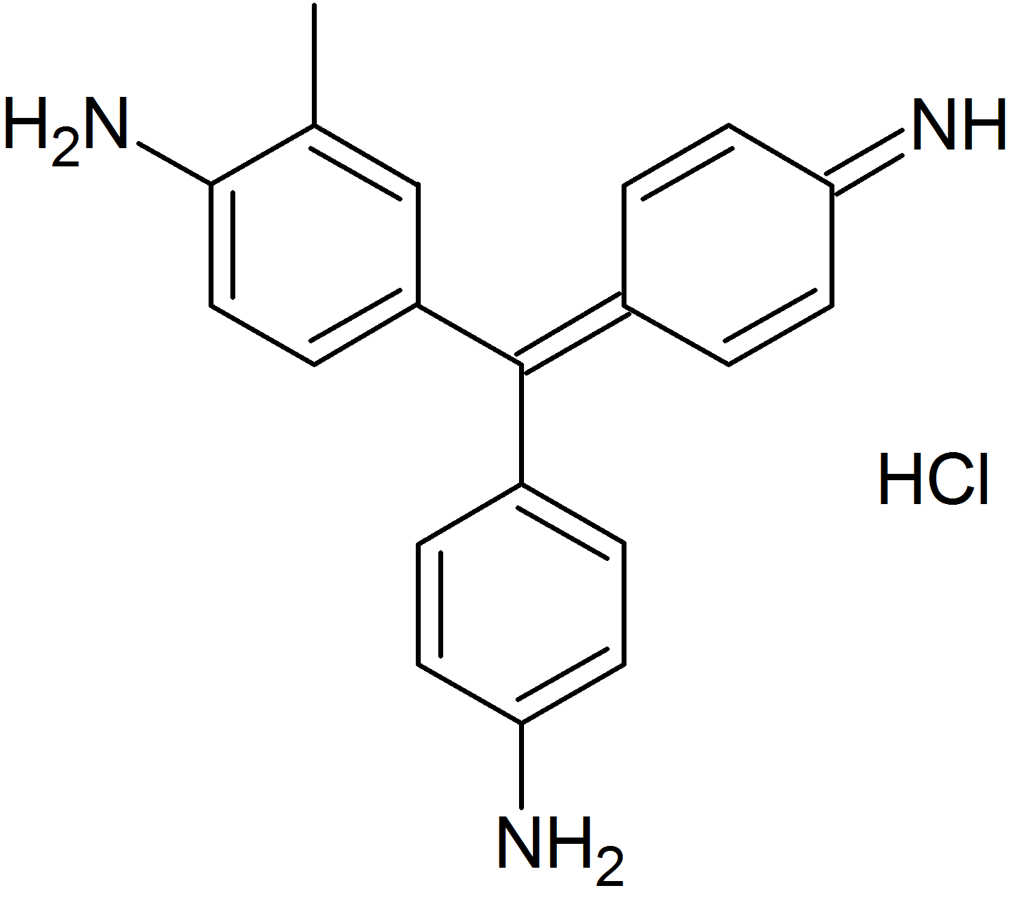
Molecular structure of carbol fuschin
- Interpret the results using the protocol below.
Interpretation
- Acid-fast bacteria are hot pink or fuschia.
- Nonacid-fast bacteria are light blue.
QUESTIONS
- What is chemically unique about the Mycobacterium genus that causes it to be acid-fast?
- How is this stain procedure similar to the spore stain procedure?
Contributors and Attributions
Jackie Reynolds, Professor of Biology (Richland College)