
16.4: Materials and Procedures
- Page ID
- 40256

\( \newcommand{\vecs}[1]{\overset { \scriptstyle \rightharpoonup} {\mathbf{#1}} } \)
\( \newcommand{\vecd}[1]{\overset{-\!-\!\rightharpoonup}{\vphantom{a}\smash {#1}}} \)
\( \newcommand{\dsum}{\displaystyle\sum\limits} \)
\( \newcommand{\dint}{\displaystyle\int\limits} \)
\( \newcommand{\dlim}{\displaystyle\lim\limits} \)
\( \newcommand{\id}{\mathrm{id}}\) \( \newcommand{\Span}{\mathrm{span}}\)
( \newcommand{\kernel}{\mathrm{null}\,}\) \( \newcommand{\range}{\mathrm{range}\,}\)
\( \newcommand{\RealPart}{\mathrm{Re}}\) \( \newcommand{\ImaginaryPart}{\mathrm{Im}}\)
\( \newcommand{\Argument}{\mathrm{Arg}}\) \( \newcommand{\norm}[1]{\| #1 \|}\)
\( \newcommand{\inner}[2]{\langle #1, #2 \rangle}\)
\( \newcommand{\Span}{\mathrm{span}}\)
\( \newcommand{\id}{\mathrm{id}}\)
\( \newcommand{\Span}{\mathrm{span}}\)
\( \newcommand{\kernel}{\mathrm{null}\,}\)
\( \newcommand{\range}{\mathrm{range}\,}\)
\( \newcommand{\RealPart}{\mathrm{Re}}\)
\( \newcommand{\ImaginaryPart}{\mathrm{Im}}\)
\( \newcommand{\Argument}{\mathrm{Arg}}\)
\( \newcommand{\norm}[1]{\| #1 \|}\)
\( \newcommand{\inner}[2]{\langle #1, #2 \rangle}\)
\( \newcommand{\Span}{\mathrm{span}}\) \( \newcommand{\AA}{\unicode[.8,0]{x212B}}\)
\( \newcommand{\vectorA}[1]{\vec{#1}} % arrow\)
\( \newcommand{\vectorAt}[1]{\vec{\text{#1}}} % arrow\)
\( \newcommand{\vectorB}[1]{\overset { \scriptstyle \rightharpoonup} {\mathbf{#1}} } \)
\( \newcommand{\vectorC}[1]{\textbf{#1}} \)
\( \newcommand{\vectorD}[1]{\overrightarrow{#1}} \)
\( \newcommand{\vectorDt}[1]{\overrightarrow{\text{#1}}} \)
\( \newcommand{\vectE}[1]{\overset{-\!-\!\rightharpoonup}{\vphantom{a}\smash{\mathbf {#1}}}} \)
\( \newcommand{\vecs}[1]{\overset { \scriptstyle \rightharpoonup} {\mathbf{#1}} } \)
\(\newcommand{\longvect}{\overrightarrow}\)
\( \newcommand{\vecd}[1]{\overset{-\!-\!\rightharpoonup}{\vphantom{a}\smash {#1}}} \)
\(\newcommand{\avec}{\mathbf a}\) \(\newcommand{\bvec}{\mathbf b}\) \(\newcommand{\cvec}{\mathbf c}\) \(\newcommand{\dvec}{\mathbf d}\) \(\newcommand{\dtil}{\widetilde{\mathbf d}}\) \(\newcommand{\evec}{\mathbf e}\) \(\newcommand{\fvec}{\mathbf f}\) \(\newcommand{\nvec}{\mathbf n}\) \(\newcommand{\pvec}{\mathbf p}\) \(\newcommand{\qvec}{\mathbf q}\) \(\newcommand{\svec}{\mathbf s}\) \(\newcommand{\tvec}{\mathbf t}\) \(\newcommand{\uvec}{\mathbf u}\) \(\newcommand{\vvec}{\mathbf v}\) \(\newcommand{\wvec}{\mathbf w}\) \(\newcommand{\xvec}{\mathbf x}\) \(\newcommand{\yvec}{\mathbf y}\) \(\newcommand{\zvec}{\mathbf z}\) \(\newcommand{\rvec}{\mathbf r}\) \(\newcommand{\mvec}{\mathbf m}\) \(\newcommand{\zerovec}{\mathbf 0}\) \(\newcommand{\onevec}{\mathbf 1}\) \(\newcommand{\real}{\mathbb R}\) \(\newcommand{\twovec}[2]{\left[\begin{array}{r}#1 \\ #2 \end{array}\right]}\) \(\newcommand{\ctwovec}[2]{\left[\begin{array}{c}#1 \\ #2 \end{array}\right]}\) \(\newcommand{\threevec}[3]{\left[\begin{array}{r}#1 \\ #2 \\ #3 \end{array}\right]}\) \(\newcommand{\cthreevec}[3]{\left[\begin{array}{c}#1 \\ #2 \\ #3 \end{array}\right]}\) \(\newcommand{\fourvec}[4]{\left[\begin{array}{r}#1 \\ #2 \\ #3 \\ #4 \end{array}\right]}\) \(\newcommand{\cfourvec}[4]{\left[\begin{array}{c}#1 \\ #2 \\ #3 \\ #4 \end{array}\right]}\) \(\newcommand{\fivevec}[5]{\left[\begin{array}{r}#1 \\ #2 \\ #3 \\ #4 \\ #5 \\ \end{array}\right]}\) \(\newcommand{\cfivevec}[5]{\left[\begin{array}{c}#1 \\ #2 \\ #3 \\ #4 \\ #5 \\ \end{array}\right]}\) \(\newcommand{\mattwo}[4]{\left[\begin{array}{rr}#1 \amp #2 \\ #3 \amp #4 \\ \end{array}\right]}\) \(\newcommand{\laspan}[1]{\text{Span}\{#1\}}\) \(\newcommand{\bcal}{\cal B}\) \(\newcommand{\ccal}{\cal C}\) \(\newcommand{\scal}{\cal S}\) \(\newcommand{\wcal}{\cal W}\) \(\newcommand{\ecal}{\cal E}\) \(\newcommand{\coords}[2]{\left\{#1\right\}_{#2}}\) \(\newcommand{\gray}[1]{\color{gray}{#1}}\) \(\newcommand{\lgray}[1]{\color{lightgray}{#1}}\) \(\newcommand{\rank}{\operatorname{rank}}\) \(\newcommand{\row}{\text{Row}}\) \(\newcommand{\col}{\text{Col}}\) \(\renewcommand{\row}{\text{Row}}\) \(\newcommand{\nul}{\text{Nul}}\) \(\newcommand{\var}{\text{Var}}\) \(\newcommand{\corr}{\text{corr}}\) \(\newcommand{\len}[1]{\left|#1\right|}\) \(\newcommand{\bbar}{\overline{\bvec}}\) \(\newcommand{\bhat}{\widehat{\bvec}}\) \(\newcommand{\bperp}{\bvec^\perp}\) \(\newcommand{\xhat}{\widehat{\xvec}}\) \(\newcommand{\vhat}{\widehat{\vvec}}\) \(\newcommand{\uhat}{\widehat{\uvec}}\) \(\newcommand{\what}{\widehat{\wvec}}\) \(\newcommand{\Sighat}{\widehat{\Sigma}}\) \(\newcommand{\lt}{<}\) \(\newcommand{\gt}{>}\) \(\newcommand{\amp}{&}\) \(\definecolor{fillinmathshade}{gray}{0.9}\)Materials
- Day 1-
- 200 ul micropipette (P200)
- 1000 ul micropipette (P1000)
- Pipette tips
- Microfuge tubes
- E. coli culture (HB101 strain)
- Ice bath
- 42C water bath
- 37C water bath
- 50mM CaCl2 (keep ice-cold)
- 10 ul pGLO (plasmid concentration 0.2 µg/ µl)
- Sterile LB broth
- 1 LB agar plates
- 2 LB+AMP
- 1 LB+AMP+Arabinose agar plates
- 2 Sterile Spreaders-small
- Day 2-
- Your inoculated plates from Day 1
- UV lights
Safety Note
Wear gloves while handling the transformed bacteria. You are genetically engineering an antibiotic resistant bacterium.
Procedures
- Label two microfuge tubes with the following: one as P+ (plasmid) and the other, P- (no plasmid, control).
- Using a P1000 micropipette, add 500 ul of ice-cold 50mM CaCl₂ to one of the tubes; you will split this volume into both tubes later so it doesn’t matter which tube you start with.
- Using a sterile inoculating loop, transfer one to three isolated colonies of the starter E.coli colonies (a colony approximately the size of an o) into the tube.
- Using a P200 micropipette and a clean pipette tip, suspend the cells by gently pipetting the solution in an out several times. Hold the tube up to the light to check for any cell clumps.
- Now you will split this volume, half in each tube. Using a P1000 micropipette, remove 250 ul of the suspension and place it in the second tube so that you now have 250 ul of the suspension in both the P+ and P- tubes.
- Replace the cap on the tubes and return them to the ice bath.
- Incubate both tubes (P+ and P-) in ice for at least one minute.
- Add 10 ul of pGLO (plasmid concentration 0.2ug/ ul) to the re-suspended cells in the P+ tube only. Mix the cells with the plasmid by gently and slowly pumping the solution in and out using a clean pipette tip. Return the P+ tube to the ice bath.
- Incubate both tubes in ice for 20 minutes.
- While the tubes are incubating obtain 1 LB, 2 LB+AMP, and 1 LB+AMP+Arabinose agar plates and label them:
- Label the bottom of the plates of one set LB and LB+AMP with P-
- Label the bottom of the plates of the other set LB+AMP and LB+AMP+Arabinose with P+
- Following the 20-minute incubation in ice, carry your ice bath with the cells to the 42C water bath. Take the tubes directly from the ice bath and place them in the 42C water bath for 90 seconds to heat-shock the cells. Immediately afterward place the cells back into the ice bath for 2 more minutes.
- Using a P1000 micropipette, and a clean pipette tip add 500 ul of sterile LB to the P+ and then with a new pipette tip, add 500ul of sterile LB to the P- tube. Gently, finger vortex (flick the tube with your finger) the tubes to mix the cells and broth together. Incubate the cells in the 37C water bath for 10 minutes.
- After the 37C incubation period the cells are ready to be plated out onto the agars.
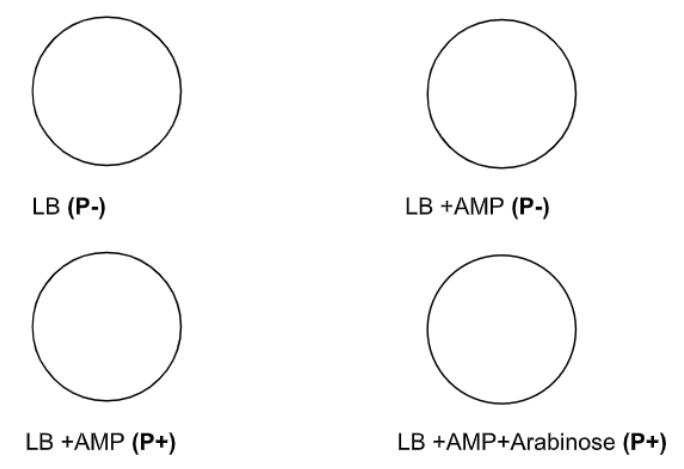
- Line up the two P- plates as shown in the above picture. Using a P200 micropipette, transfer 50 ul of cells from the P- tube to each of the two plates. Make sure to evenly spread the drops across the agar surface
- Use a sterile cell spreader and rotate the plates to evenly distribute the culture across the agar. Place the contaminated cell spreaders in the pipette disposal trays.
- Using a new micropipette tips and a sterile cell spreader, repeat the above procedure for the P+ plates by inoculating with the cells from the P+ tube.
- Allow plates to dry, right side up, for about 5 minutes. Then place inverted plates in your incubator. Discard tubes in the biohazard containers.
Results
- After incubation, count and record the number of colonies on each plate.
- While wearing safety goggles and in a dark place, illuminate the plates with the hand held UV light to determine which colonies are producing the Green Fluorescent Protein. Record results.
|
CELLS |
PLATE |
Growth? Y/N |
Glowing? Y/N |
AGAR PLATE Growth? |
Growth? Y/N |
Glowing? Y/N |
|---|---|---|---|---|---|---|
|
Plasmid - (P-) |
LB # of colonies: |
|
|
LB +Amp # of colonies: |
|
|
|
Plasmid + (P+) |
LB +AMP # of colonies: |
|
|
LB +Amp + Arabinose # of colonies: |
|
|
- Transformation Efficiency (TE): Show all your work. Include word equations, show substitutions, and include units!
- What was the total mass (µg) of plasmid DNA added to the cell suspensions? Note in the experiment you used 10 µl of plasmid (step 8) that had a concentration of 0.2 µg /ul.
- Using the information from a) and your colony count of transformed cells, calculate the number of colonies transformed per microgram of plasmid DNA. This is the Transformation Efficiency of your transformation event.\[TE=\dfrac{\# \text{ of transformed colonies}}{\mu g \text{DNA}} \nonumber \]
Contributors and Attributions
Kelly C. Burke (College of the Canyons)