

17.2: Oxidation of Fatty Acids
- Page ID
- 15026


\( \newcommand{\vecs}[1]{\overset { \scriptstyle \rightharpoonup} {\mathbf{#1}} } \)
\( \newcommand{\vecd}[1]{\overset{-\!-\!\rightharpoonup}{\vphantom{a}\smash {#1}}} \)
\( \newcommand{\id}{\mathrm{id}}\) \( \newcommand{\Span}{\mathrm{span}}\)
( \newcommand{\kernel}{\mathrm{null}\,}\) \( \newcommand{\range}{\mathrm{range}\,}\)
\( \newcommand{\RealPart}{\mathrm{Re}}\) \( \newcommand{\ImaginaryPart}{\mathrm{Im}}\)
\( \newcommand{\Argument}{\mathrm{Arg}}\) \( \newcommand{\norm}[1]{\| #1 \|}\)
\( \newcommand{\inner}[2]{\langle #1, #2 \rangle}\)
\( \newcommand{\Span}{\mathrm{span}}\)
\( \newcommand{\id}{\mathrm{id}}\)
\( \newcommand{\Span}{\mathrm{span}}\)
\( \newcommand{\kernel}{\mathrm{null}\,}\)
\( \newcommand{\range}{\mathrm{range}\,}\)
\( \newcommand{\RealPart}{\mathrm{Re}}\)
\( \newcommand{\ImaginaryPart}{\mathrm{Im}}\)
\( \newcommand{\Argument}{\mathrm{Arg}}\)
\( \newcommand{\norm}[1]{\| #1 \|}\)
\( \newcommand{\inner}[2]{\langle #1, #2 \rangle}\)
\( \newcommand{\Span}{\mathrm{span}}\) \( \newcommand{\AA}{\unicode[.8,0]{x212B}}\)
\( \newcommand{\vectorA}[1]{\vec{#1}} % arrow\)
\( \newcommand{\vectorAt}[1]{\vec{\text{#1}}} % arrow\)
\( \newcommand{\vectorB}[1]{\overset { \scriptstyle \rightharpoonup} {\mathbf{#1}} } \)
\( \newcommand{\vectorC}[1]{\textbf{#1}} \)
\( \newcommand{\vectorD}[1]{\overrightarrow{#1}} \)
\( \newcommand{\vectorDt}[1]{\overrightarrow{\text{#1}}} \)
\( \newcommand{\vectE}[1]{\overset{-\!-\!\rightharpoonup}{\vphantom{a}\smash{\mathbf {#1}}}} \)
\( \newcommand{\vecs}[1]{\overset { \scriptstyle \rightharpoonup} {\mathbf{#1}} } \)
\( \newcommand{\vecd}[1]{\overset{-\!-\!\rightharpoonup}{\vphantom{a}\smash {#1}}} \)
\(\newcommand{\avec}{\mathbf a}\) \(\newcommand{\bvec}{\mathbf b}\) \(\newcommand{\cvec}{\mathbf c}\) \(\newcommand{\dvec}{\mathbf d}\) \(\newcommand{\dtil}{\widetilde{\mathbf d}}\) \(\newcommand{\evec}{\mathbf e}\) \(\newcommand{\fvec}{\mathbf f}\) \(\newcommand{\nvec}{\mathbf n}\) \(\newcommand{\pvec}{\mathbf p}\) \(\newcommand{\qvec}{\mathbf q}\) \(\newcommand{\svec}{\mathbf s}\) \(\newcommand{\tvec}{\mathbf t}\) \(\newcommand{\uvec}{\mathbf u}\) \(\newcommand{\vvec}{\mathbf v}\) \(\newcommand{\wvec}{\mathbf w}\) \(\newcommand{\xvec}{\mathbf x}\) \(\newcommand{\yvec}{\mathbf y}\) \(\newcommand{\zvec}{\mathbf z}\) \(\newcommand{\rvec}{\mathbf r}\) \(\newcommand{\mvec}{\mathbf m}\) \(\newcommand{\zerovec}{\mathbf 0}\) \(\newcommand{\onevec}{\mathbf 1}\) \(\newcommand{\real}{\mathbb R}\) \(\newcommand{\twovec}[2]{\left[\begin{array}{r}#1 \\ #2 \end{array}\right]}\) \(\newcommand{\ctwovec}[2]{\left[\begin{array}{c}#1 \\ #2 \end{array}\right]}\) \(\newcommand{\threevec}[3]{\left[\begin{array}{r}#1 \\ #2 \\ #3 \end{array}\right]}\) \(\newcommand{\cthreevec}[3]{\left[\begin{array}{c}#1 \\ #2 \\ #3 \end{array}\right]}\) \(\newcommand{\fourvec}[4]{\left[\begin{array}{r}#1 \\ #2 \\ #3 \\ #4 \end{array}\right]}\) \(\newcommand{\cfourvec}[4]{\left[\begin{array}{c}#1 \\ #2 \\ #3 \\ #4 \end{array}\right]}\) \(\newcommand{\fivevec}[5]{\left[\begin{array}{r}#1 \\ #2 \\ #3 \\ #4 \\ #5 \\ \end{array}\right]}\) \(\newcommand{\cfivevec}[5]{\left[\begin{array}{c}#1 \\ #2 \\ #3 \\ #4 \\ #5 \\ \end{array}\right]}\) \(\newcommand{\mattwo}[4]{\left[\begin{array}{rr}#1 \amp #2 \\ #3 \amp #4 \\ \end{array}\right]}\) \(\newcommand{\laspan}[1]{\text{Span}\{#1\}}\) \(\newcommand{\bcal}{\cal B}\) \(\newcommand{\ccal}{\cal C}\) \(\newcommand{\scal}{\cal S}\) \(\newcommand{\wcal}{\cal W}\) \(\newcommand{\ecal}{\cal E}\) \(\newcommand{\coords}[2]{\left\{#1\right\}_{#2}}\) \(\newcommand{\gray}[1]{\color{gray}{#1}}\) \(\newcommand{\lgray}[1]{\color{lightgray}{#1}}\) \(\newcommand{\rank}{\operatorname{rank}}\) \(\newcommand{\row}{\text{Row}}\) \(\newcommand{\col}{\text{Col}}\) \(\renewcommand{\row}{\text{Row}}\) \(\newcommand{\nul}{\text{Nul}}\) \(\newcommand{\var}{\text{Var}}\) \(\newcommand{\corr}{\text{corr}}\) \(\newcommand{\len}[1]{\left|#1\right|}\) \(\newcommand{\bbar}{\overline{\bvec}}\) \(\newcommand{\bhat}{\widehat{\bvec}}\) \(\newcommand{\bperp}{\bvec^\perp}\) \(\newcommand{\xhat}{\widehat{\xvec}}\) \(\newcommand{\vhat}{\widehat{\vvec}}\) \(\newcommand{\uhat}{\widehat{\uvec}}\) \(\newcommand{\what}{\widehat{\wvec}}\) \(\newcommand{\Sighat}{\widehat{\Sigma}}\) \(\newcommand{\lt}{<}\) \(\newcommand{\gt}{>}\) \(\newcommand{\amp}{&}\) \(\definecolor{fillinmathshade}{gray}{0.9}\)Introduction
Fatty acids, esterified to glycerol in triacylglycerols, are the major source of stored energy in organisms. As we burn fossil fuels to produce energy to drive our society, so can we "burn" fatty acids to indirectly produce for heat, ATP to drive biosynthetic reactions, and to do work. As discussed in an earlier chapter, fatty acids are highly reduced so their oxidation by dioxygen is highly favored both enthalpically (exothermic reaction) and entropically. Of course, the biological oxidation reactions occur in a stepwise fashion using not O2 directly, but rather less potent oxidizing agents like NAD+ and FAD. We'll focus first on fatty acid oxidation in animals (humans).
As we discussed in the previous section, fatty acids released from triglyceride stores on signaling by epinephrine and glucagon in exercise and between meals are used for energy when glycogen stores are low and without breaking down protein in muscles to produce energy. Some fatty acids are broken in the normal process of membrane turnover and removal of xenobiotic lipids.
Most fatty acids are oxidized in the mitochondria, where the oxidation reaction occurs at the beta-carbon of the acyl chain, as shown in Figure \(\PageIndex{1}\).

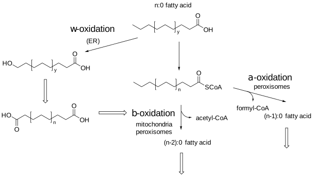
This pathway is called β-oxidation. Fatty acids are oxidized in a step-wise fashion by this pathway. In each repetitive cycle of this pathway, acetyl-CoA and one CO2 are released. In addition, oxidation can occur at both the alpha- and beta carbons when oxidized in an organelle called the peroxisome. The α-oxidation pathway is used for fatty acid branched at the beta carbon 3 releasing one CO2 until the beta-oxidation pathway can be used. The peroxisome degrades fatty acids that can't be oxidized in the mitochondria. These include very long-chain fatty acids (VLCFAs) like 24:0 and 26:0, and in addition, branched-chain fatty acids (BRCHAs) including some fatty acids from dietary sources such as pristanic acid (an odd-chain 15:0 fatty acid methylated at carbons 2, 6, 10, and 14). The α-oxidation pathway can't be used to completely oxidized fatty acids in the peroxisome. At some point in the oxidative stepwise pathway, the resulting shorter fatty acids are exported to the mitochondria for β-oxidation. Also, enzymes in the endoplasmic reticulum have the ω-oxidization pathway which oxidizes fatty acids at the omega or terminal carbon. The enzyme used is the monooxygenase cytochrome P450 which uses one oxygen from O2 to hydroxylate the ω-carbon.
These organelles, initially called microbodies, are vital to cellular metabolism and health. In people with Zellweger syndrome spectrum, there is a severe disorder in the formation of peroxisomes, which is often lethal. They are important metabolically in lipid metabolism, synthesis of myelin sheath lipids, and metabolism of reactive oxygen species like peroxides. The enzymes catalase and urate oxidase are found in such high concentrations they often form crystal "bodies" in the matrix of the peroxisome. Additional roles include responses to pathogens and viruses. Effectively they are a protective organelle.
In contrast to mitochondria, peroxisomes, like most other organelles, have a single bilayer and no DNA, from which transcription of RNA and translation of proteins occur. All proteins are hence imported from the cytoplasm after synthesis on free ribosomes. Imported proteins have a peroxisome targeting sequence (PTS) of serine-lysine and leucine (SKL) near their C-terminus which facilitates the binding of the proteins to a PTS receptor in the peroxisome membrane. These organelles oxidize very long-chain fatty acids (VLFA), make and break down hydrogen peroxide (hence the name), and also synthesize plasmalogens. The enzymes involved in the stepwise cycle of reactions in the peroxisome β-oxidation pathway use enzymes different from those used in the mitochondria for β-oxidation.
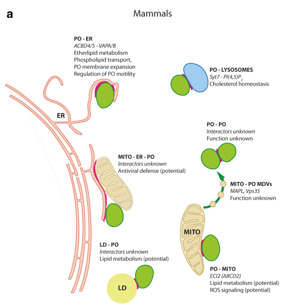
For those more inclined towards chemistry than biology, yet another organelle with its structures and function may seem like one too many. However, this less-discussed organelle is critically important in its own right. Figure \(\PageIndex{2}\) shows features of peroxisomes and their proteins.
 |
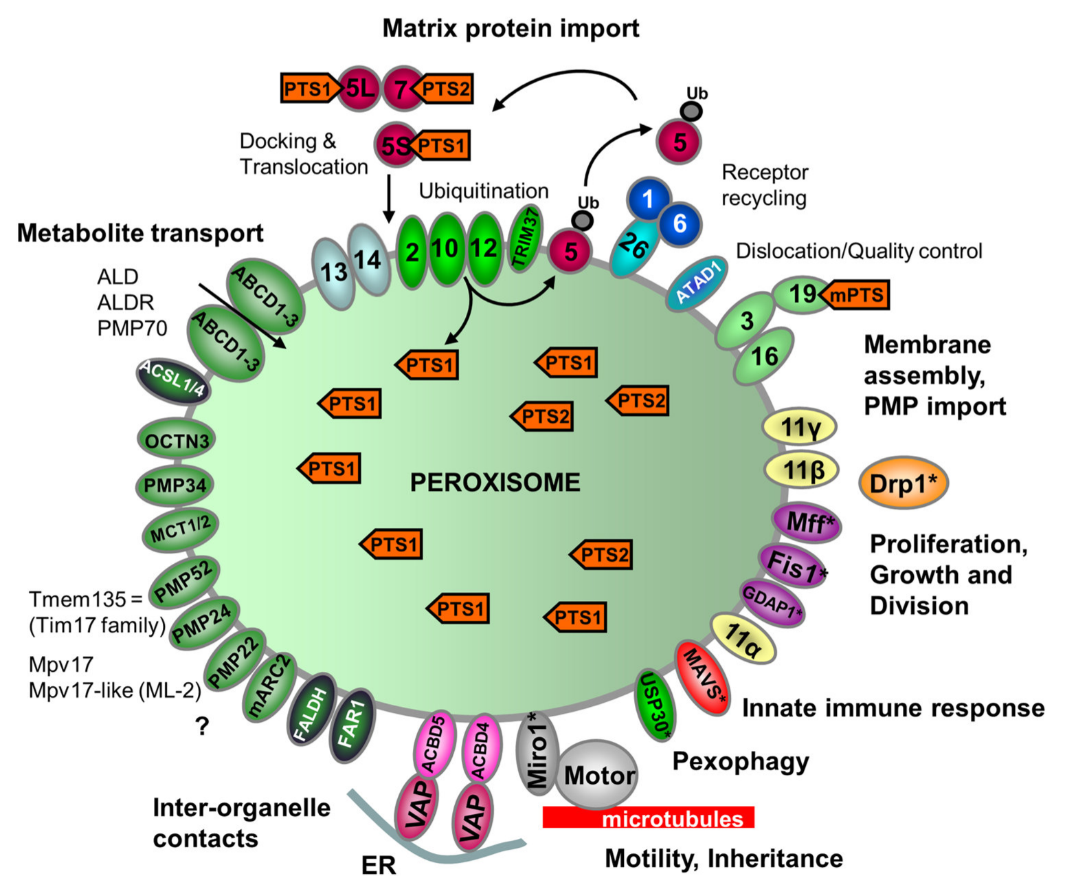 |
One interesting feature is its relationship with different organelles in cells, as shown in the left panel of Figure 2. Some proteins involved in organelle functions are shown as well (right panel).
The peroxisome (PO, green) has binding interactions (red interfaces ) with the endoplasmic reticulum (ER) lysosomes, mitochondria, lipid droplets (a pseudo-organelle), and also itself (left figure). Some of the key membrane proteins (which we have discussed previously) involved in peroxisome function include the ABC transporter proteins ABCD1-3 for fatty acids transport, OCTN3 for organic and cation/carnitine transport, and MCT1/2 for monocarboxylate transport. In addition, peroxisomes have receptors for protein import mediated by PTSs and for peroxisome movement along microtubules in the cell.
Mitochondrial β-Oxidation
Mitochondrial β-oxidation in muscle generates acetyl-CoA, which enters the citric acid cycle for subsequent production of ATP through mitochondrial electron transport and oxidative phosphorylation. In the liver, the generated acetyl-CoA is used for ketone body production under fasting states. Figure \(\PageIndex{3}\) shows the β-oxidation pathway for palmitic acid (16:0), a saturated fatty acid, starting with its import from the cytoplasm.
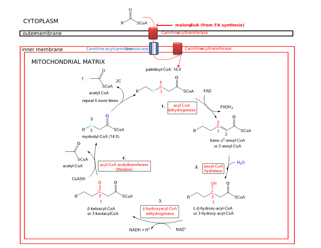
The pathway involved cyclic removal of 2C unit until 16:0 is cleaved 7 times producing 8 2C acetyl-CoAs. The net chemical equation of beta-oxidation of 16:0 is shown in the equation below.
\begin{equation}
\mathrm{C}_{16}-\mathrm{CoA}+7 \mathrm{NAD}^{+}+7 \mathrm{FAD}+7 \mathrm{CoASH}+7 \mathrm{H}_{2} \mathrm{O} \rightarrow 8 \mathrm{Acetyl}-\mathrm{CoA}+7 \mathrm{NADH}+7 \mathrm{FADH}_{2}+7 \mathrm{H}^{+}
\end{equation}
Figure \(\PageIndex{4}\) shows an abbreviated comparison of the β-oxidation pathways in the mitochondria and peroxisomes. Peroxisomal beta-oxidation is used to metabolize very-long-chain fatty acids (VLCFAs), which are composed of 24-26 carbon units as well as branched-chain fatty acids (BRCHAs).
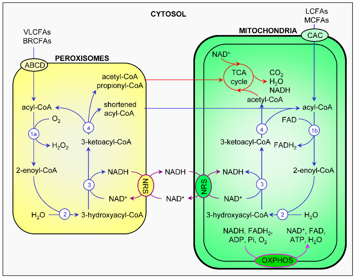
Figure \(\PageIndex{4}\): Comparison and interplay of peroxisomal and mitochondrial fatty acid β-oxidation. Fatty acid β-oxidation, the NAD(H) redox shuttles, the tricarboxylic acid cycle, and the electron transfer chain are respectively depicted in blue, purple, red, and pink. 1a, acyl-CoA oxidase; 1b, acyl-CoA dehydrogenase; 2, enoyl-CoA hydratase; 3, 3-hydroxyacyl-CoA dehydrogenase; 4, 3-ketoacyl-CoA thiolase. ABCD, ATP-binding cassette transporters of subfamily D; ADP, adenine dinucleotide phosphate; BRCFA, branched-chain fatty acid; CAC, carnitine-acylcarnitine carrier; FAD, flavin adenine dinucleotide; FADH2, reduced FAD; LCFA, long-chain fatty acid; MCFA, medium-chain fatty acid; NAD, nicotinamide adenine dinucleotide; NADH, reduced NAD; NRS, NAD(H) redox shuttles; OXPHOS, oxidative phosphorylation; TCA, tricarboxylic acid; VLCFA, very-long-chain fatty acid. Fransen et al. International Journal of Molecular Sciences. 18. 1126. 10.3390/ijms18061126. DOI: 10.3390/ijms18061126. Commons Attribution (CC BY) license (http://creativecommons.org/licenses/by/4.0/).
It should be noted that a likely NAD+/NADH mitochondria transporter, a multi-pass inner mitochondrial membrane protein has just been identified The transporter, MCART1, is also called SLC25A51,
β-Oxidation - Mechanisms
Fatty acids are imported into the matrix from the cytoplasm through their acyl-CoA derivatives. Two different proteins are required for their import. One is carnitine palmitoyltransferase-1(CPT-1), which transfers the acyl group from CoASH to a carrier protein carnitine. The acylcarnitine is translocated through the inner membrane by the carrier protein carnitine-acylcarnitine translocase (CACT). Once inside the matrix, the acyl group is transferred back to CoASH by carnitine palmitoyltransferase-2 (CPT-2). This carnitine cycle is illustrated in Figure \(\PageIndex{5}\).
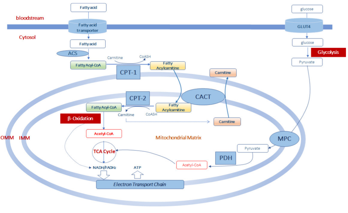
Figure \(\PageIndex{5}\): Carnitine cycle and the connections between major catabolic pathways.
At the outer mitochondrial membrane (OMM), fatty acyl-CoAs become linked to carnitine through carnitine palmitoyltransferase-1 (CPT-1). The complex is translocated across the inner mitochondrial membrane (IMM) via carnitine-acylcarnitine translocase (CACT). In the mitochondrial matrix, CPT-2 converts fatty acylcarnitines back to fatty acyl-CoAs, which enter the β-oxidation pathway. Free carnitine moves back into the cytoplasm through exchange with acyl-carnitines with CACT. β-oxidation in the matrix produces acetyl-CoA, which is also made from glycolytic pyruvate through pyruvate dehydrogenase. Hence acetyl-CoA links both glycolysis and fatty acid oxidation. The resulting acetyl-CoA can enter the TCA cycle when energy is needed.
The mitochondrial carnitine/acylcarnitine carrier protein helps transport acylcarnitines of different lengths across the mitochondrial inner membrane for β-oxidation into the mitochondrial matrix. Figure \(\PageIndex{6}\) shows an interactive iCn3D model of the human mitochondrial carnitine/acylcarnitine carrier protein AlphaFold model (O43772)
.png?revision=1&size=bestfit&width=186&height=301)
 Figure \(\PageIndex{6}\): Mitochondrial carnitine/acylcarnitine carrier protein AlphaFold model (O43772). (Copyright; author via source). Click the image for a popup or use this external link: https://structure.ncbi.nlm.nih.gov/i...DdDNu9xVJGg2i9
The transmembrane helices are shown in gray. The N- (Met 1) and C-terminal (Leu 301) amino acids are shown in spacefill color CPK.
Malonyl-CoA produced in the first committed step in fatty acids synthesis inhibits CPT1. This should make biological sense since fatty acid oxidation should not occur as fatty acids are synthesized. Palmitoyltransferase II (CPT II), which serves to convert acylcarnitine to fatty acyl CoA, traps the molecules within the mitochondrial matrix.
In contrast to this regulated transport mechanism, very long chain fatty acids (VLCFAs) and branched-chain fatty acids are transported into peroxisomes by the ABCD1-3 transporters through an ATP-dependent process
Mitochondrial β-oxidation of fatty acids has four steps that occur in the mitochondrial matrix. In those steps, a 16:0 fatty acid (for example) is converted to a (14):0 fatty acid and the 2C molecule acetyl-CoA. The (14):0 fatty acid undergoes 6 more rounds of the β-oxidation cycle until the entire 16:0 fatty acid is fully converted to 8 acetyl-CoAs.
Step 1: Acyl-CoA dehydrogenase
There are long- (LCAD), medium-(MCAD), and short-chain acyl-CoA dehydrogenases (SCAD) which catalyze the first oxidative step in the β-oxidation pathway. These enzymes catalyze the formation of a trans double bond between the α and β carbons (C2 and C3) on the acyl-CoA substrates. A stronger oxidizing agent than NAD+ is required to form an alkene between the two methylene groups, so FAD is used. Eventually the reduced FADH2 produced will lead to the production of 1.5 equivalents of ATP in the mitochondrial electron transport chain/oxidative phosphorylation.
Figure \(\PageIndex{7}\) shows the key oxidative step in the mechanism of acyl-CoA dehydrogenase to 2-enoyl-CoA by acyl-CoA dehydrogenases
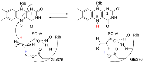
Figure \(\PageIndex{8}\) shows an interactive iCn3D model of the medium-chain acyl-CoA dehydrogenase from pig liver mitochondria with octanoyl-CoA, a substrate (3MDE)
.png?revision=1&size=bestfit&width=490&height=282)
Figure \(\PageIndex{8}\): Medium-chain acyl-CoA dehydrogenase from pig liver mitochondria with octanoyl-CoA substrate (3MDE). (Copyright; author via source). Click the image for a popup or use this external link: https://structure.ncbi.nlm.nih.gov/i...SiAZExwXVV2TG6
Just two subunits of the biologically active tetramer are shown (dark gray and cyan). FAD is shown in each subunit (sticks, CPK colors, labeled). A bound substrate, octanoyl-CoA (spacefill, CPK colors, labeled CO8) is also shown in each subunit. The catalytic base, Glu 376, is shown in sticks, CPK colors, and labeled.
The structures of the unliganded and acyl-CoA forms of the enzymes are very similar, so there are no large conformational changes on binding octanoyl-CoA. The ligand binds to the enzyme at the rectus (re) face of the FAD with the acyl chain buried. The fatty acyl chain of the thioester substrate is buried inside of the polypeptide and the 3'-AMP moiety is close to the surface of the tetrameric enzyme molecule. The carbonyl oxygen of octanoyl-CoA interacts with the ribityl 2'-hydroxyl group of the FAD and the main-chain carbonyl oxygen of Glu-376. Glu-376 acts as a general base as it removed the alpha proton in the reaction.
Step 2. Enoyl CoA hydratase
This enzyme catalyzes a hydration step of the double bond between the α and β carbons (C2 and C3), adding an OH group to the β carbon, in a reaction that poses little energy barrier. A likely mechanism for enoyl-CoA hydratase is shown in Figure \(\PageIndex{9}\).
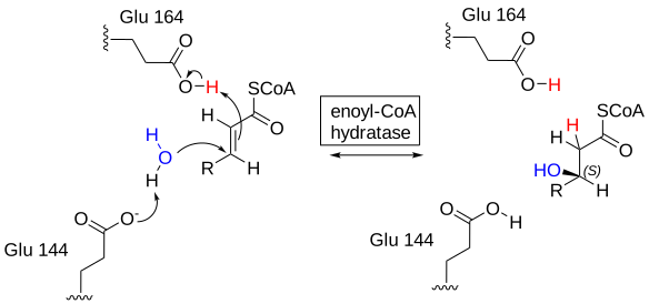
Figure \(\PageIndex{10}\) shows an interactive iCn3D model of the rat enoyl-CoA hydratase in complex with hexadienoyl-CoA (1MJ3)
.png?revision=1&size=bestfit&width=261&height=313)
Figure \(\PageIndex{10}\): Rat enoyl-CoA hydratase in complex with hexadienoyl-CoA (1MJ3). (Copyright; author via source). Click the image for a popup or use this external link: https://structure.ncbi.nlm.nih.gov/i...byx3eEKewEeEz9
Only one subunit of the biological hexamer is shown for clarity. Two glutamic acids (141 and 164) appear to activate a water molecule for the hydration reaction. Alanine 98 and Gly 141 appear also to be situated in an oxyanion hole which stabilizes the transition state and intermediate.
The addition of the water is syn since the proton and OH group are added to the same size of the double bond. The glycine amide NH provides a strong hydrogen bond to the carbonyl of the substrate, hexadienoyl-CoA, helping to polarize the ene-one. The substrate trans-2-crotonyl-CoA is converted to the 3(S) alcohol instead of the 3(R) alcohol by a huge factor. Both the cis and trans isomers of a substrate analog (hexadienoyl-CoA) can bind to the enzyme, but only the cis isomer is polarized. Since the transition state is polarized as well, it would appear the bound cis isomer is strained and destabilized, suggesting that its binding is an example of transition state binding catalysis.
Step 3. Beta-hydroxyl acyl CoA dehydrogenase
After the addition of the OH on C3 (beta) OH during the hydration reaction, the resulting ROH is oxidized to a ketone, β-ketoacyl-CoA, by the oxidizing agent NAD+ using the enzyme β-hydroxyl acyl CoA dehydrogenase. The resulting NADH is reoxidized to NAD+ through the mitochondrial electron transport chain, which leads to the formation of 2.5 molecules of ATP for each NADH. Figure \(\PageIndex{11}\) shows a plausible mechanism for the beta-hydroxyl acyl CoA dehydrogenase-catalyzed reaction.
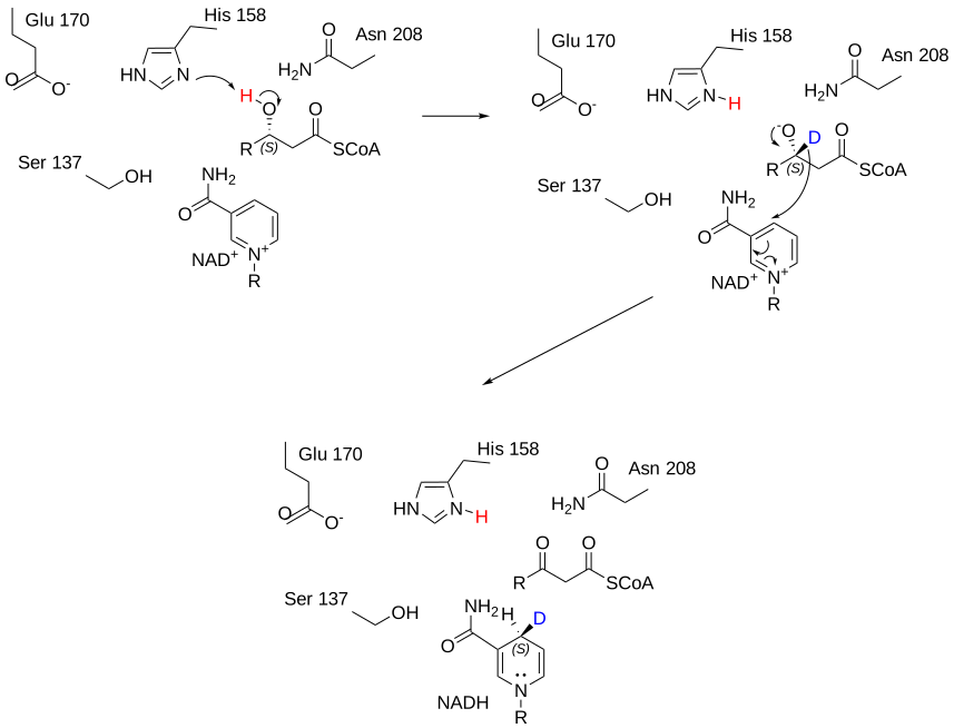
His 158 acts as a general base. Glu 170 increases the basicity of His 158. The other group is involved in H-bond and electronic stabilization interactions.
Step 4. Acetyl-CoA acetyltransferase, mitochondrial - ACAT1 (also called 3-ketoacyl-CoA thiolase)_
The final step in the beta-oxidation pathway involves cleavage of the bond between the alpha and beta carbon by CoASH. This step is catalyzed by beta-keto thiolase and is a thiolytic (as opposed to cleavage by water - a hydrolysis) reaction. The reaction produces one molecule of acetyl CoA and a fatty acyl CoA that is two carbons shorter. The process repeats until the even chain fatty acid is completely converted into acetyl CoA. The activity of the enzyme is reversible and it can also catalyze the Claisen condensation of two acetyl-CoA molecules into acetoacetyl-CoA, as we will see in the synthesis of ketone bodies in the next chapter section.
The reaction starts with the acylation reaction of the nucleophilic Cys 89 with the carbonyl at the 3-oxoacyl-CoA, with the concomitant release of acetyl-CoA. This forms a Cys 89-acyl covalent intermediate. In the next step, Cys 378 acts as a general base to facilitate the nucleophilic attack of free CoASH on the acyl-intermediate. His 348 acting as a general acid protonates the thiolate leaving group. The amino acids (Cys89, Cys378, and His348) are generally conserved in thiolases (Bhaskar et al. 2020). Kinetically this mechanism is a ping-pong reaction. A reaction mechanism is shown in Figure \(\PageIndex{12}\).
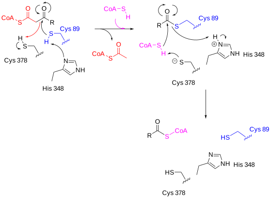
Figure \(\PageIndex{13}\) shows an interactive iCn3D model of Human Mitochondrial 3-Ketoacyl-Coa Thiolase (T1) (4C2J)
_(4C2J).png?revision=1&size=bestfit&width=421&height=296)
Figure \(\PageIndex{13}\): Human Mitochondrial 3-Ketoacyl-Coa Thiolase (T1) (4C2J). (Copyright; author via source). Click the image for a popup or use this external link: https://structure.ncbi.nlm.nih.gov/i...dzAHKEY77WpNi7
Two subunits in the biological function dimer are shown (cyan and gray). The active site is shown in the gray subunit as CPK-colored stick and labeled. The numbers are a bit different than shown in the mechanistic figure. CoASH is shown in each subunit as sticks. The fatty-acyl tail appears to bind in a tunnel.
A few more enzymes are needed
Steps 1 through 4 outlined above apply to the beta-oxidation of a saturated fatty acid with an even-numbered carbon skeleton. Unsaturated fatty acids, such as oleate (18:1) and linoleate (18:2), contain cis double bonds that must be isomerized to the trans configuration by the enzyme enoyl CoA isomerase or reduced by the enzyme NADPH (2,4-dienoyl CoA reductase or 24DCR), using NADPH.
Enoyl CoA isomerase
This enzyme catalyzes the isomerization of cis double bounds to the trans form which mimics those formed by acyl-CoA dehydrogenase by FAD in step 1. Figure \(\PageIndex{14}\) shows a plausible mechanism for the conversion of cis double bonds to their trans isomer.
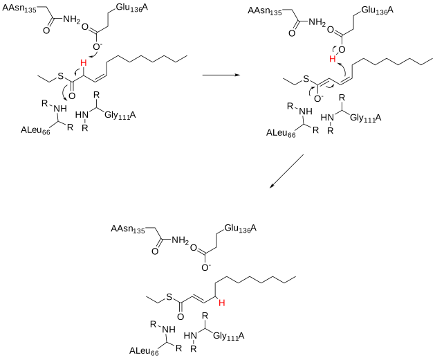
Glu 136 acts as a general base, while amide Hs of Leu 66 and Gly 111 stabilized the intermediate oxyanion and hence the developing charge in the transition state. They are optimally situated in the oxyanion hole.
Figure \(\PageIndex{15}\) shows an interactive iCn3D model of the Human mitochondrial Δ3-Δ2-enoyl-CoA isomerase (1SG4)
.png?revision=1&size=bestfit&width=403&height=371)
Figure \(\PageIndex{15}\): Human mitochondrial Δ3-Δ2-enoyl-CoA isomerase (1SG4) (Copyright; author via source). Click the image for a popup or use this external link:https://structure.ncbi.nlm.nih.gov/i...H6LxEzBKajq8b7
The three subunits are shown in different colors. The substrate analog octanoyl-CoA is shown in spacefill CPK colors bound to the gray subunit. The catalytic residues are shown in sticks with CPK colors and labeled. The distal omega end binds in a hydrophobic tunnel.
2,4-dienoyl CoA reductase or 24DCR
An alternative way to deal with the cis double bond is simply to reduce it, in this case, NADPH. This enzyme is used on all C=C at the even-number position and more at odd-numbered positions. A mechanism for the reduction is shown in Figure \(\PageIndex{16}\).
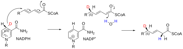
Figure \(\PageIndex{17}\) shows an interactive iCn3D model of a monomer of the homotetrameric human mitochondrial 2,4-dienoyl-Coa reductase (1W6U)
.png?revision=1&size=bestfit&width=381&height=295)
Figure \(\PageIndex{17}\): Monomer of the homotetrameric human Mitochondrial 2,4-Dienoyl-Coa Reductase (1W6U) (Copyright; author via source). Click the image for a popup or use this external link: https://structure.ncbi.nlm.nih.gov/i...pQueKc84sFqzt7
The model shows bound NADP+ and the substrate trans-2,trans-4-dienoyl-CoA. The active site is open enough to accommodate fatty acids of different lengths. Tyr-199 and Asn-148 stabilize the enolate and the oxidized nicotinamide.
For odd-number chain fatty acids, propionyl-CoA to succinyl-CoA
Although most fatty acids of biological origin have even numbers of carbons, not all of them do. Oxidation of fatty acids with odd numbers of carbons ultimately produces an intermediate with three carbons, propionyl-CoA, which cannot be oxidized further in the beta-oxidation pathway. These additional steps are necessary:
- carboxylation to make (S)-methylmalonyl-CoA;
- isomerization to (R)-methylmalonyl-CoA;
- rearrangement to form succinyl-CoA. The last step of the process utilizes the enzyme methylmalonyl-CoA mutase, which uses the B12 coenzyme in its catalytic cycle. Succinyl-CoA can then be metabolized in the citric acid cycle.
Figure \(\PageIndex{18}\) shows the pathway for the conversion of propionyl-CoA to succinyl-CoA.
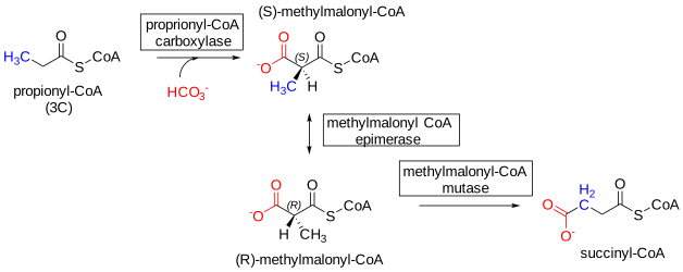
Figure \(\PageIndex{18}\): Conversion of propionyl-CoA to succinyl-CoA
Recent Updates: 7/24/23 Mechanisms for conversion of propionyl-CoA to succinyl-CoA.
We will look at each of the enzymes in turn.
Propionyl-CoA carboxylase
The net reaction for this enzyme is shown in Figure \(\PageIndex{19}\) below.
Propionyl-CoA + ATP + HCO3- → (S)-methylmalonyl-CoA + ADP + Pi + H+
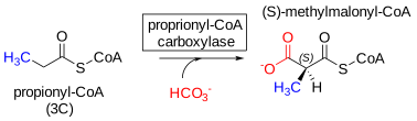
Figure \(\PageIndex{19}\): Propionyl-CoA carboxylase reaction
The enzyme has 3 subunits. The alpha subunits has biotin carboxyl carrier protein and biotin carboxylase domains. The beta subunit has carboxytransferase activity. The mechanism for the Streptomyces coelicolor propionyl-CoA carboxylase (PCC) is shown below in two parts.
In Part 1, biotin is carboxylated by HCO3- to form an activated bicarbonate derivative (similar to an anhydride) that is high energy in comparison to its hydrolysis product, as shown in Figure \(\PageIndex{20}\) below. This reaction takes place in the alpha subunit.
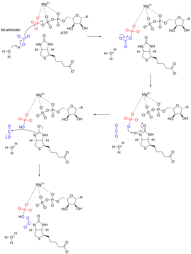
Figure \(\PageIndex{20}\): Formation of carboxylated biotin by propionyl-CoA carboxylase - Part 1. M-CSA. Gemma L. Holliday, Daniel E. Almonacid, Jonathan T. W. Ng. Creative Commons Attribution 4.0 International (CC BY 4.0) License
In Part 2, shown in Figure \(\PageIndex{21}\) below, the carboxyl group on biotin is transferred to propionyl-CoA to form methymalonyl-CoA. This reaction occurs in the beta subunit.
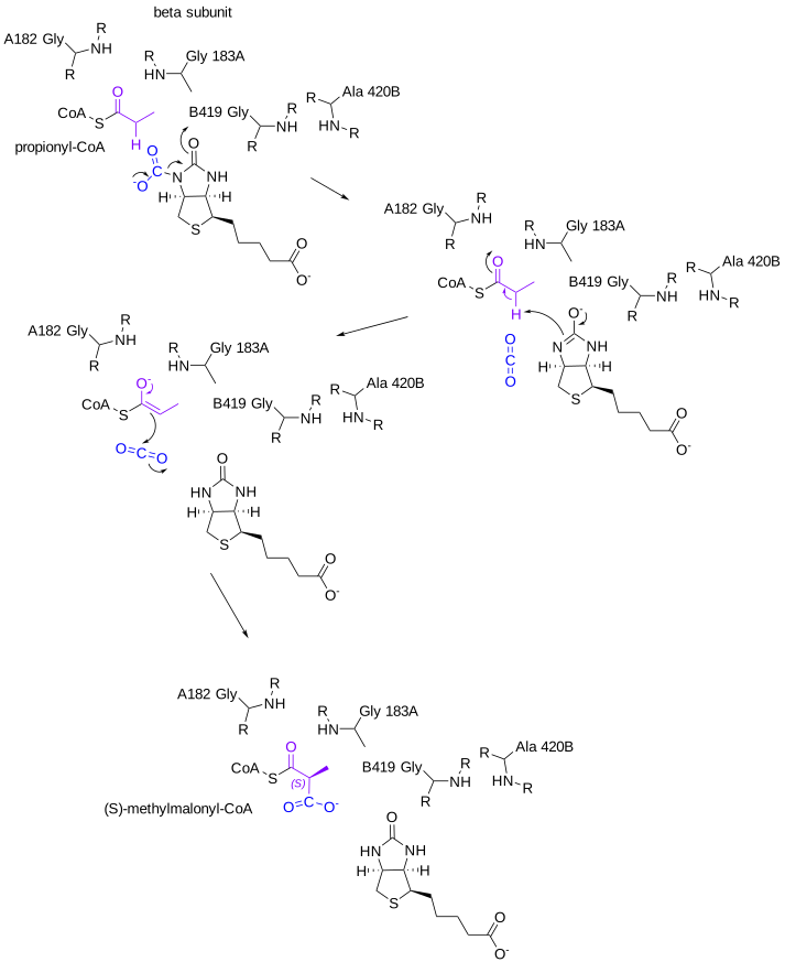
Figure \(\PageIndex{21}\): Part 2 - Formation of (S)-methylmalonyl-CoA by activated CO2 transfer from carboxybiotin by propionyl-CoA carboxylase. Holliday et al., ibid.
The roles of the main chain atoms of Ala 182 and Gly 183 in the alpha subunit are to stabilize the propionyl-CoA while the backbone atoms of Gly 429 and Ala 430 in the beta subunit are to stabilize the oxyanion of carboxylated biotin.
Figure \(\PageIndex{22}\) shows an interactive iCn3D model of biotin and propionyl-CoA bound to Acyl-CoA Carboxylase Beta Subunit from S. coelicolor (1XNY)
.png?revision=1)
Figure \(\PageIndex{22}\): Monomer of the biotin and propionyl-CoA bound to Acyl-CoA Carboxylase Beta Subunit from S. coelicolor (1XNY). (Copyright; author via source). Click the image for a popup or use this external link: https://structure.ncbi.nlm.nih.gov/i...K46r5EdZezF4o6
Biotin (labeled BTN) and propionyl-CoA are shown in spacefill in CPK colors.
Methylmalonyl-CoA Epimerase
The net reaction is shown in Figure \(\PageIndex{23}\) below.
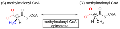
Figure \(\PageIndex{23}\): Methylmalonyl-CoA Epimerase reaction
This reaction proceeds through an enolate intermediate after an abstraction of a proton from the chiral center of the methylmalonyl-CoA, as shown in the reaction mechanism for the enzyme from propionibacterium freudenreichii subsp. shermanii is shown Figure \(\PageIndex{24}\) below. The reaction is readily reversible.
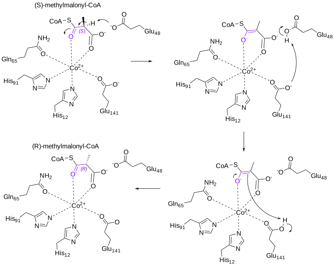
Figure \(\PageIndex{24}\): Mechanism for methylmalonyl-CoA Epimerase. Gemma L. Holliday et al. https://www.ebi.ac.uk/thornton-srv/m-csa/entry/33/. Creative Commons Attribution 4.0 International (CC BY 4.0) License
Note the presence of a Co2+ ion in the active site.
Figure \(\PageIndex{25}\) shows an interactive iCn3D model of Methylmalonyl-CoA epimerase in complex with 2-nitronate-propionyl-CoA from S. coelicolor (6WFI)
.png?revision=1&size=bestfit&width=439&height=389)
Figure \(\PageIndex{25}\): Methylmalonyl-CoA epimerase in complex with 2-nitronate-propionyl-CoA from S. coelicolor (6WFI). (Copyright; author via source). Click the image for a popup or use this external link: https://structure.ncbi.nlm.nih.gov/i...Sqc98CCjAvzkp7
The enzyme is a dimer with two identical subunits and catalytic. One of the subunits is shown with a blue transparent surface with the bound 2-nitronate-propionyl-CoA inhibitor. The other is shown in gray cartoon with the key side chains involved in substrate binding and catalysis shown as color sticks and labeled.
Methylmalonyl-CoA Mutase
In this reaction, a methyl group is removed from (R)-methylmalonyl-CoA to form succinyl-CoA, a shown in Figure \(\PageIndex{26}\) below.
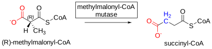
Figure \(\PageIndex{26}\): Methylmalonyl-CoA reaction
This reaction going in the opposite direction is an example of a methyltransferase. Another similar enzyme (which we will see in Chapter 18.4 - Amino Acid Degradation) catalyzes a methyl transfer from homocysteine to a new cofactor, cobalamin, which transfers it to cysteine to form methionine in a reaction catalyzed by methionine synthase. Cobalamine and its methylated form are derivatives of Vitamin B12. We will leave details of cobalamin biochemistry to the next chapter and will present a mechanism for methylmalonyl-CoA mutase here.
We present the reaction for the reverse reaction, the conversion of succinyl-CoA to (R)-methylmalonyl-CoA by the enzyme from Propionibacterium freudenreichii subsp. shermanii in two parts below. The enzyme is a heterodimer of an alpha and beta subunit and has a cofactor, adenosylcobalamin (coenzyme B12). In contrast, the human mutase, a homodimer, is very similar to the alpha subunit.
First, a free radical is formed from the adenosyl group on the cofactor. This promotes a free-radical rearrangement of succinyl-CoA to (R)-methylmalonyl-CoA (or the reverse for the pathway of interest here). The reaction is shown in two parts for more optimal viewing.
Figure \(\PageIndex{27}\) below the first parts of the reaction for the conversion of succinyl-CoA to (R)-methylmalonyl-CoA by the mutase from Propionibacterium freudenreichii subsp. shermanii.
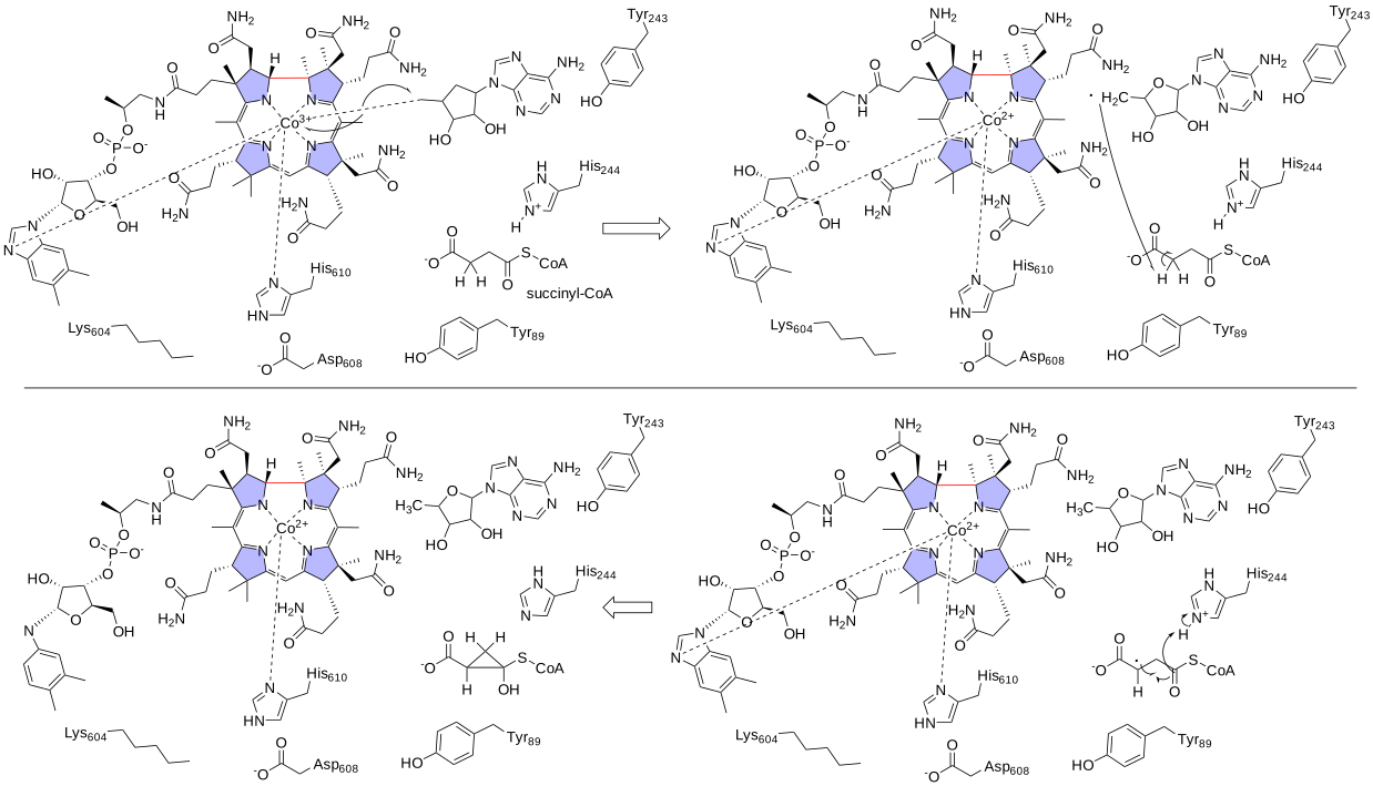
Figure \(\PageIndex{27}\): Part 1 of conversion of succinyl-CoA to (R)-methylmalonyl-CoA by methylmalonyl-CoA mutase. Gemma L. Holliday, Gail J. Bartlett, Daniel E. Almonacid. M-CSA. https://www.ebi.ac.uk/thornton-srv/m-csa/entry/62/. Creative Commons Attribution 4.0 International (CC BY 4.0) License
Figure \(\PageIndex{28}\) below shows the rest of the reaction for the conversion of succinyl-CoA to (R)-methylmalonyl-CoA.
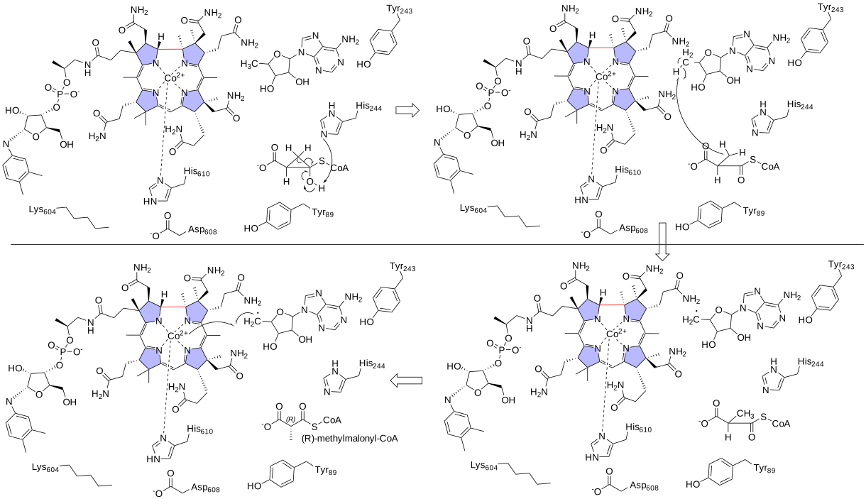
Figure \(\PageIndex{28}\): Part 2 of the conversion of succinyl-CoA to (R)-methylmalonyl-CoA by methylmalonyl-CoA mutase. Gemma L. Holliday et al., ibid.
The very last product from the bottom left reaction, the reformed active adenosylcobalamin cofactor, is not shown. The enzyme facilitates the hemolytic cleavage of the Co-C bond. This is followed by a free-radical rearrangement.
Figure \(\PageIndex{29}\) shows an interactive iCn3D model of the methylmalonyl-coenzyme A mutase from from Propionibacterium freudenreichii subsp. shermanii (1REQ)
.png?revision=1&size=bestfit&width=414&height=366)
Figure \(\PageIndex{29}\): Methylmalonyl-coenzyme A mutase from from Propionibacterium freudenreichii subsp. shermanii (1REQ). Click the image for a popup or use this external link: https://structure.ncbi.nlm.nih.gov/i...qPYm2sXjraCuN8
An analog of CoASH, desulfo-CoA (DCA) , is shown in spacefill, CPK colors and labeled. Adenosylcobalamin is shown in colored sticks and labeled B12. The alpha subunit is shown in gray with the key amino acids from the enzyme mechanism presented above shown as colored sticks and labeled. The beta subunit is shown in blue.
Propionyl-CoA is also produced as a product of the oxidation of methionine, valine, isoleucine, and threonine. (See the amino acid metabolism chapter for more details on mechanisms.)
Very long chain oxidation
In contrast to the oxidation of short and medium-chain fatty acids, which under beta-oxidation require four different discrete enzymes, the oxidation of very long-chain fatty acids (VLCFs) is carried out by two proteins with the second protein expressing three enzyme activities. The first step, analogous to step 1 in beta-oxidation described above, is carried out by a very long chain acyl-CoA dehydrogenase (VLCAD). The next three reactions are carried out by a single trifunctional protein (TFP) with two subunits. The α-subunit carries out the hydration (2-enoyl-CoA hydratase, ECH) and next oxidation step (3-hydroxyl-CoA dehydrogenase (HAD), while the β-subunit has 3-ketothiolase (KT) activity. Deficiencies of TFP can cause significant disease and even death.
TFP is a heterotetramer of two α and two β subunits with the beta subunits forming a central homodimer. The two alpha units bind at each end and the whole complex forms an arc. There appears to be a "tunnel" allowing substrate transfer after each step. This minimizes premature intermediate release. Figure \(\PageIndex{30}\) shows an interactive iCn3D model of the human mitochondrial trifunctional protein, a fatty acid beta-oxidation metabolon (6DV2)
.png?revision=1&size=bestfit&width=416&height=318)
Figure \(\PageIndex{30}\): Human mitochondrial trifunctional protein fatty acid beta-oxidation metabolon (6DV2). Click the image for a popup or use this external link: https://structure.ncbi.nlm.nih.gov/i...XiUXru4vkRVh87
- Grays: two thiolase subunits: reversible thiolytic cleavage of 3-ketoacyl-CoA into acyl-CoA and acetyl-CoA, a 2-step reaction involving a covalent intermediate formed with a catalytic cysteine. The catalytic residue (C138, C458, and H428) are shown as sticks with CPK colors.
- Cyan and magenta subunits: enoyl-CoA hydratase (EC 4.2.1.17) and 3-hydroxyacyl-CoA dehydrogenase (EC 1.1.1.35)
- The red dots represent the inner leaflet of the inner membrane so the proteins reside in the mitochondrial matrix.
Regulation of beta-oxidation
We saw that the metabolic decision to use carbohydrate energy reserves (glycogen) is a highly regulated event. Glycogen breakdown occurs during fasting and energy need. Fatty acids, our largest stores of energy, are released from triglyceride reserves in adipose cells through extracellular epinephrine and glucagon activation of pathways that activate intracellular hormone-sensitive lipase. Released fatty acids are bound to the serum protein albumin which transports them to tissue. Also as we mentioned above, malonyl-CoA inhibits the movement of fatty acids into the mitochondria. Malonyl-CoA is the first committed product of fatty acid biosynthesis. Each acyl-CoA product of each of the four enzymes engages in product inhibition for the enzyme that produced it. 3-ketoacyl-CoA also inhibits enoyl-CoA hydratase and acyl-CoA dehydrogenase [17]. The NADH/NAD+ and acetyl-CoA/CoA ratios also influence the beta oxidation pathway through allosteric regulation. For example, the ratio of acetyl-CoA/CoA ratio affects the activity ketoacyl-CoA thiolase.
Fatty acids also bind to the transcription factors called peroxisome proliferator-activated receptors (PPARs) and also coactivator PGC-1α, which regulate the transcription of enzymes in the beta-oxidation pathways. PPARs and the retinoid X receptor form heterodimers that bind to the PPAR response element in key promoters site involved in fatty acid degradation. These include CPT1, long-chain acyl-CoA dehydrogenase (LCAD), medium-chain acyl-CoA dehydrogenase (MCAD), and acyl-CoA synthetase (ACS). The PPARs have tissue specificity. We will discuss PPARs in more detail in the chapter on fatty acid synthesis.
Peroxisomal α-Oxidation
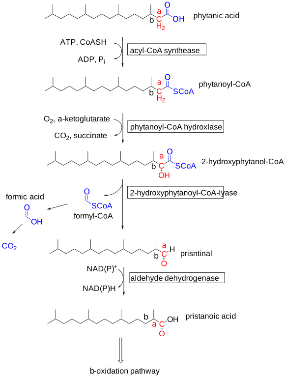
Alpha oxidation of fatty acids occurs in the peroxisome. It is used to metabolize phytanic acid (3,7,11,15-tetramethyl hexadecanoic acid), found in dairy products, animal fat, and some fish. Phytanic acid is produced in ruminants on the degradation of plant material and derives from phytol, an isoprenoid alcohol esterified to chlorophyll. Phytol is first converted to phytanic acid.
Fatty acid β-oxidation can also occur in peroxisomes. In animals, peroxisomes are believed to be important in the initial breakdown of very long-chain fatty acids and methyl-branched fatty acids [11]. The enzymes involved in fatty acid oxidation in peroxisomes are different from mitochondria. An important difference is acyl-CoA oxidase, the first enzyme in peroxisome β-oxidation, which transfers the hydrogen to oxygen producing H2O2 instead of producing FADH2. The H2O2 is broken down to water by catalase. The fatty acyl-CoA intermediates formed during β-oxidation are the same in peroxisomes and mitochondria. Peroxisomes also contain the necessary enzymes for α-oxidation, which are necessary for the oxidation of some fatty acids with methyl branches.
Branched-chain fatty acids also require additional enzymatic modification to enter the alpha-oxidation pathway within peroxisomes. Phytanic acid (3,7,11,14-tetramethylhexadecanoic acid), requires additional peroxisomal enzymes to undergo beta-oxidation. Phytanic acid initially forms phytanyl CoA which is then hydroxylated at the alpha carbon by phytanyl CoA hydroxylase (alpha-hydroxylase), encoded by the PHYH gene. The alpha carbon-hydroxyl bond then undergoes two successive rounds of oxidation to pristanic acid. Pristanic acid undergoes beta-oxidation, which produces acetyl CoA and propionyl CoA in alternative rounds. As with peroxisomal beta-oxidation of VLCFAs, this process generally ends when the carbon chain length reaches 6-8 carbons, at which point the molecule is shuttled to the mitochondria by carnitine for complete oxidation to carbon dioxide and water. Figure \(\PageIndex{31}\) shows the steps in the catabolism of phytanic acid (3,7,11,14-tetramethylhexadecanoic acid).
Omega-oxidation
The omega-oxidation pathway occurs in the endoplasmic reticulum and is used to metabolize larger fatty acids, which given their hydrophobicity might be damaging to cells in high concentrations. In this pathway the fatty acids are metabolized to dicarboxylic acids which increases their water solubility for excretion in the urine. The first step uses cytochrome P450 enzymes that are also used to modify xenobiotic compounds with dioxygen, making them more soluble as well. The omega oxidation pathway is shown in Figure \(\PageIndex{32}\).
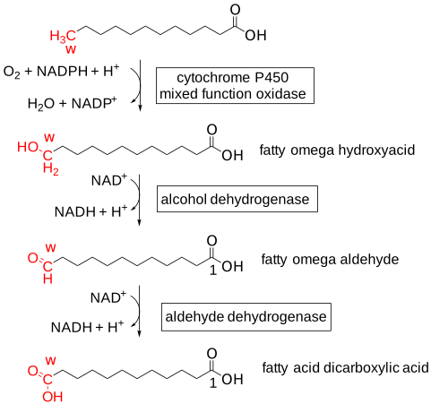
Three subfamily members of the cytochrome P450s (CYPs) show a preference for hydroxylation of short-chain fatty acids (C7-C10, CYP4B), medium-chain (C10-C16, CYP4A), and long-chain (C16-C26, CYP4F) fatty acids. which can be saturated, unsaturated and branched, fatty acids. Figure \(\PageIndex{33}\) shows a summary of the alpha, beta, and omega oxidation pathway
Diseases of fatty acid metabolism
Listed below are a few select diseases that either directly involves defective fatty acid metabolism through intrinsic enzyme deficiencies or indirectly prevent the proper functioning of fatty acid metabolism through extrinsic enzyme deficiencies. Many, but not all, deficiencies of enzymes involved in fatty acid oxidation result in abnormal neurological development and or function early in life; a brief list of signs and symptoms appears under the selected diseases mentioned.
MCAD Deficiency
Medium-chain acyl dehydrogenase is the most common inherited defect of fatty acid oxidation in humans; as one would expect, medium-chain, 6-8 carbon molecules accumulate in this disease. Clinical manifestations of MCAD deficiency primarily present during fasting conditions and include lethargy, weakness, sweating, and hypoglycemia, most commonly in children under the age of 5. Serum measurements of octanoyl carnitine are usually elevated in these patients and can aid in the diagnosis. These abundant molecules then undergo oxidation by the cytochrome P450 system involved in omega-oxidation, resulting in a dicarboxylic acidemia and dicarboxylic aciduria.
Zellweger Syndrome
Zellweger syndrome results from autosomal recessive mutations in the PEX genes, which code for peroxin proteins needed for the assembly of peroxisomes. Almost 70% of all peroxisomal biogenesis disorders (PBDs) result from a PEX1 gene mutation. Many different fatty acid compounds can accumulate without the oxidative machinery of peroxisomes, including VLCFAs and phytanic acid. Manifestations of this disease generally include the brain, kidneys, and skeleton.
X-Linked Adrenoleukodystrophy (X-ALD)
X-ALD is a genetic deficiency of the ABCD transporters in the membrane of peroxisomes, as mentioned previously, resulting in the pathological accumulation of phytanic acid and VLCFAs within cells and is most clinically significant when the ABCD1 transporter is absent. The disease presents with neurodegenerative and adrenal abnormalities.
Refsum Disease
Refsum disease results from a genetic deficiency of the enzyme phytanyl CoA 2-hydroxylase, which, as previously mentioned, is involved in the alpha-oxidation of phytanic acid, a breakdown product of chlorophyll. Notable clinical manifestations of Refsum disease include cardiac malfunction and defective functioning of the olfactory and auditory nerves due to the accumulation of phytanic acid.