
19.2: Helminths

( \newcommand{\kernel}{\mathrm{null}\,}\)
Helminthology is the study of worms, or helminths. Over one billion people worldwide are infected with intestinal helminths alone. Helminths are multicellular, often macroscopic worms having both rudimentary organs and organ systems. We will look at three groups of pathogenic helminths: nematodes, cestodes, and trematodes.
1. The Nematodes (Roundworms)
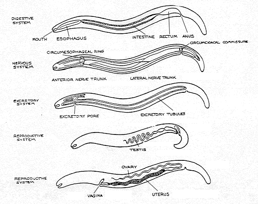
Nematodes are elongated, unsegmented, cylindrical worms having separate sexes. The various systems of the roundworms can be seen in Fig. 19.2.1, and a generalized drawing of a nematode is shown in Fig. 19.2.2. We will look at several pathogenic nematodes.
|
Fig. 19.2.1: Organ Systems of Nematodes |
Fig. 19.2.2A: Morphology of Nematodes |
Fig. 19.2.2B: Ascaris lumbricoides |
|---|---|---|
 |
 |
 |
| Copyright; Gary E. Kaiser, Ph.D. The Community College of Baltimore County, Catonsville Campus CC-BY-3.0 | ||
a. Ascaris lumbricoides
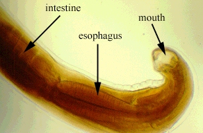
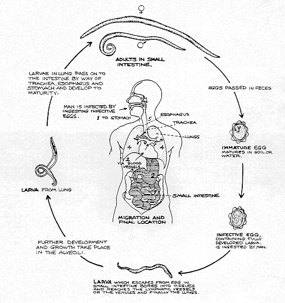
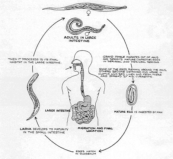
These worms range from 20-45 cm long and are 5 mm in diameter in the adult form, the female being larger than the male. The Ascaris life cycle is seen in Fig. 19.2.3. The disease is called ascariasis. This is the most common helminth in humans worldwide. In the U.S., it infects over 4,000,000 people. Humans become infected by ingesting water or food contaminated with feces that containsAscaris ova or from fingers contaminated with polluted soil. The disease is diagnosed by microscopically looking for Ascaris ova in a fecal smear (see Fig. 19.2.4). A similar roundworm, Toxocara, parasitizes dogs and cats. Visceral larva migrans is the migration of larvae of these worms in human tissues such as lung, liver, and brain, where they may cause tissue damage and allergic reactions.
|
Fig. 19.2.3: Life cycle of the nematode Ascaris lumbricoides |
Fig. 19.2.4: Ova of Ascaris lumbricoides |
|---|---|
 |
 |
| Copyright; Gary E. Kaiser, Ph.D. The Community College of Baltimore County, Catonsville Campus CC-BY-3.0 | |
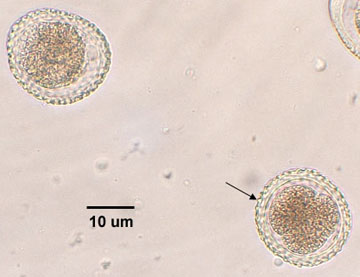
b. Enterobius vermicularis (pinworms)
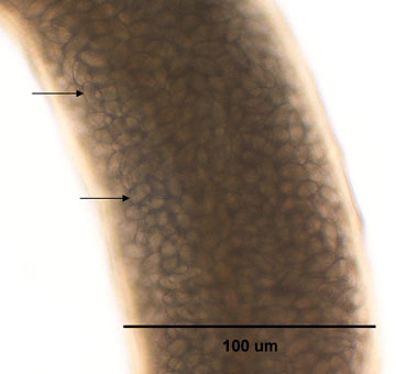
E. vermicularis is a small worm, the female being 8-13 mm long and 0.3-0.5 mm wide; the male being 2-5 mm long and 0.1 mm wide. The life cycle of Enterobiusis shown in Fig. 19.2.5. Pinworms are the most common helminth infection in the U.S. with as many as 50,000,000 people infected. Humans, frequently children, become infected by inhaling E. vermicularis ova or from transfer of ova to the mouth from fecally-contaminated fingers. The female worm migrates to the perianal region of the infected individual, releasing masses of ova and causing an itching sensation. Each pregnant worm produces between 4600 and 16,000 eggs (see Fig. 19.2.6). The disease is diagnosed by applying tape to the perianal region and microscopically looking for pinworm ova that have stuck to the tape (see Fig. 19.2.75).
|
Fig. 19.2.5: Organ Systems of Nematodes |
Fig. 19.2.6: Morphology of Nematodes |
Fig. 19.2.7: Ascaris lumbricoides |
|---|---|---|
 |
 |
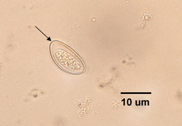 |
| Central view of the nematode Enterobius vermicularis. Note that this female is filled with ova (arrows). | Ova of Enterobius vermicularis. Note "smooth edge" of ova (arrows). | |
| Copyright; Gary E. Kaiser, Ph.D. The Community College of Baltimore County, Catonsville Campus CC-BY-3.0 | ||
Video of Pinworms

Video of Enterobius vermicularis

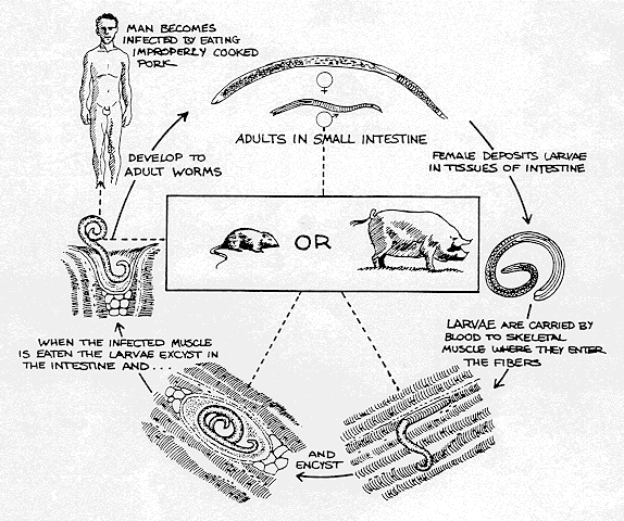
c. Trichinella spiralis
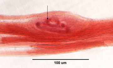
T. spiralis causes a disease called trichinosis. Humans become infected mainly by eating poorly cooked infected pork containing the encysted larva (1-2 mm long). The larvae excyst and develop into adult worms in the intestines. After mating, the female releases larvae which enter the blood and are distributed throughout the body where they become encysted in muscle tissue. The life cycle of Trichinella spiralis is seen in Fig. 19.2.8. The disease is diagnosed by serological tests and microscopic examination of biopsy specimens (see Fig. 19.2.9).
|
Fig. 19.2.8: Life cycle of the nematode Trichinella spiralis |
Fig. 19.2.9: Trichinella spiralis Encysted in Muscle Tissue |
|---|---|
 |
 |
| Note spiraled larva of Trichinella (arrow) encysted in the muscle. | |
| Copyright; Gary E. Kaiser, Ph.D. The Community College of Baltimore County, Catonsville Campus CC-BY-3.0 | |
Video of Larvae of Trichinella spiralis

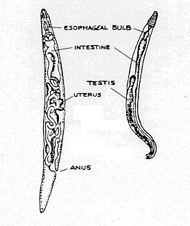
2. The Cestodes (Tapeworms)
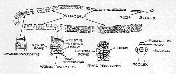
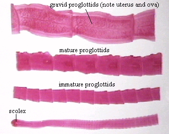
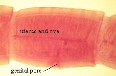
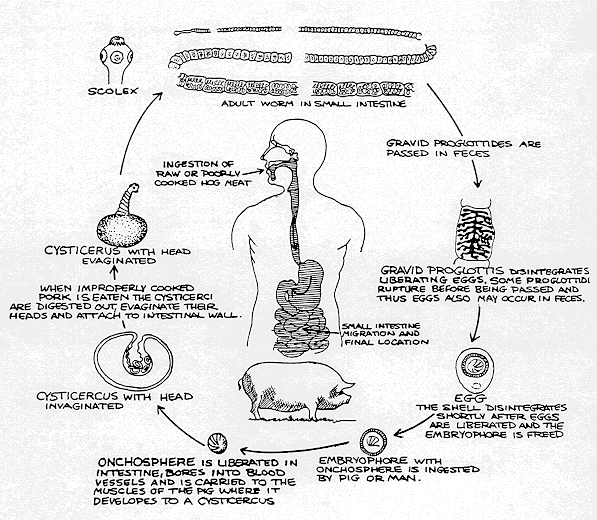
Tapeworms are flat segmented worms which are hermaphroditic (contain both male and female sexual organs). Adult tapeworms have several distinct regions (see Fig. 19.2.11). The scolex (see Fig. 19.2.12) is a head-like structure with distinct suckers and possibly hooks used for attachment to the intestinal wall. Behind the scolex is a constricted neck region consisting of germinative tissue from which new segments, or proglottids, are formed. Finally, there is a long strobila or chain of proglottids of varying stages of maturity. Proglottids containing a uterus and thousands of ova are excreted in the feces (see Fig. 19.2.13). When ingested by intermediate hosts (such as cattle, pigs, and fish), the larva hatch from the ingested ova and migrate to muscle where they encyst as cysticerci. The life cycle of the pork tapeworm Taenia solium is shown in Fig. 19.2.14.
|
Fig. 19.2.10: Organ Systems of Nematodes |
Fig. 19.2.11: Morphology of Nematodes |
Fig. 19.2.12: Ascaris lumbricoides |
|---|---|---|
 |
 |
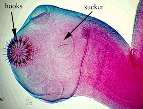 |
| Note the attachment structure or scolex, immature, mature, and gravid proglottids. | Note hooks and suckers for attachment to intestinal wall. | |
| Copyright; Gary E. Kaiser, Ph.D. The Community College of Baltimore County, Catonsville Campus CC-BY-3.0 | ||
|
Fig. 19.2.13: Gravid Proglottis of theTapeworm Taenia pisiformis (Cestode) |
Fig. 19.2.14: Life cycle of the cestode Taenia solium (pork tapeworm) |
|---|---|
 |
 |
| The gravid proglottids are basically segments containing a uterus filled with ova and a genital pore for release of ova | |
| Copyright; Gary E. Kaiser, Ph.D. The Community College of Baltimore County, Catonsville Campus CC-BY-3.0 | |
Humans become infected with tapeworms by eating poorly cooked infected beef, pork, or fish containing cysticerci. Taenia saginata, the beef tapeworm often reaches 6 meters in length; Taenia solium, the pork tapeworm is normally 2-7 meters in length; and Diphyllobothrium latum, the fish tapeworm may reach 3-6 meters in length. These tapeworms are diagnosed by looking for proglottids and ova in the feces.
When humans ingest tapeworm eggs instead of cysts, embryos are released, penetrate the intestinal wall, and enter the blood. The embryos migrate to various tissues (frequently the brain) and develop into cysticerci. Humans also act as intermediate hosts for Echinococcus granulosus found in dogs and cats. Larva hatch from ingested ova and migrate to the liver and lungs and form hydatid cysts.
Video of Tapeworm growing in the intestines.
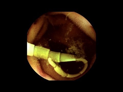
Video of a Colonoscopy showing Diphyllobothrium latum (fish tapeworm)
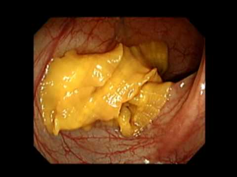
Video of a broken Tapeworm

- Video of a Taenia solium (pork tapeworm)
3. The Trematodes (Flukes)
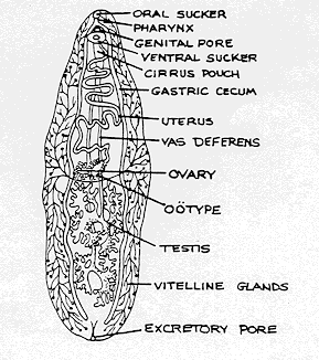
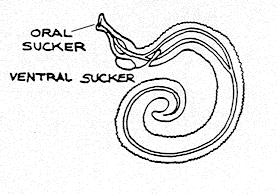
Flukes are unsegmented, flat, leaf-shaped worms having a variety of organ systems (see Fig. 19.2.15). Most flukes are hermaphroditic. They attach to the host by means of an oral sucker and a ventral sucker. Flukes, as adults, may infect either the portal blood vessels, intestines, liver, or lungs of humans and are named according to the tissue they infect. Humans become infected with liver flukes, lung flukes, and intestinal flukes by ingesting poorly cooked fish, crayfish, crabs, snails, or water vegetables infested with flukes. Blood flukes directly penetrate the skin.
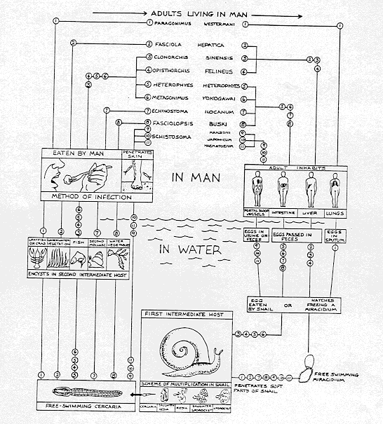
In the life cycle of flukes (see Fig. 19.2.16), ova leave the body of the infected human or animal by means of feces, urine, or sputum (depending on the type of fluke). The ova enter water and infect the first intermediate host, certain species of water snails. A free swimming form of the fluke called the cercaria, then leaves the snail and infects second intermediate hosts (fish, crayfish, water vegetables, etc.) which are ingested by humans. The cercariae of the blood fluke Schistosoma (see Fig.19.2.17) can directly penetrate the skin of humans and cause schistosomiasis, a major problem in Africa, South America, and Asia.
|
Fig. 19.2.15: Organ Systems of Nematodes |
Fig. 19.2.16: Morphology of Nematodes |
Fig. 19.2.17: Ascaris lumbricoides |
|---|---|---|
 |
 |
 |
| Copyright; Gary E. Kaiser, Ph.D. The Community College of Baltimore County, Catonsville Campus CC-BY-3.0 | ||
Contributors and Attributions
Dr. Gary Kaiser (COMMUNITY COLLEGE OF BALTIMORE COUNTY, CATONSVILLE CAMPUS)