
19.1: Protozoans
- Page ID
- 122783

\( \newcommand{\vecs}[1]{\overset { \scriptstyle \rightharpoonup} {\mathbf{#1}} } \)
\( \newcommand{\vecd}[1]{\overset{-\!-\!\rightharpoonup}{\vphantom{a}\smash {#1}}} \)
\( \newcommand{\id}{\mathrm{id}}\) \( \newcommand{\Span}{\mathrm{span}}\)
( \newcommand{\kernel}{\mathrm{null}\,}\) \( \newcommand{\range}{\mathrm{range}\,}\)
\( \newcommand{\RealPart}{\mathrm{Re}}\) \( \newcommand{\ImaginaryPart}{\mathrm{Im}}\)
\( \newcommand{\Argument}{\mathrm{Arg}}\) \( \newcommand{\norm}[1]{\| #1 \|}\)
\( \newcommand{\inner}[2]{\langle #1, #2 \rangle}\)
\( \newcommand{\Span}{\mathrm{span}}\)
\( \newcommand{\id}{\mathrm{id}}\)
\( \newcommand{\Span}{\mathrm{span}}\)
\( \newcommand{\kernel}{\mathrm{null}\,}\)
\( \newcommand{\range}{\mathrm{range}\,}\)
\( \newcommand{\RealPart}{\mathrm{Re}}\)
\( \newcommand{\ImaginaryPart}{\mathrm{Im}}\)
\( \newcommand{\Argument}{\mathrm{Arg}}\)
\( \newcommand{\norm}[1]{\| #1 \|}\)
\( \newcommand{\inner}[2]{\langle #1, #2 \rangle}\)
\( \newcommand{\Span}{\mathrm{span}}\) \( \newcommand{\AA}{\unicode[.8,0]{x212B}}\)
\( \newcommand{\vectorA}[1]{\vec{#1}} % arrow\)
\( \newcommand{\vectorAt}[1]{\vec{\text{#1}}} % arrow\)
\( \newcommand{\vectorB}[1]{\overset { \scriptstyle \rightharpoonup} {\mathbf{#1}} } \)
\( \newcommand{\vectorC}[1]{\textbf{#1}} \)
\( \newcommand{\vectorD}[1]{\overrightarrow{#1}} \)
\( \newcommand{\vectorDt}[1]{\overrightarrow{\text{#1}}} \)
\( \newcommand{\vectE}[1]{\overset{-\!-\!\rightharpoonup}{\vphantom{a}\smash{\mathbf {#1}}}} \)
\( \newcommand{\vecs}[1]{\overset { \scriptstyle \rightharpoonup} {\mathbf{#1}} } \)
\( \newcommand{\vecd}[1]{\overset{-\!-\!\rightharpoonup}{\vphantom{a}\smash {#1}}} \)
\(\newcommand{\avec}{\mathbf a}\) \(\newcommand{\bvec}{\mathbf b}\) \(\newcommand{\cvec}{\mathbf c}\) \(\newcommand{\dvec}{\mathbf d}\) \(\newcommand{\dtil}{\widetilde{\mathbf d}}\) \(\newcommand{\evec}{\mathbf e}\) \(\newcommand{\fvec}{\mathbf f}\) \(\newcommand{\nvec}{\mathbf n}\) \(\newcommand{\pvec}{\mathbf p}\) \(\newcommand{\qvec}{\mathbf q}\) \(\newcommand{\svec}{\mathbf s}\) \(\newcommand{\tvec}{\mathbf t}\) \(\newcommand{\uvec}{\mathbf u}\) \(\newcommand{\vvec}{\mathbf v}\) \(\newcommand{\wvec}{\mathbf w}\) \(\newcommand{\xvec}{\mathbf x}\) \(\newcommand{\yvec}{\mathbf y}\) \(\newcommand{\zvec}{\mathbf z}\) \(\newcommand{\rvec}{\mathbf r}\) \(\newcommand{\mvec}{\mathbf m}\) \(\newcommand{\zerovec}{\mathbf 0}\) \(\newcommand{\onevec}{\mathbf 1}\) \(\newcommand{\real}{\mathbb R}\) \(\newcommand{\twovec}[2]{\left[\begin{array}{r}#1 \\ #2 \end{array}\right]}\) \(\newcommand{\ctwovec}[2]{\left[\begin{array}{c}#1 \\ #2 \end{array}\right]}\) \(\newcommand{\threevec}[3]{\left[\begin{array}{r}#1 \\ #2 \\ #3 \end{array}\right]}\) \(\newcommand{\cthreevec}[3]{\left[\begin{array}{c}#1 \\ #2 \\ #3 \end{array}\right]}\) \(\newcommand{\fourvec}[4]{\left[\begin{array}{r}#1 \\ #2 \\ #3 \\ #4 \end{array}\right]}\) \(\newcommand{\cfourvec}[4]{\left[\begin{array}{c}#1 \\ #2 \\ #3 \\ #4 \end{array}\right]}\) \(\newcommand{\fivevec}[5]{\left[\begin{array}{r}#1 \\ #2 \\ #3 \\ #4 \\ #5 \\ \end{array}\right]}\) \(\newcommand{\cfivevec}[5]{\left[\begin{array}{c}#1 \\ #2 \\ #3 \\ #4 \\ #5 \\ \end{array}\right]}\) \(\newcommand{\mattwo}[4]{\left[\begin{array}{rr}#1 \amp #2 \\ #3 \amp #4 \\ \end{array}\right]}\) \(\newcommand{\laspan}[1]{\text{Span}\{#1\}}\) \(\newcommand{\bcal}{\cal B}\) \(\newcommand{\ccal}{\cal C}\) \(\newcommand{\scal}{\cal S}\) \(\newcommand{\wcal}{\cal W}\) \(\newcommand{\ecal}{\cal E}\) \(\newcommand{\coords}[2]{\left\{#1\right\}_{#2}}\) \(\newcommand{\gray}[1]{\color{gray}{#1}}\) \(\newcommand{\lgray}[1]{\color{lightgray}{#1}}\) \(\newcommand{\rank}{\operatorname{rank}}\) \(\newcommand{\row}{\text{Row}}\) \(\newcommand{\col}{\text{Col}}\) \(\renewcommand{\row}{\text{Row}}\) \(\newcommand{\nul}{\text{Nul}}\) \(\newcommand{\var}{\text{Var}}\) \(\newcommand{\corr}{\text{corr}}\) \(\newcommand{\len}[1]{\left|#1\right|}\) \(\newcommand{\bbar}{\overline{\bvec}}\) \(\newcommand{\bhat}{\widehat{\bvec}}\) \(\newcommand{\bperp}{\bvec^\perp}\) \(\newcommand{\xhat}{\widehat{\xvec}}\) \(\newcommand{\vhat}{\widehat{\vvec}}\) \(\newcommand{\uhat}{\widehat{\uvec}}\) \(\newcommand{\what}{\widehat{\wvec}}\) \(\newcommand{\Sighat}{\widehat{\Sigma}}\) \(\newcommand{\lt}{<}\) \(\newcommand{\gt}{>}\) \(\newcommand{\amp}{&}\) \(\definecolor{fillinmathshade}{gray}{0.9}\)Parasitic Protozoa
Protozoans are unicellular eukaryotic microorganisms belonging to the Kingdom Protista. They reproduce asexually by fission (one cell splits into two), schizogony (multiple fission; the nucleus divides many times and the nuclei are separated into daughter cells), or budding (pinching off of a bud from a parent cell). Some protozoans also reproduce sexually by fusion of haploid sex cells called gametes.
The vegetative form (motile, feeding, reproducing form) of a protozoan is called a trophozoite. Under certain conditions, some protozoans produce a protective form called a cyst that enables them to survive harsh environments. Cysts allow some pathogens to survive outside their host. Favorable conditions in a new host result in excystation that once again produces a trophozoite.
The parasitic protozoans can be divided into groups based primarily on their means of locomotion.
1. The Sarcomastigophora (Amoeboflagellates)
a. The amoebas (subphylum Sarcodina) move by extending lobelike projections of their cytoplasm called pseudopodia. Food is obtained by phagocytosis.
Video of an amoeba feeding
Video of an amoeba moving by forming pseodopodia.
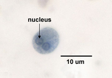
An important pathogen in this group is Entamoeba histolytica, the causative agent of amoebic dysentery. The organism is transmitted by the fecal-oral route. Cysts are excreted in the feces of an infected individual or carrier and ingested through fecally-contaminated food, water, objects, etc. After excystation, the trophozoites penetrate the walls of the large intestines causing ulceration and frequently causing the symptoms of dysentery. Involvement of the liver and other organs may occur if the protozoan invades the blood. The disease is diagnosed by microscopically looking for cysts of E. histolytica in a fecal smear (see Fig. \(\PageIndex{2}\).
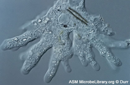
Acanthamoeba can infect the eye, blood, spinal cord, and brain and is transmitted by waterborne cysts picked up while swimming in contaminated water, crossing the mucous membranes.
Video of Acanthamoeba
b. The flagellates (subphylum Mastigophora) move by means of flagella. Some also have an undulating membrane.
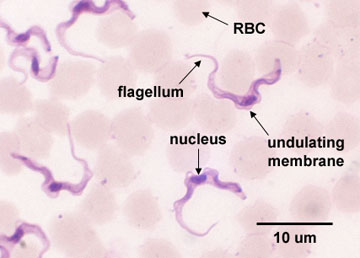
1. T. gambiense and T. rhodesiense cause the disease African sleeping sickness or African trypanosomiasis. They are transmitted to humans by the bite of an infected tsetse fly (a vector). The disease primarily involves the lymphatic and nervous systems of humans and is diagnosed by microscopically looking for Trypanosoma in the blood (see Fig. \(\PageIndex{3}\)), in aspirated fluid from lymph nodes, or in spinal fluid.
Video of Trypanosoma
T. cruzi causes South American sleeping sickness or Chagas' disease and is transmitted by infected Triatomid bugs (kissing bugs).
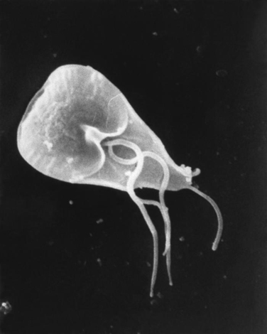
2. Giardia lamblia
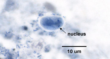
Giardia lamblia (G. intestinalis) causes a gastroenteritis-type of disease called giardiasis. Giardiasis is the most common protozoan intestinal disease in the U.S. and is transmitted by the fecal-oral route. Cysts of the organism are ingested through fecally-contaminated food, water, etc. Giardiasis is diagnosed by microscopically looking for cysts of G. lamblia in fecal smears (see Fig. \(\PageIndex{4}\)).
|
Fig. \(\PageIndex{5A}\) Scanning Electron Micrograph of Giardia and several Bacteria. Adhering to the Wall of the Small Intestine of a Mouse |
Fig. \(\PageIndex{5B}\): Production of Monoclonal Antibodies, Step-2 |
|---|---|
 |
|
|
© Ken Rozee, author. Licensed for use, ASM MicrobeLibrary. |
Image provided by Janice Haney Carr. Courtesy of the Centers for Disease Control and Prevention. Public Domain |
Video illustrating giardiasis
3. Trichomonas vaginalis
This protozoan causes genitourinary trichomoniasis. There are an estimated 2.5 million cases per year in the U.S. In females, it usually appears as vaginitis with itching and a white discharge. In males it is often asymptomatic but may cause urethritis. It is transmitted mainly by venereal contact and is diagnosed by microscopically looking for T. vaginalis trophozoites in vaginal discharge and urine (see Fig. \(\PageIndex{6}\)).

Video showing motility of Trichomonas vaginalis.
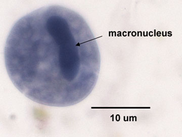
2. The Ciliophora
This group of protozoans is characterized by a covering of cilia used for motility and direction of food particles into the mouth or cytosome. In the trophozoite, a large macronucleus, small micronucleus, cilia, and contractile vacuoles may be seen.
The only pathogen in this group is Balantidium coli, which causes a diarrhea-type infection called balantidiasis. The protozoan is transmitted to humans by the fecal-oral route and invades the large intestines causing ulceration. It is diagnosed by microscopically looking for B. coli in a fecal smear (Fig. \(\PageIndex{7}\)).
Video showing motility of Balantidium coli.
3. The Apicomplexans
The sporozoa are not motile in their mature forms, reproduce both asexually and sexually, and often have complex life cycles for transmission from host to host. They possess a complex of organelles at their apex (apical complexes) that contain enzymes used in penetrating host tissues.
a. Toxoplasma gondii
This protozoan causes the disease toxoplasmosis. In adults, the disease is usually mild and resembles infectious mononucleosis. However, new-born infants who contracted toxoplasmosis in utero commonly have severe central nervous system damage. It also causes severe disease in immunosuppressed individuals such as people with AIDS. Domestic cats, that pick up the organism from eating infected rodents, may act as carriers of T. gondii, and their feces may contain oocysts of the protozoan. However, the organism may be found in practically every mammal. The disease is transmitted to humans by ingesting raw meat of an infected mammal or by inhaling or ingesting cysts of T. gondii from cat feces. Pregnant women should be especially careful to avoid raw meat and cat feces. The disease is diagnosed by serologic testing and by growing the organism in cell culture.
Video of Toxoplasma gondii
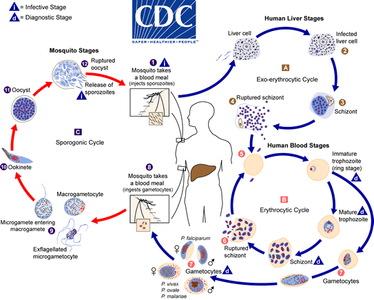
b. Plasmodium
Four Plasmodium species, P. falciparum, P. malariae, P. ovale, and P. vivax cause malaria. The vector involved in the transmission of the disease from human to human or from animal to human is an infected female Anopheles mosquito.
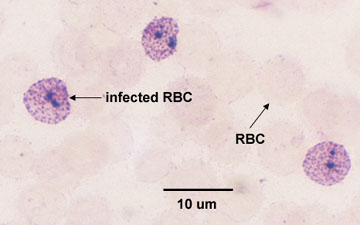
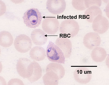
Asexual reproduction (or schizogony) of the Plasmodium occurs within liver cells and red blood cells of the infected human. With malaria caused by P. vivax and P. ovale, a dormant form or hypnozoite remains in the liver and may cause later relapses. The infected cells in which the organism is reproducing by schizogony are called schizonts. The sexual cycle (or sporogeny) occurs in the mosquito (see Fig. 6, the life cycle of Plasmodium). The typical recurring malarial fever is a result of the lysis of the infected red blood cells, causing release of merozoites and their metabolic by-products. Fever cycles of 24, 48, or 72 hours usually occur depending on the infecting species. Malaria is diagnosed by microscopically looking for the parasite within infected red blood cells (schizonts). See Fig. 7A and Fig 7B.
|
Fig. \(\PageIndex{8}\): Life cycle of Plasmodium, the protozoan that causes malaria |
Fig. \(\PageIndex{9A}\): Plasmodium malariae infecting red blood cells |
Fig. \(\PageIndex{9B}\): Plasmodium malariae infecting red blood cells |
|---|---|---|
 |
 |
 |
| By Content Providers: CDC [Public domain]. Courtesy of the Centers for Disease Control and Prevention. |
Copyright; Gary E. Kaiser, Ph.D. The Community College of Baltimore County, Catonsville Campus CC-BY-3.0 | Copyright; Gary E. Kaiser, Ph.D. The Community College of Baltimore County, Catonsville Campus CC-BY-3.0 |
c. Cryptosporidium
Cryptosporidium is an intracellular parasite that causes diarrhea, although in people who are immunosuppressed it can also cause respiratory and gallbladder infections. It is transmitted by the fecal-oral route.
Video of Cryptosporidium
Contributors and Attributions
Dr. Gary Kaiser (COMMUNITY COLLEGE OF BALTIMORE COUNTY, CATONSVILLE CAMPUS)