
10.4: Dimorphic Fungi
- Page ID
- 123398
Dimorphic fungi may exhibit two different growth forms. Outside the body they grow as a mold, producing hyphae and asexual reproductive spores, but inside the body they grow as a yeast-like form. Dimorphic fungi may cause systemic mycoses which usually begin by inhaling spores from the mold form. After germination in the lungs, the fungus grows as a yeast. Factors such as body temperature, osmotic stress, oxidative stress, and certain human hormones activate a dimorphism-regulating histidine kinase enzyme in dimorphic molds, causing them to switch from their avirulent mold form to their more virulent yeast form.
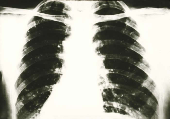
The infection usually remains localized in the lungs and characteristic lesions called granuloma (see Fig. \(\PageIndex{1}\)) may be formed to wall-off and localize the organism. In rare cases, usually in an immunosuppressed host, the organism may disseminate to other areas of the body and be life threatening. Examples of dimorphic fungi include Coccidioides immitis, Histoplasma capsulatum, and Blastomyces dermatitidis.
1. Coccidioides immitis
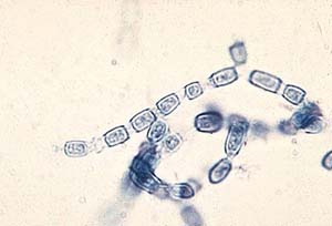
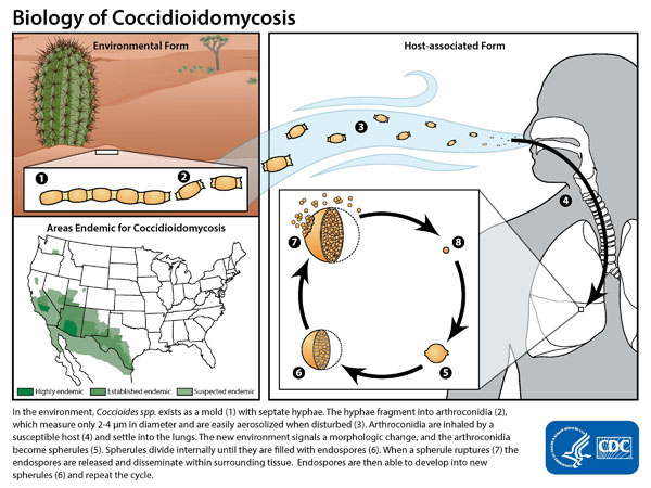
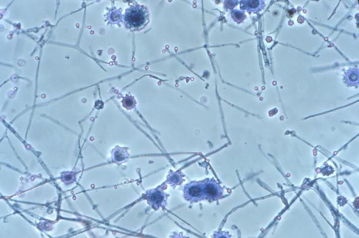
Coccidioides immitis (see Fig. \(\PageIndex{2A}\)) and Coccidioides posadasii are dimorphic fungi that causes coccidioidomycosis, a disease endemic to areas of the southwestern United States where there is a semiarid climate, an alkaline soil, and hot summers. An estimated 150,000 infections occur annually in the United States, but two thirds of these cases are sub-clinical. The mold form of the fungus grows in arid soil and produces thick-walled, barrel-shaped asexual spores called arthroconidia by a fragmentation of its vegetative hyphae (see Fig. \(\PageIndex{2B}\)).
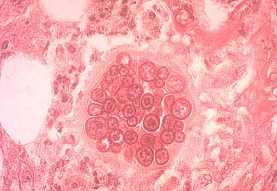
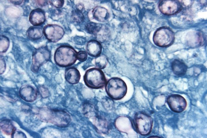
After inhalation, the arthroconidia sheds its outer coating, swells, and becomes an endosporulating spherule in the terminal bronchioles of the lungs (see Fig. \(\PageIndex{2C}\) and Fig. \(\PageIndex{2D}\)). The spherule contains hundreds to thousands of yeast-like endospores. The spherule subsequently ruptures and releases endospores that develop into new spherules.

|
Fig \(\PageIndex{2B}\): Arthroconidia of Coccidioides immitis |
Fig. \(\PageIndex{2C}\): Coccidioides immitis in the Lung: A Spherule Containing Endospores |
Fig. \(\PageIndex{2D}\): Coccidioides immitis in the Lung: Spherules Containing Endospores |
|---|---|---|
 |
 |
 |
| By Content Providers: CDC [Public domain]. Courtesy of the Centers for Disease Control and Prevention. |
By Content Providers: CDC [Public domain]. Courtesy of the Centers for Disease Control and Prevention. |
By Content Providers: CDC/Dr. Edwin P. Ewing, Jr. [Public domain]. Courtesy of the Centers for Disease Control and Prevention. |
Coccidioidomycosis can be diagnosed by culture, by a coccidioidin skin test, and by indirect serologic tests (discussed in Lab 18).
2.Histoplasma capsulatum
Histoplasma capsulatum (see Fig. \(\PageIndex{3A}\)) is a dimorphic fungus that causes histoplasmosis, a disease that is endemic to the Ohio, Missouri, and Mississippi River valleys in the United States. Approximately 250,000 people are thought to be infected annually in the US, but clinical symptoms of histoplasmosis occur in less than 5% of the population. Most individuals with histoplasmosis are asymptomatic. Those who develop clinical symptoms are typically either immunocompromised or are exposed to a large quantity of fungal spores.
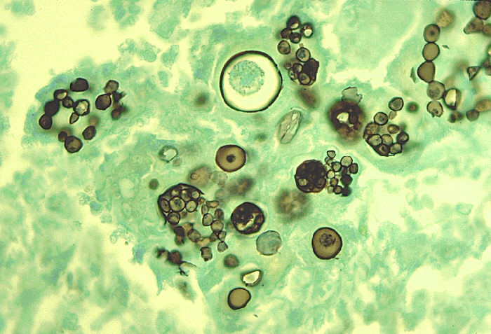
The mold form of the fungus often grows in acidic, damp soil with high organic content, especially near areas inhabited by bats and birds and produces large tuberculate macroconidia and small microconidia (see Fig. \(\PageIndex{1B}\)). Although birds cannot be infected by the fungus and do not transmit the disease, bird excretions contaminate the soil and enrich it for mycelial growth. Bats, however, can become infected and transmit histoplasmosis through their droppings. After inhalation of the fungal spores and their germination in the lungs, the fungus grows as a budding, encapsulated yeast (see Fig. \(\PageIndex{3C}\)).
Courtesy of the Centers for Disease Control and Prevention.)
|
Fig. \(\PageIndex{3B}\): Mold Phase of Histoplasma capsulatum Showing Tuberculate Macroconidia and Microconidia |
Fig. \(\PageIndex{3C}\): Yeast Phase of Histoplasma capsulatum in the Lungs |
|---|---|
 |
 |
| By Content Providers: CDC/Dr. Libero Ajello [Public domain]. Courtesy of the Centers for Disease Control and Prevention. |
By Content Providers: CDC/Dr. Libero Ajello [Public domain]. Courtesy of the Centers for Disease Control and Prevention. |
Histoplasmosis can be diagnosed by culture, by a histoplasmin skin test, and by indirect serologic tests (discussed in Lab 18).
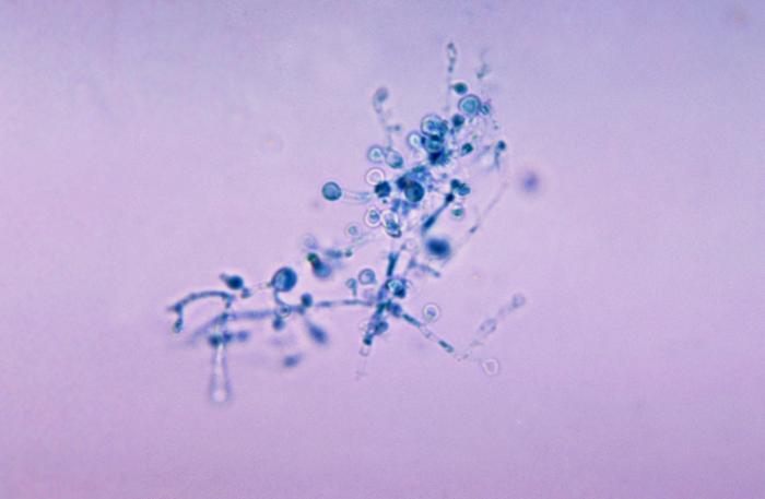
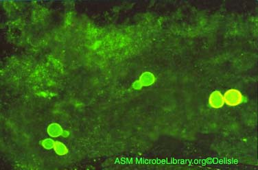
3. Blastomyces dermatitidis
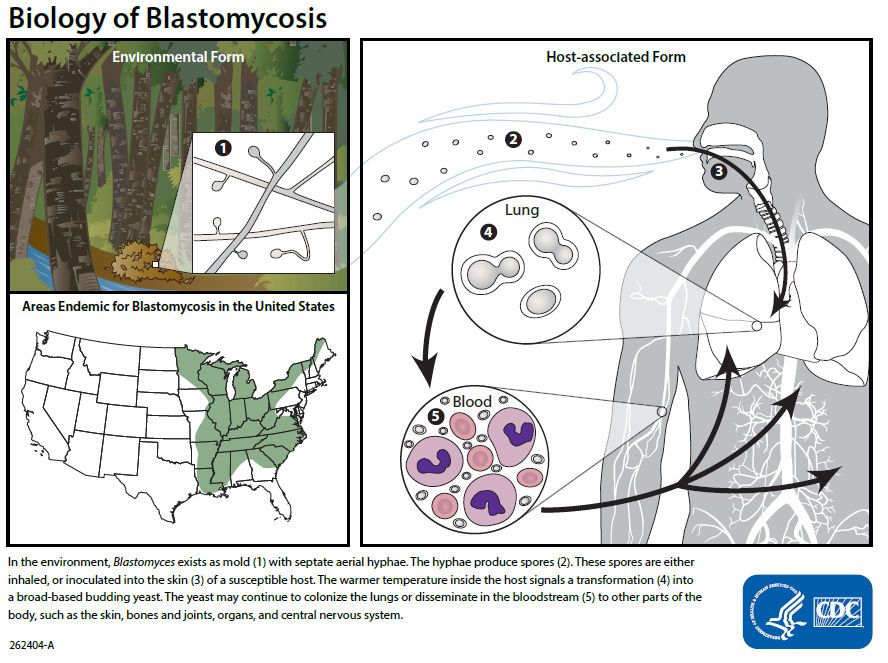
Blastomycosis, caused by Blastomyces dermatitidis, is common around the Great Lakes region and the Mississippi and Ohio River valleys.Infection can range from an asymptomatic, self-healing pulmonary infection to widely disseminated and potentially fatal disease. Pulmonary infection may be asymptomatic in nearly 50% of patients. Blastomyces dermatitidis can also sometimes infect the skin.
Blastomyces dermatitidis produces a mycelium with small conidiospores and grows actively in bird droppings and contaminated soil. When spores are inhaled or enter breaks in the skin, they germinate and the fungus grows as a yeast having a characteristic thick cell wall. It is diagnosed by culture and by biopsy examination.
Courtesy of the Centers for Disease Control and Prevention.)
|
Fig. \(\PageIndex{5B}\): Mold Form of Blastomyces dermatitidis with Hyphae and Conidiospores |
Fig. \(\PageIndex{5C}\): Yeast Form of Blastomyces dermatitidis in the Lung |
 |
 |
| By Content Providers: CDC/Dr. Leanor Haley [Public domain]. Courtesy of the Centers for Disease Control and Prevention. |
Blastomyces dermatitidis. Gloria Delisle and Lewis Tomalty, authors. Licensed for use, ASM MicrobeLibrary. |
Medscape articles on infections associated with organisms mentioned in this lab exercise. Registration to access this website is free.
Contributors and Attributions
Dr. Gary Kaiser (COMMUNITY COLLEGE OF BALTIMORE COUNTY, CATONSVILLE CAMPUS)