
10.3: Dermatophytes
( \newcommand{\kernel}{\mathrm{null}\,}\)
The dermatophytes are a group of molds that cause superficial mycoses of the hair, skin, and nails and utilize the protein keratin, that is found in hair, skin, and nails, as a nitrogen and energy source. Infections are commonly referred to as ringworm or tinea infections and include:
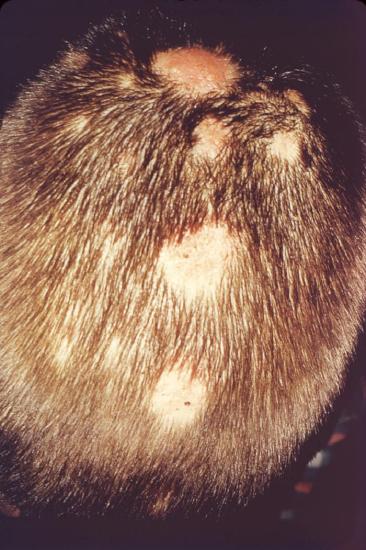
- tinea capitis (infection of the skin of the scalp, eyebrows, and eyelashes) See Fig. 10.3.1A.
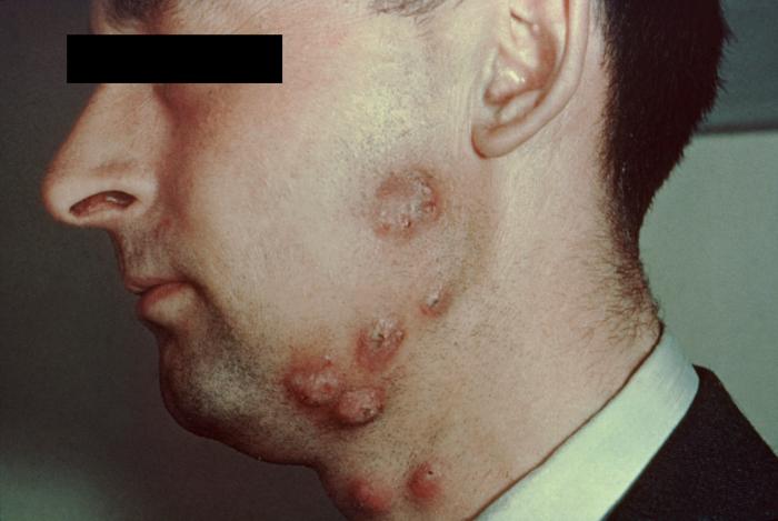
- tinea barbae (infection of the bearded areas of the face and neck) See Fig. \(\PageIndex{1B}\).
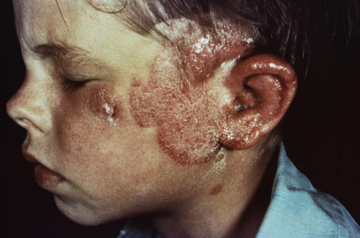
- tinea faciei (infection of the skin of the face) See Fig. 10.3.1C.
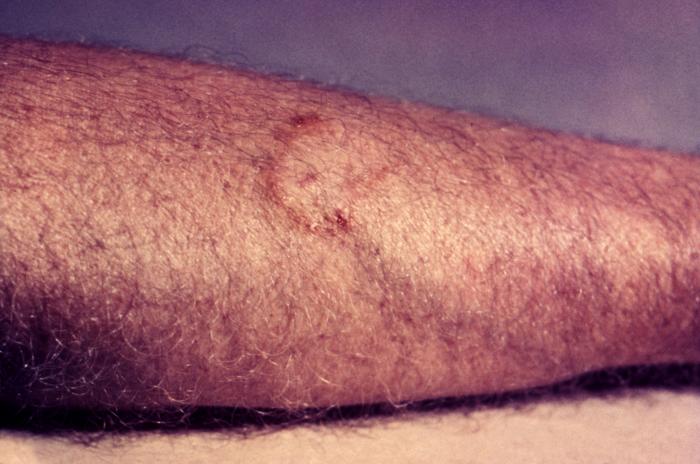
- tinea corporis (infection of the skin regions other than the scalp, groin, palms, and soles) See Fig. 10.3.1D.
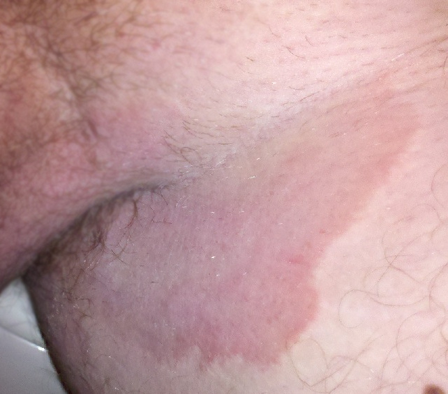
- tinea cruris (infection of the groin; jock itch) See Fig. 10.3.1E.
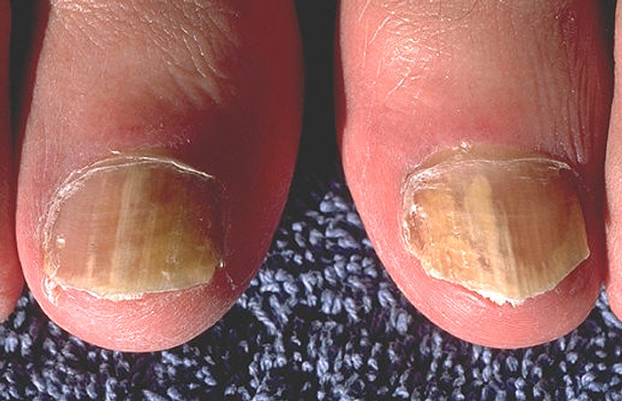
- tinea unguium (onychomycosis; infection of the fingernails and toenails) See Fig. \(\PageIndex{1F}\).
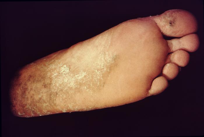
- tinea pedis (athlete's foot; infection of the soles of the feet and between the toes) See Fig. 10.3.1G.
|
Fig 10.3.1A: Tinea Capitis |
Fig. \(\PageIndex{1B}\): Tinea Barbae |
Fig. 10.3.1C: Tinea Faciei |
Fig 10.3.1D: Tinea Corporis |
|---|---|---|---|
 |
 |
 |
 |
| By Content Providers: CDC/Dr. Lucille K. Georg [Public domain]. Courtesy of the Centers for Disease Control and Prevention. |
By Content Providers: CDC [Public domain]. Courtesy of the Centers for Disease Control and Prevention. |
By Content Providers: CDC [Public domain]. Courtesy of the Centers for Disease Control and Prevention. |
By Content Providers: CDC [Public domain]. Courtesy of the Centers for Disease Control and Prevention |
|
Fig 10.3.1E: Tinea Capitis |
Fig. \(\PageIndex{1F}\): Tinea Barbae |
Fig. 10.3.1G: Tinea Faciei |
|---|---|---|
 |
 |
 |
| By Content Providers: CDC/Dr. Lucille K. Georg [Public domain]. Courtesy of the Centers for Disease Control and Prevention. |
By Content Providers: CDC [Public domain]. Courtesy of the Centers for Disease Control and Prevention. |
By Content Providers: CDC [Public domain]. Courtesy of the Centers for Disease Control and Prevention. |
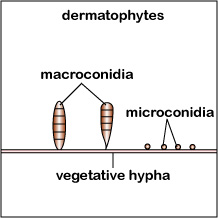
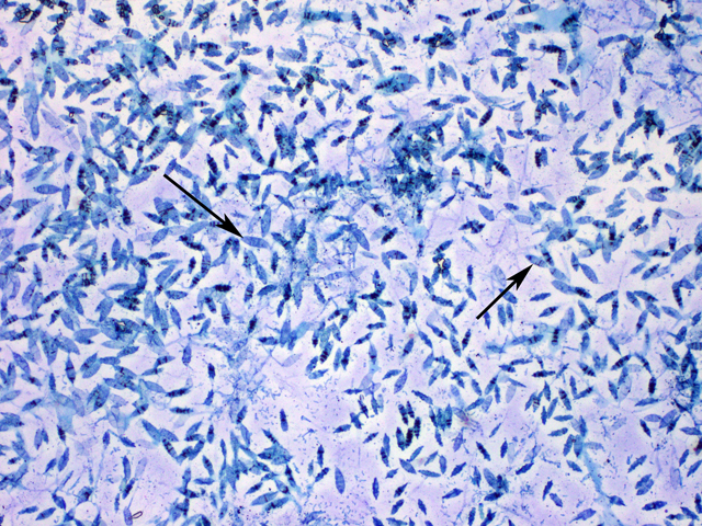
The three common dermatophytes are Microsporum, Trichophyton, and Epidermophyton. These organisms grow well at 25°C. They may produce large leaf or club-shaped asexual spores called macroconidia (see Fig. 10.3.2A) as well as small spherical asexual spores called microconidia, both from vegetative hyphae (see Fig. \(\PageIndex{2B}\)).
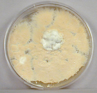
Microsporum (see Fig. \(\PageIndex{2C}\)) commonly infects the skin and hair, Epidermophyton, the skin and nails, and Trichophyton, the hair, skin, and nails. Dermatophytic infections are acquired by contact with fungal spores from infected humans, animals, or objects. On the skin, the dermatophytes typically cause reddening, itching, edema, and necrosis of tissue. This is a result of fungal growth and a hypersensitivity of the host to the fungus and its products. Frequently there is secondary bacterial or Candida invasion of the traumatized tissue.
|
Fig 10.3.2A: Macroconidia and Microconidia of Dermatophytes |
Fig. \(\PageIndex{2B}\): Macroconidia of the Dermatophyte Microsporum |
Fig. 10.3.2C: The Dermatophyte Microsposum Growing on Saboraud Dextrose Agar |
|---|---|---|
 |
 |
 |
| Copyright; Gary E. Kaiser, Ph.D. The Community College of Baltimore County, Catonsville Campus CC-BY-3.0 | ||
To diagnose dermatophytic infections, tissue scrapings can be digested with 10% potassium hydroxide (which causes lysis of the human cells but not the fungus) and examined microscopically for the presence of fungal hyphae and spores. To establish the specific cause of the infection, fungi from the affected tissue can be cultured on Dermatophyte Test Medium (DTM) and Saboraud Dextrose agar (SDA).
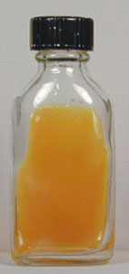
Dermatophyte Test Medium (DTM) has phenol red as a pH indicator with the medium yellow (acid) prior to inoculation. As the dermatophytes utilize the keratin in the medium, they produce alkaline end products that raise the pH, thus turning the phenol red in the medium from yellow or acid to red or alkaline. (See Fig. (\PageIndex{3B}\) and (\PageIndex{3B}\).).
The types of macroconidia and microconidia (see Fig. (\PageIndex{2B}\) above) can be observed by growing the mold on SDA and observing under a microscope. In addition, many dermatophyte species produce yellow to red-pigmented colonies on SDA and the most common species of Microsporum fluoresce under ultraviolet light.
|
Fig 10.3.3A: An Uninoculated Flask of Dermatophyte Test Medium (DTM) |
Fig. \(\PageIndex{3B}\): A Dermatophyte Growing on Dermatophyte Test Medium (DTM |
|---|---|
 |
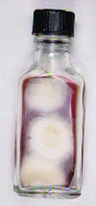 |
| An uninoculated flask of DTM with its acidic pH (yellow). | A dermatophyte growing on DTM. Note alkaline end products. |
| Copyright; Gary E. Kaiser, Ph.D. The Community College of Baltimore County, Catonsville Campus CC-BY-3.0 | |
Contributors and Attributions
Dr. Gary Kaiser (COMMUNITY COLLEGE OF BALTIMORE COUNTY, CATONSVILLE CAMPUS)