
10.2: Common Molds
- Page ID
- 123396
\( \newcommand{\vecs}[1]{\overset { \scriptstyle \rightharpoonup} {\mathbf{#1}} } \)
\( \newcommand{\vecd}[1]{\overset{-\!-\!\rightharpoonup}{\vphantom{a}\smash {#1}}} \)
\( \newcommand{\dsum}{\displaystyle\sum\limits} \)
\( \newcommand{\dint}{\displaystyle\int\limits} \)
\( \newcommand{\dlim}{\displaystyle\lim\limits} \)
\( \newcommand{\id}{\mathrm{id}}\) \( \newcommand{\Span}{\mathrm{span}}\)
( \newcommand{\kernel}{\mathrm{null}\,}\) \( \newcommand{\range}{\mathrm{range}\,}\)
\( \newcommand{\RealPart}{\mathrm{Re}}\) \( \newcommand{\ImaginaryPart}{\mathrm{Im}}\)
\( \newcommand{\Argument}{\mathrm{Arg}}\) \( \newcommand{\norm}[1]{\| #1 \|}\)
\( \newcommand{\inner}[2]{\langle #1, #2 \rangle}\)
\( \newcommand{\Span}{\mathrm{span}}\)
\( \newcommand{\id}{\mathrm{id}}\)
\( \newcommand{\Span}{\mathrm{span}}\)
\( \newcommand{\kernel}{\mathrm{null}\,}\)
\( \newcommand{\range}{\mathrm{range}\,}\)
\( \newcommand{\RealPart}{\mathrm{Re}}\)
\( \newcommand{\ImaginaryPart}{\mathrm{Im}}\)
\( \newcommand{\Argument}{\mathrm{Arg}}\)
\( \newcommand{\norm}[1]{\| #1 \|}\)
\( \newcommand{\inner}[2]{\langle #1, #2 \rangle}\)
\( \newcommand{\Span}{\mathrm{span}}\) \( \newcommand{\AA}{\unicode[.8,0]{x212B}}\)
\( \newcommand{\vectorA}[1]{\vec{#1}} % arrow\)
\( \newcommand{\vectorAt}[1]{\vec{\text{#1}}} % arrow\)
\( \newcommand{\vectorB}[1]{\overset { \scriptstyle \rightharpoonup} {\mathbf{#1}} } \)
\( \newcommand{\vectorC}[1]{\textbf{#1}} \)
\( \newcommand{\vectorD}[1]{\overrightarrow{#1}} \)
\( \newcommand{\vectorDt}[1]{\overrightarrow{\text{#1}}} \)
\( \newcommand{\vectE}[1]{\overset{-\!-\!\rightharpoonup}{\vphantom{a}\smash{\mathbf {#1}}}} \)
\( \newcommand{\vecs}[1]{\overset { \scriptstyle \rightharpoonup} {\mathbf{#1}} } \)
\(\newcommand{\longvect}{\overrightarrow}\)
\( \newcommand{\vecd}[1]{\overset{-\!-\!\rightharpoonup}{\vphantom{a}\smash {#1}}} \)
\(\newcommand{\avec}{\mathbf a}\) \(\newcommand{\bvec}{\mathbf b}\) \(\newcommand{\cvec}{\mathbf c}\) \(\newcommand{\dvec}{\mathbf d}\) \(\newcommand{\dtil}{\widetilde{\mathbf d}}\) \(\newcommand{\evec}{\mathbf e}\) \(\newcommand{\fvec}{\mathbf f}\) \(\newcommand{\nvec}{\mathbf n}\) \(\newcommand{\pvec}{\mathbf p}\) \(\newcommand{\qvec}{\mathbf q}\) \(\newcommand{\svec}{\mathbf s}\) \(\newcommand{\tvec}{\mathbf t}\) \(\newcommand{\uvec}{\mathbf u}\) \(\newcommand{\vvec}{\mathbf v}\) \(\newcommand{\wvec}{\mathbf w}\) \(\newcommand{\xvec}{\mathbf x}\) \(\newcommand{\yvec}{\mathbf y}\) \(\newcommand{\zvec}{\mathbf z}\) \(\newcommand{\rvec}{\mathbf r}\) \(\newcommand{\mvec}{\mathbf m}\) \(\newcommand{\zerovec}{\mathbf 0}\) \(\newcommand{\onevec}{\mathbf 1}\) \(\newcommand{\real}{\mathbb R}\) \(\newcommand{\twovec}[2]{\left[\begin{array}{r}#1 \\ #2 \end{array}\right]}\) \(\newcommand{\ctwovec}[2]{\left[\begin{array}{c}#1 \\ #2 \end{array}\right]}\) \(\newcommand{\threevec}[3]{\left[\begin{array}{r}#1 \\ #2 \\ #3 \end{array}\right]}\) \(\newcommand{\cthreevec}[3]{\left[\begin{array}{c}#1 \\ #2 \\ #3 \end{array}\right]}\) \(\newcommand{\fourvec}[4]{\left[\begin{array}{r}#1 \\ #2 \\ #3 \\ #4 \end{array}\right]}\) \(\newcommand{\cfourvec}[4]{\left[\begin{array}{c}#1 \\ #2 \\ #3 \\ #4 \end{array}\right]}\) \(\newcommand{\fivevec}[5]{\left[\begin{array}{r}#1 \\ #2 \\ #3 \\ #4 \\ #5 \\ \end{array}\right]}\) \(\newcommand{\cfivevec}[5]{\left[\begin{array}{c}#1 \\ #2 \\ #3 \\ #4 \\ #5 \\ \end{array}\right]}\) \(\newcommand{\mattwo}[4]{\left[\begin{array}{rr}#1 \amp #2 \\ #3 \amp #4 \\ \end{array}\right]}\) \(\newcommand{\laspan}[1]{\text{Span}\{#1\}}\) \(\newcommand{\bcal}{\cal B}\) \(\newcommand{\ccal}{\cal C}\) \(\newcommand{\scal}{\cal S}\) \(\newcommand{\wcal}{\cal W}\) \(\newcommand{\ecal}{\cal E}\) \(\newcommand{\coords}[2]{\left\{#1\right\}_{#2}}\) \(\newcommand{\gray}[1]{\color{gray}{#1}}\) \(\newcommand{\lgray}[1]{\color{lightgray}{#1}}\) \(\newcommand{\rank}{\operatorname{rank}}\) \(\newcommand{\row}{\text{Row}}\) \(\newcommand{\col}{\text{Col}}\) \(\renewcommand{\row}{\text{Row}}\) \(\newcommand{\nul}{\text{Nul}}\) \(\newcommand{\var}{\text{Var}}\) \(\newcommand{\corr}{\text{corr}}\) \(\newcommand{\len}[1]{\left|#1\right|}\) \(\newcommand{\bbar}{\overline{\bvec}}\) \(\newcommand{\bhat}{\widehat{\bvec}}\) \(\newcommand{\bperp}{\bvec^\perp}\) \(\newcommand{\xhat}{\widehat{\xvec}}\) \(\newcommand{\vhat}{\widehat{\vvec}}\) \(\newcommand{\uhat}{\widehat{\uvec}}\) \(\newcommand{\what}{\widehat{\wvec}}\) \(\newcommand{\Sighat}{\widehat{\Sigma}}\) \(\newcommand{\lt}{<}\) \(\newcommand{\gt}{>}\) \(\newcommand{\amp}{&}\) \(\definecolor{fillinmathshade}{gray}{0.9}\)To illustrate how morphological characteristics such as the type and form of asexual reproductive spores and the appearance of the mycelium may be used in identification, we will look at three common non-pathogenic molds.
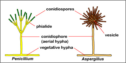
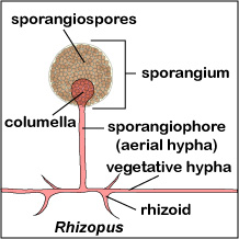
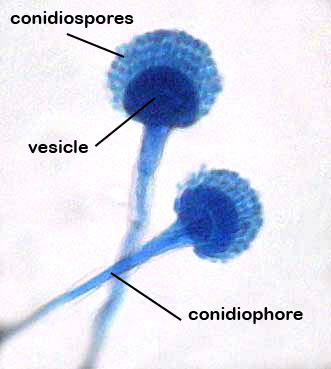
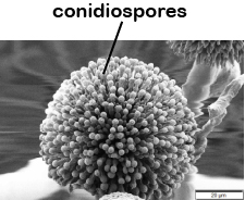
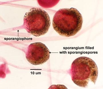
The two most common types of asexual reproductive spores produced by molds are conidiospores and sporangiospores. Conidiospores are borne externally in chains on an aerial hypha called a conidiophore. (See Fig. \(\PageIndex{1}\).); sporangiospores are produced within a sac or sporangium on an aerial hypha called a sporangiophore. (See Fig. \(\PageIndex{2}\).)
|
Fig \(\PageIndex{1}\): Fungal Conidiospores |
Fig. \(\PageIndex{2}\): Fungal Sporangiospores within a Sporangium |
|---|---|
 |
 |
| Copyright; Gary E. Kaiser, Ph.D. The Community College of Baltimore County, Catonsville Campus CC-BY-3.0 | |
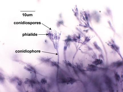
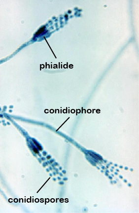
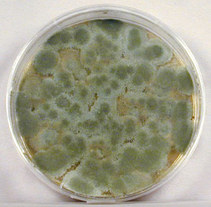
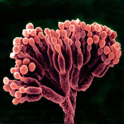
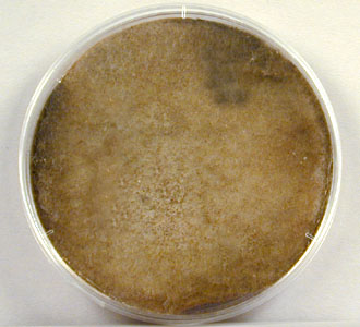
Penicillium and Aspergillus are examples of molds that produce conidiospores. Penicillium is one of the most common household molds and is a frequent food contaminant. The conidiospores of Penicillium (see Fig. \(\PageIndex{3A}\) and Fig. \(\PageIndex{3B}\)) usually appear grey, green, or blue (See Fig. \(\PageIndex{3C}\)) and are produced in chains on finger-like projections called phialides coming off of the conidiophore. Fig. \(\PageIndex{3D}\) shows conidiospores of Penicillium as seen with a scanning electron microscope.
|
Fig \(\PageIndex{3A}\): Conidiospores of Penicillium |
Fig. \(\PageIndex{3B}\): Conidiospores of Penicillium |
Fig. \(\PageIndex{3C}\): Penicillium Growing on Saboraud Dextrose Agar |
Fig \(\PageIndex{3D}\): Scanning Electron Micrograph of Penicillium showing Conidiospores |
|---|---|---|---|
 |
 |
 |
 |
| Copyright; Gary E. Kaiser, Ph.D. The Community College of Baltimore County, Catonsville Campus CC-BY-3.0 | Copyright; Gary E. Kaiser, Ph.D. The Community College of Baltimore County, Catonsville Campus CC-BY-3.0 | Copyright; Gary E. Kaiser, Ph.D. The Community College of Baltimore County, Catonsville Campus CC-BY-3.0 | By AJC1 [CC BY-SA 4.0-3.0-2.5-2.0-1.0 (https://creativecommons.org/licenses....0-2.5-2.0-1.0)], via Wikimedia Commons |
Aspergillus is another common contaminant. Although usually non-pathogenic, it may become opportunistic in the respiratory tract of a compromised host and, in certain foods, can produce mycotoxins. The conidiophore terminates in a ball-like structure called a vesicle. Its conidiospores, which typically appear brown to black (see Fig. \(\PageIndex{4A}\)), are produced in chains on phialides coming off of the vesicle. (See Fig. \(\PageIndex{4B}\) and Fig. \(\PageIndex{4C}\)). Fig. \(\PageIndex{4D}\) shows conidiospores of Aspergillus as seen with a scanning electron microscope
|
Fig \(\PageIndex{4A}\): Conidiospores of Penicillium |
Fig. \(\PageIndex{4B}\): Conidiospores of Penicillium |
Fig. \(\PageIndex{4C}\): Penicillium Growing on Saboraud Dextrose Agar |
Fig \(\PageIndex{4D}\): Scanning Electron Micrograph of Penicillium showing Conidiospores |
|---|---|---|---|
 |
 |
 |
 |
| Copyright; Gary E. Kaiser, Ph.D. The Community College of Baltimore County, Catonsville Campus CC-BY-3.0 | Copyright; Gary E. Kaiser, Ph.D. The Community College of Baltimore County, Catonsville Campus CC-BY-3.0 | By Carlos de Paz (https://www.flickr.com/photos/cdepaz/2421827982/) [CC BY-SA 2.0 (https://creativecommons.org/licenses/by-sa/2.0)], via Wikimedia Commons | By Mogana Das Murtey and Patchamuthu Ramasamy ([1]) [CC BY-SA 3.0 (https://creativecommons.org/licenses/by-sa/3.0)], via Wikimedia Commons |
Although generally harmless in most healthy individuals, Aspergillus species do cause allergic bronchopulmonary aspergillosis (ABPA), chronic necrotizing Aspergillus pneumonia (or chronic necrotizing pulmonary aspergillosis [CNPA]), aspergilloma (a mycetoma or fungus ball in a body cavity such as the lung), and invasive aspergillosis. In highly immunosuppressed individuals, however, Aspergillus may disseminate beyond the lung via the blood.
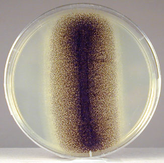
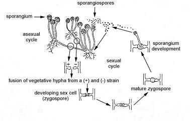
Rhizopus is an example of a mold that produces sporangiospores. Although usually non-pathogenic, it sometimes causes opportunistic wound and respiratory infections in the compromised host. At the end of its sporangiophore is dome-shaped end called a columella that extends into a sac-like structure called a sporangium. Its sporangiospores, typically brown or black (see Fig. \(\PageIndex{5A}\)), are produced within the sporangium (see Fig. \(\PageIndex{5B}\)). Anchoring structures called rhizoids are also produced on the vegetative hyphae.
Mucormycoses are infections caused by fungi belonging to the order of Mucorales. Rhizopus species are the most common causative organisms. The most common infection is a severe infection of the facial sinuses, which may extend into the brain. Other mycoses include pulmonary, cutaneous, and gastrointestinal.
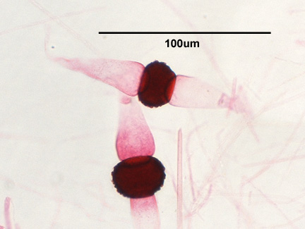
Rhizopus can also reproduce sexually. During sexual reproduction (see Fig. \(\PageIndex{5C}\)), hyphal tips of (+) and (-) mating type join and their nuclei fuse to form a sexual spore called a zygospore (see Fig.\(\PageIndex{5D}\)). This gives rise to a new sporangium producing sporangiospores having DNA that is a recombination of the two parent strain's DNA.
|
Fig \(\PageIndex{5A}\): Rhizopus Growing on SDA |
Fig \(\PageIndex{5B}\): Sporangiospores of Rhizopus |
Fig \(\PageIndex{5C}\): Sexual reproduction of Rhizopus |
Fig \(\PageIndex{5D}\): Zygospore of Rhizopus |
|---|---|---|---|
 |
 |
 |
 |
| Copyright; Gary E. Kaiser, Ph.D. The Community College of Baltimore County, Catonsville Campus CC-BY-3.0 | |||
Molds are commonly cultured on fungal-selective or enriched media such as Saboraud Dextrose agar (SDA), Corn Meal agar, and Potato Dextrose agar.
Contributors and Attributions
Dr. Gary Kaiser (COMMUNITY COLLEGE OF BALTIMORE COUNTY, CATONSVILLE CAMPUS)