Lab 8-11: Fetal Pig Dissection
- Page ID
- 24397

\( \newcommand{\vecs}[1]{\overset { \scriptstyle \rightharpoonup} {\mathbf{#1}} } \)
\( \newcommand{\vecd}[1]{\overset{-\!-\!\rightharpoonup}{\vphantom{a}\smash {#1}}} \)
\( \newcommand{\dsum}{\displaystyle\sum\limits} \)
\( \newcommand{\dint}{\displaystyle\int\limits} \)
\( \newcommand{\dlim}{\displaystyle\lim\limits} \)
\( \newcommand{\id}{\mathrm{id}}\) \( \newcommand{\Span}{\mathrm{span}}\)
( \newcommand{\kernel}{\mathrm{null}\,}\) \( \newcommand{\range}{\mathrm{range}\,}\)
\( \newcommand{\RealPart}{\mathrm{Re}}\) \( \newcommand{\ImaginaryPart}{\mathrm{Im}}\)
\( \newcommand{\Argument}{\mathrm{Arg}}\) \( \newcommand{\norm}[1]{\| #1 \|}\)
\( \newcommand{\inner}[2]{\langle #1, #2 \rangle}\)
\( \newcommand{\Span}{\mathrm{span}}\)
\( \newcommand{\id}{\mathrm{id}}\)
\( \newcommand{\Span}{\mathrm{span}}\)
\( \newcommand{\kernel}{\mathrm{null}\,}\)
\( \newcommand{\range}{\mathrm{range}\,}\)
\( \newcommand{\RealPart}{\mathrm{Re}}\)
\( \newcommand{\ImaginaryPart}{\mathrm{Im}}\)
\( \newcommand{\Argument}{\mathrm{Arg}}\)
\( \newcommand{\norm}[1]{\| #1 \|}\)
\( \newcommand{\inner}[2]{\langle #1, #2 \rangle}\)
\( \newcommand{\Span}{\mathrm{span}}\) \( \newcommand{\AA}{\unicode[.8,0]{x212B}}\)
\( \newcommand{\vectorA}[1]{\vec{#1}} % arrow\)
\( \newcommand{\vectorAt}[1]{\vec{\text{#1}}} % arrow\)
\( \newcommand{\vectorB}[1]{\overset { \scriptstyle \rightharpoonup} {\mathbf{#1}} } \)
\( \newcommand{\vectorC}[1]{\textbf{#1}} \)
\( \newcommand{\vectorD}[1]{\overrightarrow{#1}} \)
\( \newcommand{\vectorDt}[1]{\overrightarrow{\text{#1}}} \)
\( \newcommand{\vectE}[1]{\overset{-\!-\!\rightharpoonup}{\vphantom{a}\smash{\mathbf {#1}}}} \)
\( \newcommand{\vecs}[1]{\overset { \scriptstyle \rightharpoonup} {\mathbf{#1}} } \)
\(\newcommand{\longvect}{\overrightarrow}\)
\( \newcommand{\vecd}[1]{\overset{-\!-\!\rightharpoonup}{\vphantom{a}\smash {#1}}} \)
\(\newcommand{\avec}{\mathbf a}\) \(\newcommand{\bvec}{\mathbf b}\) \(\newcommand{\cvec}{\mathbf c}\) \(\newcommand{\dvec}{\mathbf d}\) \(\newcommand{\dtil}{\widetilde{\mathbf d}}\) \(\newcommand{\evec}{\mathbf e}\) \(\newcommand{\fvec}{\mathbf f}\) \(\newcommand{\nvec}{\mathbf n}\) \(\newcommand{\pvec}{\mathbf p}\) \(\newcommand{\qvec}{\mathbf q}\) \(\newcommand{\svec}{\mathbf s}\) \(\newcommand{\tvec}{\mathbf t}\) \(\newcommand{\uvec}{\mathbf u}\) \(\newcommand{\vvec}{\mathbf v}\) \(\newcommand{\wvec}{\mathbf w}\) \(\newcommand{\xvec}{\mathbf x}\) \(\newcommand{\yvec}{\mathbf y}\) \(\newcommand{\zvec}{\mathbf z}\) \(\newcommand{\rvec}{\mathbf r}\) \(\newcommand{\mvec}{\mathbf m}\) \(\newcommand{\zerovec}{\mathbf 0}\) \(\newcommand{\onevec}{\mathbf 1}\) \(\newcommand{\real}{\mathbb R}\) \(\newcommand{\twovec}[2]{\left[\begin{array}{r}#1 \\ #2 \end{array}\right]}\) \(\newcommand{\ctwovec}[2]{\left[\begin{array}{c}#1 \\ #2 \end{array}\right]}\) \(\newcommand{\threevec}[3]{\left[\begin{array}{r}#1 \\ #2 \\ #3 \end{array}\right]}\) \(\newcommand{\cthreevec}[3]{\left[\begin{array}{c}#1 \\ #2 \\ #3 \end{array}\right]}\) \(\newcommand{\fourvec}[4]{\left[\begin{array}{r}#1 \\ #2 \\ #3 \\ #4 \end{array}\right]}\) \(\newcommand{\cfourvec}[4]{\left[\begin{array}{c}#1 \\ #2 \\ #3 \\ #4 \end{array}\right]}\) \(\newcommand{\fivevec}[5]{\left[\begin{array}{r}#1 \\ #2 \\ #3 \\ #4 \\ #5 \\ \end{array}\right]}\) \(\newcommand{\cfivevec}[5]{\left[\begin{array}{c}#1 \\ #2 \\ #3 \\ #4 \\ #5 \\ \end{array}\right]}\) \(\newcommand{\mattwo}[4]{\left[\begin{array}{rr}#1 \amp #2 \\ #3 \amp #4 \\ \end{array}\right]}\) \(\newcommand{\laspan}[1]{\text{Span}\{#1\}}\) \(\newcommand{\bcal}{\cal B}\) \(\newcommand{\ccal}{\cal C}\) \(\newcommand{\scal}{\cal S}\) \(\newcommand{\wcal}{\cal W}\) \(\newcommand{\ecal}{\cal E}\) \(\newcommand{\coords}[2]{\left\{#1\right\}_{#2}}\) \(\newcommand{\gray}[1]{\color{gray}{#1}}\) \(\newcommand{\lgray}[1]{\color{lightgray}{#1}}\) \(\newcommand{\rank}{\operatorname{rank}}\) \(\newcommand{\row}{\text{Row}}\) \(\newcommand{\col}{\text{Col}}\) \(\renewcommand{\row}{\text{Row}}\) \(\newcommand{\nul}{\text{Nul}}\) \(\newcommand{\var}{\text{Var}}\) \(\newcommand{\corr}{\text{corr}}\) \(\newcommand{\len}[1]{\left|#1\right|}\) \(\newcommand{\bbar}{\overline{\bvec}}\) \(\newcommand{\bhat}{\widehat{\bvec}}\) \(\newcommand{\bperp}{\bvec^\perp}\) \(\newcommand{\xhat}{\widehat{\xvec}}\) \(\newcommand{\vhat}{\widehat{\vvec}}\) \(\newcommand{\uhat}{\widehat{\uvec}}\) \(\newcommand{\what}{\widehat{\wvec}}\) \(\newcommand{\Sighat}{\widehat{\Sigma}}\) \(\newcommand{\lt}{<}\) \(\newcommand{\gt}{>}\) \(\newcommand{\amp}{&}\) \(\definecolor{fillinmathshade}{gray}{0.9}\)Dissection is a powerful tool that provides us a profound understanding of our own anatomy and physiology as living, breathing creatures and also helps us to develop a stronger understanding of evolutionary relationships between taxonomic groups. What sort of dissection experiences do you already have? As you begin this sequence of dissections, please keep in mind several things. Dissection should be done thoughtfully and respectfully. It is important to take your time carefully and to think about what you are doing. This care will help you to preserve structures for the next several weeks and will make review easier. Also, merely identifying structures is insufficient to develop fully an appreciation for how these structures work. You must think about the function of these structures. How do they work?! Also, please do these dissections respectfully. These organisms were euthanized so that we might have a wonderful opportunity to learn something greater about the living world. Please keep that in mind as you are working.
Objectives:
- Develop dissecting skills.
- Learn anatomy and physiology of key vertebrate and mammalian systems. You will be using this handout, your dissected specimen, and the dissection key on your bench.
External Anatomy:
Determine the Sex of Your Pig:
1. Before you start dissecting, examine the outside of the pig and determine its sex. Look for these features:
- Males: The urogenital opening is located near the umbilicus; the penis is hidden inside. The scrotal sac may be visible as a swelling just ventral to the anus, depending on the age of the fetus. The testes are still deep inside the body cavity; they don't descend into the scrotal sac until later.
- Females: Look for the urogenital papilla (a nipple-looking structure), located just below the anus.
Also, both males and females have nipples, just as in humans.
What sex is your pig? ______________
2. Make sure you are familiar with anatomical terms of reference: anterior (front), posterior (back), dorsal (above), ventral (below).
In addition, you’ll need to know the following terms:
- Medial: toward the midline or middle of the body
- Lateral: toward the outside of the body
- Proximal: close to a point of reference
- Distal: farther from a point of reference
*label the sides on the pig picture above*
3. Open the pig’s mouth and locate the hard and soft palate on the roof of the mouth. Can you feel your own hard and soft palates with your tongue?
- Note the taste buds (also known as sensory papillae) on the side of the tongue. Locate the esophagus at the back of the mouth. Feel the edge of the mouth for teeth. Does the fetal pig have teeth? ____________
- To access the following structures, you will have to cut down either side of the jaw and pry the jaw down. This can be difficult and requires some force. You, essentially, must break the jaw, and it will make a cracking sound. Once you do this locate the epiglottis, a cone-shaped structure at the back of the mouth, a flap of skin helps to cover the trachea when a pig swallows. The pharynx (throat) is the cavity in the back of the mouth – it is the junction for food (esophagus) and air (trachea).
4. Observe the toes of the pig. How many toes are on the feet? ___________________ Do they have an odd or even number of toes? ___________________
Note
Make sure you know the locations of all the bold words on this handout.
You will now work on opening the abdominal and thoracic cavities of the pig and identify structures. Remember, that to dissect means to "cut into pieces" from Latin 'dessicare '- a careful dissection will make it easier for you to find the organs and structures. Be sure to follow all directions.
The Incision: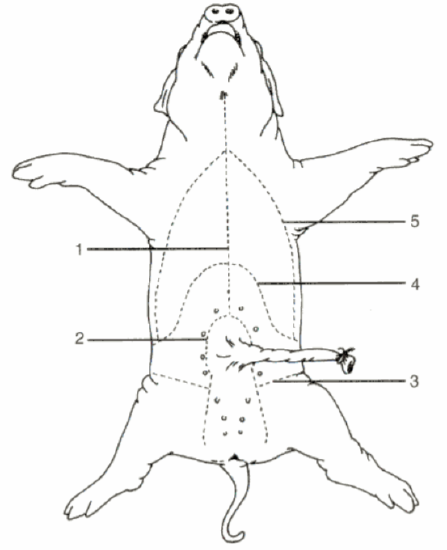
Place your fetal pig in the dissecting pan ventral side up. Use string to tie the legs behind the back of the pan. Use scissors to cut through the skin and muscles according to the diagram. Do not remove the umbilical cord.
After completing the cuts, locate the umbilical vein that leads from the umbilical cord to the liver. You will need to cut this vein in order to open up the abdominal cavity.
Your pig may be filled with water and preservative, drain over the sink if necessary.
Neck Region:
Using the diagram to the right begin your dissection in the neck region. Try to cut as little as possible. Once you open the body cavity, you will generally be able to separate the different organs by simply pulling them apart with your fingers, forceps, or a probe. The more you cut things up, the harder it will be to figure out what you’re looking at. Cut midline on the ventral surface of the neck to expose the underlying muscles. Carefully separate the muscles to observe the underlying structures. Locate and understand the functions of the following structures:
- Larynx: an enlarged structure on the trachea. If you cut it open, you can see the vocal cords inside.
- Thymus Gland: an endocrine (hormone-secreting) gland that helps regulate the immune system. It’s a large, spongy structure covering the ventral surface of the trachea, extending up along either side, and also resides over the heart. It is easy to cut so be extra careful.
- Thyroid Gland: another endocrine gland; it’s a small bilobed (two parts) structure just posterior to the larynx. The thyroid secretes hormones that help regulate metabolism.
- Trachea: the airway; it's reinforced with rings of cartilage so it does not collapse.
- Esophagus: carries food from mouth to stomach; soft and muscular so it can move a food bolus by peristalsis. It is located dorsal to the trachea (but appears behind it because the specimen is upside down).
Thoracic Cavity:
Vertebrates have true coeloms (a body cavity). In mammals, the coelom is divided into two main cavities: the thoracic cavity, which contains the lungs, and the abdominal cavity, which contains the digestive system. The thoracic cavity and the abdominal cavity are separated by the diaphragm. Note the many membranes lining the coelom and holding the organs in place. Look for these structures in the thoracic cavity:
- Lungs: they have several lobes. Note how spongy the tissue is.
- Heart: muscular and easy to find. The heart is surrounded by a pericardial sac. Note the aorta, where high-pressure blood leaves the heart on its way to the systemic circulation. You may also see the right and left carotid arteries, which supply blood to the head. For now, don’t spend too much time on the various lobes of the heart and the many blood vessels. Come back to these later.
- Diaphragm: a sheet of muscle and connective tissue that helps in breathing and divides the two cavities described here.
Abdominal Cavity (Digestion & Absorption):
Locate and understand the functions of the following structures:
- Liver: very large and dark. It has several lobes. You’ll need to lift it out of the way to see the organs beneath. The liver produces bile that is stored in the gall bladder. The gall bladder is a small organ attached to the underside of the liver; it's usually greenish due to the bile. It connects to the small intestine by the bile duct.
- Stomach: a muscular, sac-like organ that sits posterior to and to the left of the liver. Here is where the gastric juices released by glands continue enzymatic digestion started in the mouth. In particular, proteins are hydrolyzed via pepsin. At each end of the stomach are valves that regulate food entering and leaving the stomach. At the esophagus is the cardiac sphincter valve, and at the duodenum is the pyloric sphincter valve. View the inside of the stomach by slicing it open lengthwise.
- Small & Large Intestine: tube-like structures that continue the movement of food (now called chyme). The small intestine is first. The initial part of the small intestine (duodenum) is responsible for the final steps of enzymatic digestion and then eventually absorption of the degraded molecules. The large intestine primarily functions to compact the remaining waste material by absorbing water invested in the digestion and lubrication process. Also absorption of vitamins completes here.
- Rectum: the final portion of the large intestine where wastes are stored before elimination through the anus.
- Mesenteries: thin, transparent sheets of connective tissue containing blood vessels connecting the intestine and other organs.
- Pancreas: white and looks a little bit like cauliflower and located along the underside of the stomach, a pancreatic duct leads to the duodenum – first part of the small intestine. The pancreas also makes insulin, which is necessary for the proper uptake of sugars from the blood. It secretes digestive enzymes and buffers as well that contribute to the digestion of material within the small intestine.
- Spleen: The spleen is a flat organ located near the stomach. It performs several functions related to producing and maturing new blood cells and eliminating old ones. Blood passes through open sinuses in the spleen, rather than being confined to narrow blood vessels.
Identify the Organ (or Structure):
1. _____________________________ Opening (valve) between stomach and small intestine.
2. _____________________________ Stores bile, lies underneath the liver.
3. _____________________________ Separates the thoracic and abdominal cavity; aids breathing.
4. _____________________________ Membrane that holds the coils of the small intestine.
5. _____________________________ The part of the small intestine just after the stomach.
6. _____________________________ Empties bile into the duodenum from the gall bladder.
7. _____________________________ The last part of the large intestine before it exits at the anus.
8. _____________________________ Bumpy structure under the stomach; makes insulin.
Circulatory System:
Note the following features:
Arteries, which carry high-pressure blood away from the heart, are generally thicker walled than veins, which carry lower-pressure blood back to the heart. Mammalian hearts have four chambers. Each side of the heart has an atrium that receives blood from elsewhere in the body and a ventricle that pumps the blood out of the heart. The right atrium receives blood from the systemic circulation and passes it to the right ventricle, which pumps the blood to the pulmonary circuit. After the blood passes through the lungs it goes to the left atrium and then into the left ventricle, which pumps the blood into the systemic circuit. The first part of the systemic arterial circuit is the aorta, which soon branches out to supply various regions of the body.
Fetal circulation is different from adult circulation. In the fetus, blood doesn’t get oxygenated in the lungs; it gets oxygenated at the placenta. The umbilical arteries carry blood from the fetus to the placenta. The umbilical vein carries blood from the placenta back to the fetus. (In the placenta substances are exchanged between fetal and maternal blood, but the blood itself does not mix.) Therefore, the most highly oxygenated blood in the fetus is in the umbilical vein. Blood from the umbilical vein gets mixed with the rest of the systemic circulation and returns to the right atrium. The blood entering the right atrium is the most oxygenated blood in the fetal heart, but it’s the least oxygenated blood in the adult heart. The fetus has two key tricks to adapt to this fact: First, some of the blood that leaves the right ventricle bypasses the lungs. In an adult, this blood needs to go to the lungs to get oxygenated, but the fetus has a ductus arteriosus that shortcircuits this blood flow, allowing some blood to go directly into the aorta and then into the systemic circulation. Second, in the fetal heart, there is an opening between the right atrium and the left atrium. This opening is called the foramen ovale. The foramen ovale is helpful in the fetus because it lets the oxygenated blood from the placenta get circulated faster. The foramen ovale normally closes up at birth, keeping blood flow of the two sides of the heart completely separate. In some people, the foramen ovale does not close up. This condition, called patent foramen ovale, can result in serious health problems.
Dissection of the Thoracic Cavity:
Arteries:
Note
The diagram below only focuses on arteries and you do not need to know all of them.
1. Find the diaphragm again. Remember that the diaphragm separates the abdominal cavity from the thoracic cavity and it aids in breathing. Above the diaphragm, the center of the chest, is the heart.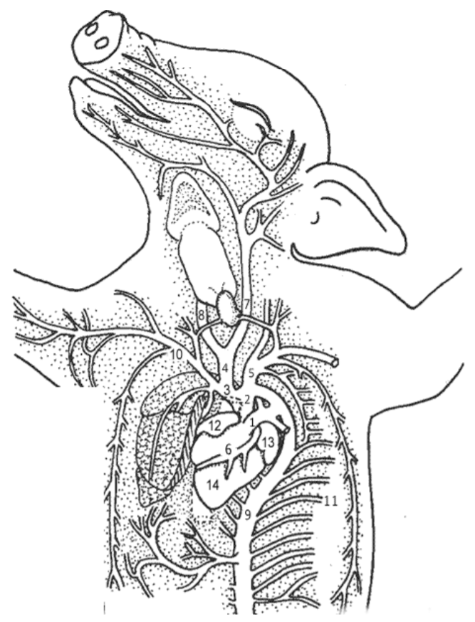
2. Remove the pericardium, which is a thin membrane that surrounds the heart.
3. The structures visible on the heart are the two atria (12,13), the ventricle (14), which has two chambers not easily visible from the outside.
4. The most obvious vessel on the front of the heart is the pulmonary trunk (1). It curves upward and joins the aorta (2) - a vessel which arches from the heart and curves around to go to the lower part of the body -where it is called the abdominal (dorsal) aorta (9). The aorta supplies the body with blood.
5. Find the vessel anterior to the heart in the base of the neck. This is the common carotid (4).
6. The common carotid will branch into the left (7) and right carotid arteries (8). The carotid arteries supply blood to the head and neck.
7. Observe the coronary vessels (6) on the outside of the heart—these vessels supply blood to the muscle of the heart.
Veins:
8. Lift the heart to look on its dorsal side (toward the back), you should be able to see the anterior and posterior vena cavae, which bring blood back to the heart from the body. In addition, you should also be able to find the left and right jugular veins that drain blood from the head and run parallel to the carotids. Veins are injected with blue latex.
Identify the structure.
1. ___________________________ Membrane over the heart.
2. ___________________________ Blood supply to head
3. ___________________________ Lower heart chambers
4. ___________________________ Blood supply to lower body
5. ___________________________ Muscle to aid breathing
6. ___________________________ Returns blood to heart
7. ___________________________ Large artery that arches over top of heart
8. ___________________________ Arteries on heart surface.
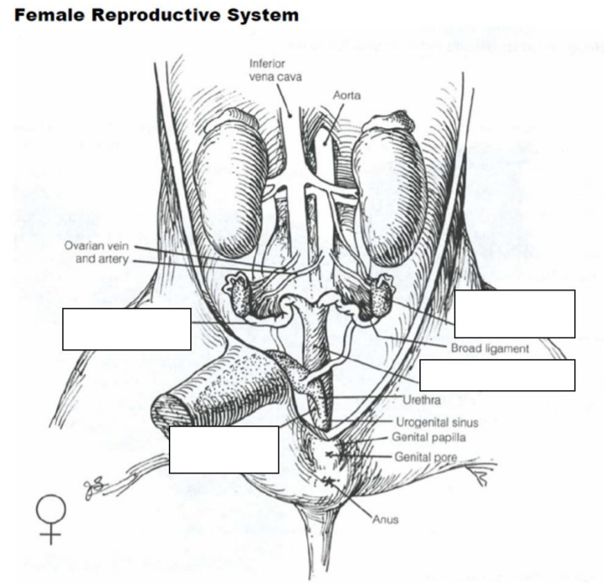
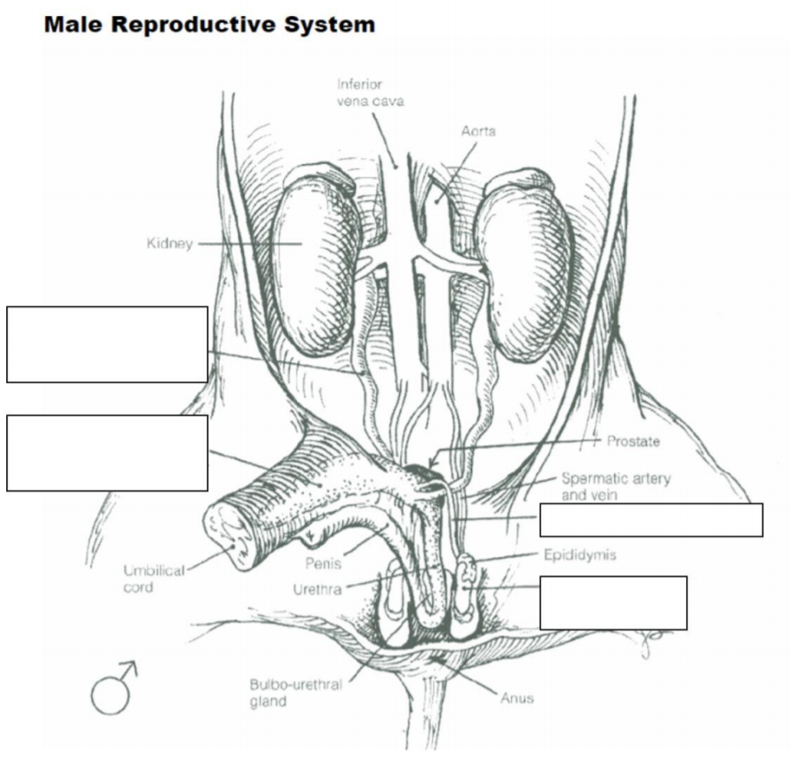
Urogenital System:
Locate and understand the functions of the following structures:
Kidneys: The two kidneys are not actually located in the abdominal cavity; they occupy another coelomic compartment dorsal to the abdominal cavity. You won't see them until you move the intestines aside. The kidneys actually are located behind the mesentery lining the abdominal cavity. Gently break through this tissue. Urine from the kidneys goes into the urinary bladder via the ureters, and then through the urethra as it is eliminated from the body.
Urinary Bladder & Urethra: The urethra is the tube that carries urine from the urinary bladder to the urinary opening. You can find the urinary bladder positioned between the two umbilical arteries.
Ovaries and Uterus (females) or Testes (males): The testes of males and the ovaries of females both arise from the same embryonic structures; however, the testes migrate during fetal development until they descend into the scrotal sac. The size of the testes varies significantly, depending on the age of the fetal pig.
- Female:
- In the female pig, locate two bean-shaped ovaries located just posterior to the kidneys and connected to the curly oviducts. These typically are quite small in the fetal pig.
- Trace the oviducts toward the posterior to find that they merge at the uterus. Trace the uterus to the vagina. The vagina actually will appear as a continuation of the uterus.
- Male:
- Find the scrotal sacs at the posterior end of the pig (between the legs), testes are located in each sac. Open the scrotal sac to locate the testis.
- On each testis, find the coiled epididymis. Sperm cells produced in the testes pass through the epididymis and into a tube called the vas deferens (in humans, a vasectomy involves cutting this tube).
- The penis can be located by cutting away the skin on the flap near the umbilical cord. This tube-like structure eventually exits out the urogenital opening, also known as the urethra.
On the next two pages label the diagrams of the female and male urogenital systems.
This lab includes material that has been adapted from http://brianmccauley.net/bio-6a/bio-...tal-piganatomy, http://www.biologycorner.com/workshe...issection.html,
https://7zscience.wikispaces.com/Fetal+Pig+Dissection, and https://designeranimal2012.wikispaces.com/Wild+Pig and is licensed under a Creative Commons Attribution-NonCommercial-ShareAlike 4.0 International License.