
Lab 5: Sensory Systems
- Page ID
- 24391

\( \newcommand{\vecs}[1]{\overset { \scriptstyle \rightharpoonup} {\mathbf{#1}} } \)
\( \newcommand{\vecd}[1]{\overset{-\!-\!\rightharpoonup}{\vphantom{a}\smash {#1}}} \)
\( \newcommand{\dsum}{\displaystyle\sum\limits} \)
\( \newcommand{\dint}{\displaystyle\int\limits} \)
\( \newcommand{\dlim}{\displaystyle\lim\limits} \)
\( \newcommand{\id}{\mathrm{id}}\) \( \newcommand{\Span}{\mathrm{span}}\)
( \newcommand{\kernel}{\mathrm{null}\,}\) \( \newcommand{\range}{\mathrm{range}\,}\)
\( \newcommand{\RealPart}{\mathrm{Re}}\) \( \newcommand{\ImaginaryPart}{\mathrm{Im}}\)
\( \newcommand{\Argument}{\mathrm{Arg}}\) \( \newcommand{\norm}[1]{\| #1 \|}\)
\( \newcommand{\inner}[2]{\langle #1, #2 \rangle}\)
\( \newcommand{\Span}{\mathrm{span}}\)
\( \newcommand{\id}{\mathrm{id}}\)
\( \newcommand{\Span}{\mathrm{span}}\)
\( \newcommand{\kernel}{\mathrm{null}\,}\)
\( \newcommand{\range}{\mathrm{range}\,}\)
\( \newcommand{\RealPart}{\mathrm{Re}}\)
\( \newcommand{\ImaginaryPart}{\mathrm{Im}}\)
\( \newcommand{\Argument}{\mathrm{Arg}}\)
\( \newcommand{\norm}[1]{\| #1 \|}\)
\( \newcommand{\inner}[2]{\langle #1, #2 \rangle}\)
\( \newcommand{\Span}{\mathrm{span}}\) \( \newcommand{\AA}{\unicode[.8,0]{x212B}}\)
\( \newcommand{\vectorA}[1]{\vec{#1}} % arrow\)
\( \newcommand{\vectorAt}[1]{\vec{\text{#1}}} % arrow\)
\( \newcommand{\vectorB}[1]{\overset { \scriptstyle \rightharpoonup} {\mathbf{#1}} } \)
\( \newcommand{\vectorC}[1]{\textbf{#1}} \)
\( \newcommand{\vectorD}[1]{\overrightarrow{#1}} \)
\( \newcommand{\vectorDt}[1]{\overrightarrow{\text{#1}}} \)
\( \newcommand{\vectE}[1]{\overset{-\!-\!\rightharpoonup}{\vphantom{a}\smash{\mathbf {#1}}}} \)
\( \newcommand{\vecs}[1]{\overset { \scriptstyle \rightharpoonup} {\mathbf{#1}} } \)
\(\newcommand{\longvect}{\overrightarrow}\)
\( \newcommand{\vecd}[1]{\overset{-\!-\!\rightharpoonup}{\vphantom{a}\smash {#1}}} \)
\(\newcommand{\avec}{\mathbf a}\) \(\newcommand{\bvec}{\mathbf b}\) \(\newcommand{\cvec}{\mathbf c}\) \(\newcommand{\dvec}{\mathbf d}\) \(\newcommand{\dtil}{\widetilde{\mathbf d}}\) \(\newcommand{\evec}{\mathbf e}\) \(\newcommand{\fvec}{\mathbf f}\) \(\newcommand{\nvec}{\mathbf n}\) \(\newcommand{\pvec}{\mathbf p}\) \(\newcommand{\qvec}{\mathbf q}\) \(\newcommand{\svec}{\mathbf s}\) \(\newcommand{\tvec}{\mathbf t}\) \(\newcommand{\uvec}{\mathbf u}\) \(\newcommand{\vvec}{\mathbf v}\) \(\newcommand{\wvec}{\mathbf w}\) \(\newcommand{\xvec}{\mathbf x}\) \(\newcommand{\yvec}{\mathbf y}\) \(\newcommand{\zvec}{\mathbf z}\) \(\newcommand{\rvec}{\mathbf r}\) \(\newcommand{\mvec}{\mathbf m}\) \(\newcommand{\zerovec}{\mathbf 0}\) \(\newcommand{\onevec}{\mathbf 1}\) \(\newcommand{\real}{\mathbb R}\) \(\newcommand{\twovec}[2]{\left[\begin{array}{r}#1 \\ #2 \end{array}\right]}\) \(\newcommand{\ctwovec}[2]{\left[\begin{array}{c}#1 \\ #2 \end{array}\right]}\) \(\newcommand{\threevec}[3]{\left[\begin{array}{r}#1 \\ #2 \\ #3 \end{array}\right]}\) \(\newcommand{\cthreevec}[3]{\left[\begin{array}{c}#1 \\ #2 \\ #3 \end{array}\right]}\) \(\newcommand{\fourvec}[4]{\left[\begin{array}{r}#1 \\ #2 \\ #3 \\ #4 \end{array}\right]}\) \(\newcommand{\cfourvec}[4]{\left[\begin{array}{c}#1 \\ #2 \\ #3 \\ #4 \end{array}\right]}\) \(\newcommand{\fivevec}[5]{\left[\begin{array}{r}#1 \\ #2 \\ #3 \\ #4 \\ #5 \\ \end{array}\right]}\) \(\newcommand{\cfivevec}[5]{\left[\begin{array}{c}#1 \\ #2 \\ #3 \\ #4 \\ #5 \\ \end{array}\right]}\) \(\newcommand{\mattwo}[4]{\left[\begin{array}{rr}#1 \amp #2 \\ #3 \amp #4 \\ \end{array}\right]}\) \(\newcommand{\laspan}[1]{\text{Span}\{#1\}}\) \(\newcommand{\bcal}{\cal B}\) \(\newcommand{\ccal}{\cal C}\) \(\newcommand{\scal}{\cal S}\) \(\newcommand{\wcal}{\cal W}\) \(\newcommand{\ecal}{\cal E}\) \(\newcommand{\coords}[2]{\left\{#1\right\}_{#2}}\) \(\newcommand{\gray}[1]{\color{gray}{#1}}\) \(\newcommand{\lgray}[1]{\color{lightgray}{#1}}\) \(\newcommand{\rank}{\operatorname{rank}}\) \(\newcommand{\row}{\text{Row}}\) \(\newcommand{\col}{\text{Col}}\) \(\renewcommand{\row}{\text{Row}}\) \(\newcommand{\nul}{\text{Nul}}\) \(\newcommand{\var}{\text{Var}}\) \(\newcommand{\corr}{\text{corr}}\) \(\newcommand{\len}[1]{\left|#1\right|}\) \(\newcommand{\bbar}{\overline{\bvec}}\) \(\newcommand{\bhat}{\widehat{\bvec}}\) \(\newcommand{\bperp}{\bvec^\perp}\) \(\newcommand{\xhat}{\widehat{\xvec}}\) \(\newcommand{\vhat}{\widehat{\vvec}}\) \(\newcommand{\uhat}{\widehat{\uvec}}\) \(\newcommand{\what}{\widehat{\wvec}}\) \(\newcommand{\Sighat}{\widehat{\Sigma}}\) \(\newcommand{\lt}{<}\) \(\newcommand{\gt}{>}\) \(\newcommand{\amp}{&}\) \(\definecolor{fillinmathshade}{gray}{0.9}\)Introduction:
In this lab, we will explore the anatomy & physiology used for interpreting the environment both within and outside our bodies. The essential component is neurons, the major functional cells in nervous tissue. In many sensory organs, additional cells and tissues will contribute to the process of signal transduction.
Signal transduction is the process of a receptor detecting specific forms of matter or energy, and activating chemical and electrical changes in neurons. The neurons can then communicate with other neurons in the nervous system via synapses and networks to coordinate responses.
Receptor is a term used for the part of a sensory organ that detects the signal. ‘Receptor’ can refer to specific protein molecules which first interact with the matter or energy, the cell(s) that contains those proteins, or an assembly of cells in the larger organ.
Sensory Organs:
The major sensory organs can be grouped based on various characteristics, i.e. what type of matter or energy they detect and subsequently ‘transduce’ to produce our perceptions (e.g. vision, taste). Eventually, there are electrical and chemical signals within our brains. Specific organs include:
| Sensory Organ (Major Receptor) | Matter/Energy Detected By Receptor | General Anatomy and Physiology | Perceived Sensation |
| Eye (Retina) | Visible light (Electromagnetic Radiation) | Multilayered nervous sheet within the eye with muscles and lenses for focusing | Vision |
| Ear (Cochlea) | Physical force (Sound) | Flexible ‘hair’ cells that release signal molecules based on waves in fluid started by the motion of the eardrum | Hearing |
| Ear (Semicircular Canals, Saccule, Utricle) | Physical Force | Flexible ‘hair’ cells that release signal molecules based on waves in fluid started by the motion of the head | Movement |
| Nose (Olfactory Epithelium) | Chemicals (‘Odorants’) | A layer of neurons at the top of the nasal cavity | Smell (’Olfaction’) |
| Tongue (Taste Buds) | Chemicals | Clusters of epithelial cells that release signals to neurons if specific chemicals are present (e.g. sodium ions) | Taste (‘Gustation’) |
| Skin (Mechanoreceptors) | Physical Force | Various neurons that respond to physical movements | Touch |
| Skin (Thermoreceptors) | Heat Transfer | Specific neurons that respond to increases in temperature | Hot |
| Skin (Thermoreceptors) | Heat Transfer | Specific neurons that respond to decreases in temperature | Cold |
| Muscles & Tendons | Physical Force | Neurons responding to stretch and contraction of muscles & tendons | Body Position (Proprioception) |
| Most of the body | Various Signals | Neurons responding to physical force, temperature, and specific chemicals to warn of (potential) damage. | Pain (‘Nociception’) |
In order to investigate and understand sensory processes, we will investigate their anatomical structures (at macro- and microscopic levels) and physiological functions. An important distinction to consider is how humans can functionally separate sensation (activation of the different receptors) as compared to perception (the conscious awareness of the sensation). This distinction reveals how sensory deficits can result from damage in brain regions, even though the sensory organ is intact. Also, we may have perceptions that are only present in the brain, even though the sensory organs are silent.
Vision:
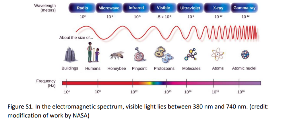
The eye can focus light images on the retina using the cornea and the lens. Visible light only occupies a small portion of the electromagnetic spectrum. Other species and artificial technologies can detect other parts of this energy spectrum.
Light passes through the eyeball via the cornea, pupil, and lens. The humors are fluids filling the anterior and posterior chambers of the eye.
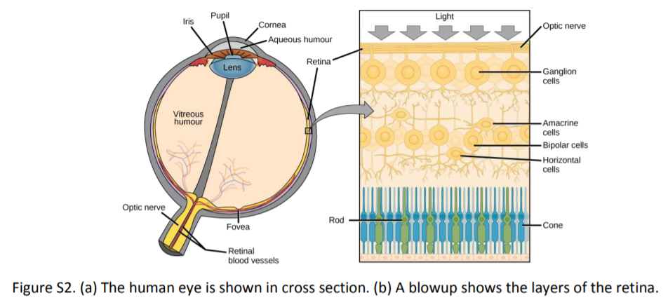
The focused image is directed toward the fovea (or fovea centralis), which contains the highest density of photoreceptor neurons. Muscles in the iris alter pupil size to vary light entering the eye. Choroid body muscles surround the lens. They alter the lens to aid focusing.
- Anatomy Terms to Know: Extra-ocular muscles, sclera, choroid, pigmented epithelium, fovea, vitreous humor, aqueous humor, iris, lens, ciliary body, cornea, conjunctiva, optic nerve, blind spot (or optic disc), retina [retinal neurons - ganglion cells, amacrine cells, bipolar cells, horizontal cells, photoreceptors (rods, cones)], and occipital (visual) cortex of the brain.
- Possible Specimens & Models for examination: (sheep or cow) eyeballs for dissection, microscope slides of the retina, and models of eyes.
Exercise 1:
You are responsible for identifying these major anatomical structures of the eye: sclera (tough, outer layer), choroid (dark, middle layer), pigmented epithelium, fovea, vitreous humor, aqueous humor, iris, lens, ciliary body, cornea, conjunctiva, optic nerve, blind spot (or optic disc), retina (thin, and pale inner layer). You will have to identify these structures using both the sheep or cow eye and the models.
Dissection:
You will work in groups of two or three to dissect an eye. There are several ways to slice through an eye. Check with your instructor to determine which way they want you to cut the eye in half. Possible cuts include:
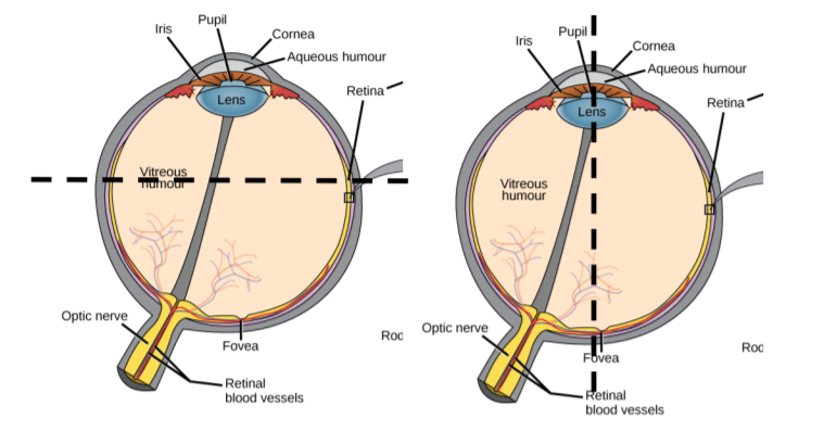
Note
To be able to best see the eye’s structures, you should work very carefully. Many internal structures are delicate and tear easily such as the retina. Some structures are quite tough including the lens and sclera (in Latin, sclera means ‘tough’!). Practicing careful dissecting is an important skill. Take your time! In order to get to the first layer, the sclera, you may have to clear away some fat and connective tissue. Ask for help if you need it.
Physiology:
Light striking photoreceptor neurons activate networks of retinal neurons. One network of neurons in the retina sends signals to one ganglion cell. Action potentials from ganglion cells, whose axons form the optic nerves, represent patterns of light. Perception of the network of interconnected neuron signals is eventually perceived in the occipital cortex.
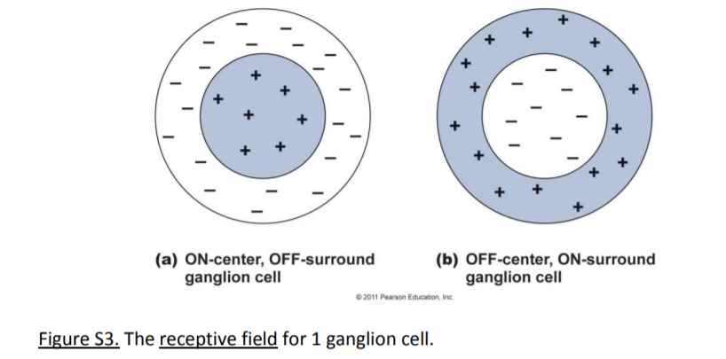
In this example, the ON-center cell will send a maximum rate of action potentials along its axon (in the optic nerve) to the brain if the brightest light is striking photoreceptors near the center of its portion of the retina and the surrounding photoreceptors in that portion are receiving minimal light. Eventually, patterns of ganglion cells signals are integrated within the brain (in the occipital cortex) to generate the perception of complex images.
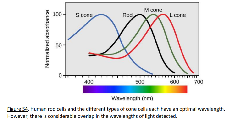
Color vision results from the interaction of 3 sub-types of cone photoreceptors. They preferentially absorb light at different wavelengths, shown in the figure below.
Exercise 2:
A: Blind Spot Test
- The optic disk, the sight where ganglion cell axons exit the eye, does not contain photoreceptors. We do not perceive the blind spot because the brain interpolates information to fill in the gaps. You can locate the blind spot by moving the image below toward your head.
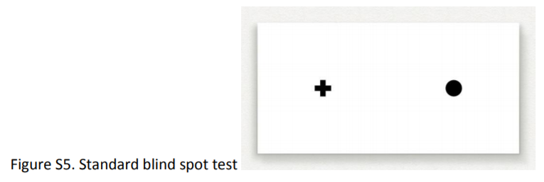
Directions:
- With your right eye (left closed), stare at the cross, and move the paper towards your eyes until the circle disappears.
- Measure the distance from the image to your eye. You can repeat this with your left eye, staring at the circle. The cross will disappear at a distance related to the physical separation of your fovea and your optic disk. You can estimate this distance (d’, in mm) of your retinal structures by recording the distance when the image disappears (x), and measuring the distance between the cross and circle in the image (d). This is actually a ‘reduced eye’ model, which involves some approximations.
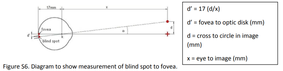
What is the distance between the fovea and optic disk in your left eye? _______________ Right eye? __________________ Average distance? _______________
Are your eyes exactly the same? __________________
Hypothesize why or why not:
______________________________________________________________________________________________________________________________________________
______________________________________________________________________________________________________________________________________________
______________________________________________________________________________________________________________________________________________
______________________________________________________________________________________________________________________________________________
B: Color-blindness Test
Color-blindness can be tested with an appropriate Standard Pseudoisochromatic plates (e.g. Ichikawa et al., ISBN 0-89640-030-1)
Directions:
Using the plates are you able to detect the image present for the presented colors?
Plate #: _______________ Image that you see: _____________
Plate #: _______________ Image that you see: _____________
Plate #: _______________ Image that you see: _____________
Plate #: _______________ Image that you see: _____________
Plate #: _______________ Image that you see: _____________
Plate #: _______________ Image that you see: _____________
If you exhibit some degree of colorblindness, what type? What may be happening with your cone cells within your retinas? Read back a little to see if you can figure it out.
______________________________________________________________________________________________________________________________________________
______________________________________________________________________________________________________________________________________________
______________________________________________________________________________________________________________________________________________
______________________________________________________________________________________________________________________________________________
C: Visual Acuity Test
Visual acuity refers to the sharpness or clarity of vision and is an indication of the focusing capacities of your eyes, especially the lens and cornea.
Directions:
Use the standard eye chart (Snellen chart) at the appropriate distances (usually 20 feet) to measure your acuity. Comparisons on the chart will refer to this standard measure. To use the chart, find the red tape on the floor. Stand there and cover one eye without squinting. Have your partner stand near the chart and tell you which line to read starting from the top. When you can no longer read a line accurately with one eye, then this is the acuity for that eye.
Write it here: _______________.
Now do the other eye the same way.
Write your acuity here: ____________________.
Do you have the same acuity in both eyes? Why or why not?
______________________________________________________________________________________________________________________________________________
______________________________________________________________________________________________________________________________________________
______________________________________________________________________________________________________________________________________________
______________________________________________________________________________________________________________________________________________
Note
Corrective lenses: If you have contacts, keep them in, but if you wear glasses you may want to try the test with and without them to see just how much your corrective lenses improve your vision!
Hearing:
Hearing involves the signal transduction of mechanical waves into neural signals in the cochlea, within the inner ear.
Anatomy:
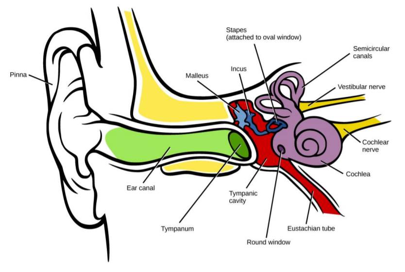
The auditory receptors for the ear (shown below) include the:
- Outer ear - from pinna (or auricle) to tympanum (also known as tympaninc membrane or eardrum)
- Middle ear - contains 3 ossicles, anchored between tympanum & oval window. The Eustachian (or auditory) tube connects the middle ear cavity to the pharynx (it is an evolutionary descendant of pharyngeal pouches).
- Inner ear – Cochlea, which contains the hair cells (receptors) within the Organ of Corti
Balance & Movement (The Vestibular System):
Anatomy:
Within the inner ear, the 3 semicircular canals are arranged at right angles to each other, and they contain hair cells and fluid similar to the cochlea. In addition, two separate clusters of hair cells – the saccule and utricle – are oriented to detect vertical and horizontal movements. Each of the hair cell clusters has a small collection of dense connective tissue attached to the hair cell membrane extensions (stereocilia) to add mass to the system.
Physiology:
Movements of the head cause dislocations of the fluid in the chambers around the hair cells. Movements generate electrical signals in hair cells, which signal sensory neurons with released chemical neurotransmitters. Patterns of signals are integrated in the cerebellum and parietal cortex.
Specimens & Models for Examination:
Models of the ear and cochlea.
Exercise 3:
A: Identifying Structures:
Using the model of the ear find the following structures: ear canal, tympanum (tympanic membrane), ossicles (malleus, incus, stapes in order moving inward into the ear), cochlea, semicircular canals.
B: Sound Localization:
Using a tuning fork, have a subject sit with their eyes closed. Strike the fork so it makes a sound and move it to front, back, side, and top of the head at a constant distance, holding it to allow the subject to point out the location. Note the accuracy at each position of their pointing, and determine the most and least accurate positions for localization. Can you explain any differences? List the positions for localization from most to least accurate:
______________________________________________________________________________________________________________________________________________
______________________________________________________________________________________________________________________________________________
______________________________________________________________________________________________________________________________________________
______________________________________________________________________________________________________________________________________________
C: Romberg Testing Involves Maintaining Balance:
- Have the subject stand with their back to the whiteboard. The board should be marked at approximately shoulder height with centimeter units covering ~1 meter.
- Note the shoulder positions of the subject.
- Have the subject stand and stare straight ahead for 2 minutes, and note the range of movement.
- Repeat with eyes closed.
- Repeat while standing with your right or left side closest to the board, and note front-to-back swaying, First with eyes open and then with eyes closed.
| Data | Eyes Open | Eyes Closed |
| Side to Side (cm.) | ||
| Front to Back (cm.) |
Describe any differences in relation to the sensory input required to maintain balance.
______________________________________________________________________________________________________________________________________________
______________________________________________________________________________________________________________________________________________
______________________________________________________________________________________________________________________________________________
______________________________________________________________________________________________________________________________________________
Smell:
Anatomy:
Sensory (olfactory) neurons are present at the top of the nasal cavity, extending their axons into the cranium. Olfactory signals are the only sensory system to send signals directly to the limbic system, which is integral to memory and emotional functions. In humans, from 100-200 different functional receptor proteins have been identified (there are over 1000 in rodents).
Physiology:
Specific molecules (odorants) bind to receptor proteins and activate neural electrical signals (action potentials). Patterns of olfactory neuron activity can code for complex odors, integrated within the olfactory bulb and temporal cortex. Olfactory neurons will undergo adaptation and decrease signals to the brain with constant exposure to a stimulus.
Exercise 4:
Humans consistently recognize certain odorants (e.g. spearmint, orange, anise). Specific oils for these are available and can be prepared as serial dilutions. Students can then test for sensitivities for each by starting with a series at the low end of the concentrations. Odorants can be detected by some sensitive individuals at concentrations below the micromolar range. Below, list the micromolar concentrations of mint and circle the one where you can begin to smell the mint.
Concentration 1: ______________ Concentration 2: ______________ Concentration 3: ______________ Concentration 4: ______________ Concentration 5: ______________
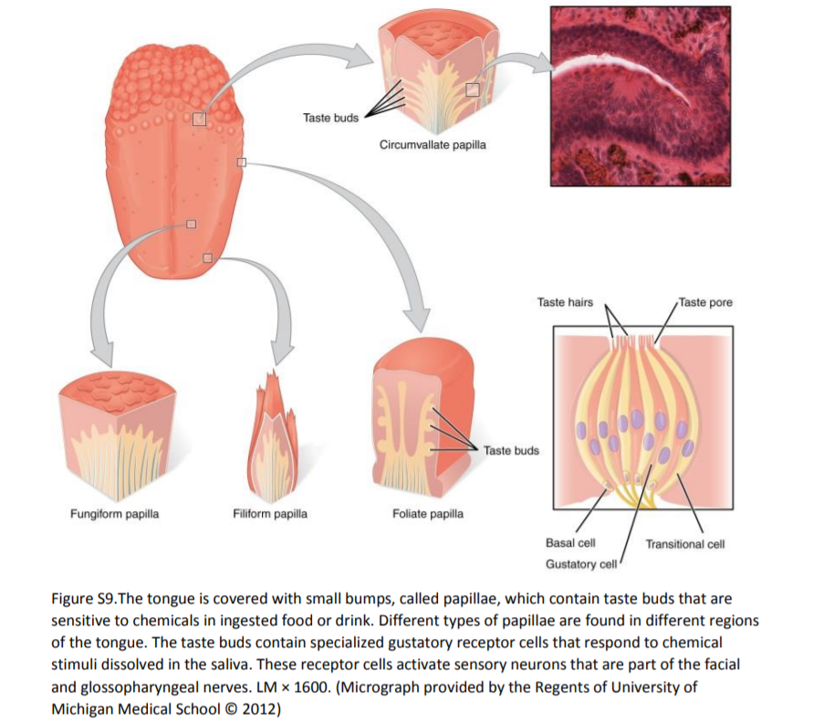
Taste:
Taste involves stimulation of receptor proteins on gustatory cells within taste buds. The perceived sensations correspond to common chemicals: Salty (Na+), Sweet (disaccharides, e.g. sucrose), Bitter (various, common test is Ca2+), sour (H+), and umami (glutamate). Also, taste is often integrated as a perception with olfactory sensory input.
Anatomy & Physiology:
Taste buds are arranged along the tongue epithelium. Sensory epithelial cells release neurotransmitter signal molecules to sensory neurons of cranial nerves. Information is integrated along the brain stem and in the temporal cortex.
Specimens & Models for Examination:
Histology slide of tongue/taste buds
Physiology:
Taste can be identified using solutions of chemicals known to stimulate distinct receptor proteins. Solutions can be prepared from common ingredients to test for sensitivity.
Exercise 5:
A: Histology Exploration
Use a microscope to explore the cellular aspect of a taste bud. Find an individual taste bud and draw it in the space provided. Can you label any structures if you use the above diagram as a guide?
B: PTC Test
One bitter taste receptor protein is encoded by the PTC gene, or TAS2R38 (discovered in 2003). There are at least 30 different genes coding for bitter taste receptors. Phenylthiocarbamide (PTC), also known as phenylthiourea (PTU), is only detected by ~70% of the population on average. Tasting PTC is correlated with the dominant genotype.
- PTC tasting test kits provide material to survey the class. Testing is a simple positive response for bitter taste, while non-tasters will report no taste.
- a. After placing the strip on your tongue do you taste anything? Yes or no? ________. If you answered YES!, then you have the dominant genotype for the PTC gene!
C: Taste Threshold Test
Similar to the olfaction tests, serial dilutions of basic chemicals can be used to test for variable sensitivity in subjects. Sucrose and NaCl are common tests for sweet and salty. Serial solutions can be applied with cotton swabs to the subject's tongue to test for sensitivity. Similar to the smell test, list the concentrations of the two substances and circle the one where you can begin to taste the substance.
Results:
Salt:
Concentration 1: _____________ Concentration 2: _____________ Concentration 3: _____________ Concentration 4: _____________ Concentration 5: _____________
Sucrose:
Concentration 1: _____________ Concentration 2: _____________ Concentration 3: _____________ Concentration 4: _____________ Concentration 5: _____________
Do you and your lab partner vary in your sensitivities? If you differ, then provide a possible explanation as to why:
______________________________________________________________________________________________________________________________________________
______________________________________________________________________________________________________________________________________________
______________________________________________________________________________________________________________________________________________
______________________________________________________________________________________________________________________________________________
- HISTORICAL NOTE: The ‘standard’ map of taste buds common in many lab manuals has been disproved by subsequent research (J. Cell Biology, 2010 vol. 190 no. 3 285-296 doi: 10.1083/jcb.201003144). There is more variability among individuals than accounted for by the original 1942 map (not shown, intentionally).
- Individuals can map their tongues for taste buds, once sensitivity thresholds have been determined.
This lab has been adapted from Rice University and is licensed under a Creative Commons Attribution License License (3.0).