
Lab 4: Nervous System
- Page ID
- 24390

\( \newcommand{\vecs}[1]{\overset { \scriptstyle \rightharpoonup} {\mathbf{#1}} } \)
\( \newcommand{\vecd}[1]{\overset{-\!-\!\rightharpoonup}{\vphantom{a}\smash {#1}}} \)
\( \newcommand{\dsum}{\displaystyle\sum\limits} \)
\( \newcommand{\dint}{\displaystyle\int\limits} \)
\( \newcommand{\dlim}{\displaystyle\lim\limits} \)
\( \newcommand{\id}{\mathrm{id}}\) \( \newcommand{\Span}{\mathrm{span}}\)
( \newcommand{\kernel}{\mathrm{null}\,}\) \( \newcommand{\range}{\mathrm{range}\,}\)
\( \newcommand{\RealPart}{\mathrm{Re}}\) \( \newcommand{\ImaginaryPart}{\mathrm{Im}}\)
\( \newcommand{\Argument}{\mathrm{Arg}}\) \( \newcommand{\norm}[1]{\| #1 \|}\)
\( \newcommand{\inner}[2]{\langle #1, #2 \rangle}\)
\( \newcommand{\Span}{\mathrm{span}}\)
\( \newcommand{\id}{\mathrm{id}}\)
\( \newcommand{\Span}{\mathrm{span}}\)
\( \newcommand{\kernel}{\mathrm{null}\,}\)
\( \newcommand{\range}{\mathrm{range}\,}\)
\( \newcommand{\RealPart}{\mathrm{Re}}\)
\( \newcommand{\ImaginaryPart}{\mathrm{Im}}\)
\( \newcommand{\Argument}{\mathrm{Arg}}\)
\( \newcommand{\norm}[1]{\| #1 \|}\)
\( \newcommand{\inner}[2]{\langle #1, #2 \rangle}\)
\( \newcommand{\Span}{\mathrm{span}}\) \( \newcommand{\AA}{\unicode[.8,0]{x212B}}\)
\( \newcommand{\vectorA}[1]{\vec{#1}} % arrow\)
\( \newcommand{\vectorAt}[1]{\vec{\text{#1}}} % arrow\)
\( \newcommand{\vectorB}[1]{\overset { \scriptstyle \rightharpoonup} {\mathbf{#1}} } \)
\( \newcommand{\vectorC}[1]{\textbf{#1}} \)
\( \newcommand{\vectorD}[1]{\overrightarrow{#1}} \)
\( \newcommand{\vectorDt}[1]{\overrightarrow{\text{#1}}} \)
\( \newcommand{\vectE}[1]{\overset{-\!-\!\rightharpoonup}{\vphantom{a}\smash{\mathbf {#1}}}} \)
\( \newcommand{\vecs}[1]{\overset { \scriptstyle \rightharpoonup} {\mathbf{#1}} } \)
\(\newcommand{\longvect}{\overrightarrow}\)
\( \newcommand{\vecd}[1]{\overset{-\!-\!\rightharpoonup}{\vphantom{a}\smash {#1}}} \)
\(\newcommand{\avec}{\mathbf a}\) \(\newcommand{\bvec}{\mathbf b}\) \(\newcommand{\cvec}{\mathbf c}\) \(\newcommand{\dvec}{\mathbf d}\) \(\newcommand{\dtil}{\widetilde{\mathbf d}}\) \(\newcommand{\evec}{\mathbf e}\) \(\newcommand{\fvec}{\mathbf f}\) \(\newcommand{\nvec}{\mathbf n}\) \(\newcommand{\pvec}{\mathbf p}\) \(\newcommand{\qvec}{\mathbf q}\) \(\newcommand{\svec}{\mathbf s}\) \(\newcommand{\tvec}{\mathbf t}\) \(\newcommand{\uvec}{\mathbf u}\) \(\newcommand{\vvec}{\mathbf v}\) \(\newcommand{\wvec}{\mathbf w}\) \(\newcommand{\xvec}{\mathbf x}\) \(\newcommand{\yvec}{\mathbf y}\) \(\newcommand{\zvec}{\mathbf z}\) \(\newcommand{\rvec}{\mathbf r}\) \(\newcommand{\mvec}{\mathbf m}\) \(\newcommand{\zerovec}{\mathbf 0}\) \(\newcommand{\onevec}{\mathbf 1}\) \(\newcommand{\real}{\mathbb R}\) \(\newcommand{\twovec}[2]{\left[\begin{array}{r}#1 \\ #2 \end{array}\right]}\) \(\newcommand{\ctwovec}[2]{\left[\begin{array}{c}#1 \\ #2 \end{array}\right]}\) \(\newcommand{\threevec}[3]{\left[\begin{array}{r}#1 \\ #2 \\ #3 \end{array}\right]}\) \(\newcommand{\cthreevec}[3]{\left[\begin{array}{c}#1 \\ #2 \\ #3 \end{array}\right]}\) \(\newcommand{\fourvec}[4]{\left[\begin{array}{r}#1 \\ #2 \\ #3 \\ #4 \end{array}\right]}\) \(\newcommand{\cfourvec}[4]{\left[\begin{array}{c}#1 \\ #2 \\ #3 \\ #4 \end{array}\right]}\) \(\newcommand{\fivevec}[5]{\left[\begin{array}{r}#1 \\ #2 \\ #3 \\ #4 \\ #5 \\ \end{array}\right]}\) \(\newcommand{\cfivevec}[5]{\left[\begin{array}{c}#1 \\ #2 \\ #3 \\ #4 \\ #5 \\ \end{array}\right]}\) \(\newcommand{\mattwo}[4]{\left[\begin{array}{rr}#1 \amp #2 \\ #3 \amp #4 \\ \end{array}\right]}\) \(\newcommand{\laspan}[1]{\text{Span}\{#1\}}\) \(\newcommand{\bcal}{\cal B}\) \(\newcommand{\ccal}{\cal C}\) \(\newcommand{\scal}{\cal S}\) \(\newcommand{\wcal}{\cal W}\) \(\newcommand{\ecal}{\cal E}\) \(\newcommand{\coords}[2]{\left\{#1\right\}_{#2}}\) \(\newcommand{\gray}[1]{\color{gray}{#1}}\) \(\newcommand{\lgray}[1]{\color{lightgray}{#1}}\) \(\newcommand{\rank}{\operatorname{rank}}\) \(\newcommand{\row}{\text{Row}}\) \(\newcommand{\col}{\text{Col}}\) \(\renewcommand{\row}{\text{Row}}\) \(\newcommand{\nul}{\text{Nul}}\) \(\newcommand{\var}{\text{Var}}\) \(\newcommand{\corr}{\text{corr}}\) \(\newcommand{\len}[1]{\left|#1\right|}\) \(\newcommand{\bbar}{\overline{\bvec}}\) \(\newcommand{\bhat}{\widehat{\bvec}}\) \(\newcommand{\bperp}{\bvec^\perp}\) \(\newcommand{\xhat}{\widehat{\xvec}}\) \(\newcommand{\vhat}{\widehat{\vvec}}\) \(\newcommand{\uhat}{\widehat{\uvec}}\) \(\newcommand{\what}{\widehat{\wvec}}\) \(\newcommand{\Sighat}{\widehat{\Sigma}}\) \(\newcommand{\lt}{<}\) \(\newcommand{\gt}{>}\) \(\newcommand{\amp}{&}\) \(\definecolor{fillinmathshade}{gray}{0.9}\)Introduction:
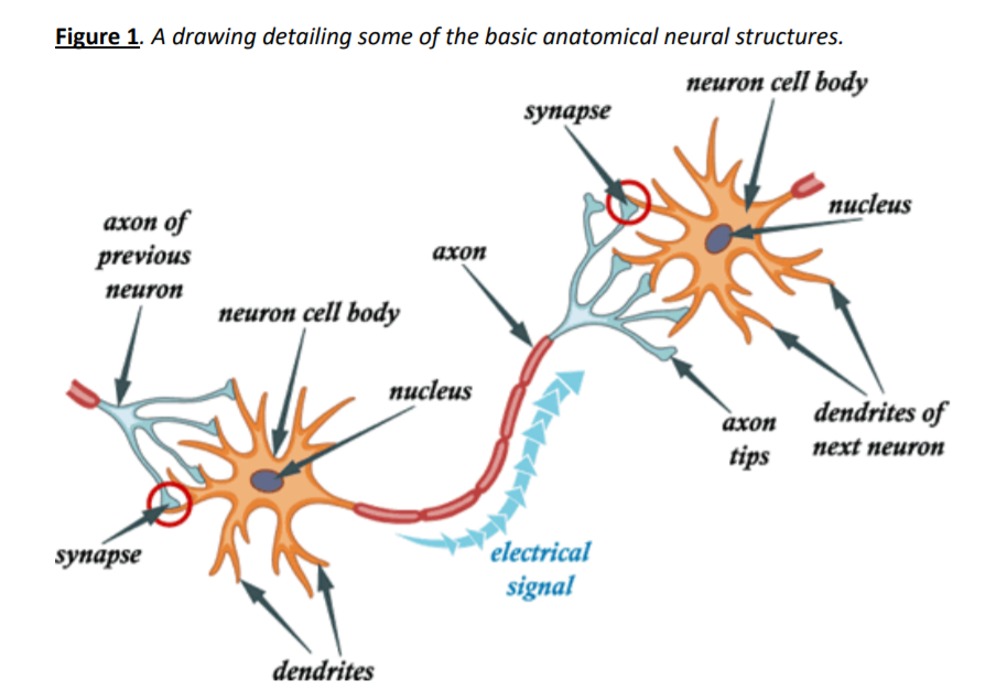
In this lab, we will explore the anatomy & physiology of the nervous system. Nervous systems are unique to animals and are critical for detecting and interpreting information, making decisions, and regulating body functions and movements. Nervous systems are constructed from neurons and glia. Neurons are the main functional cells while glia play a variety of support roles.
Nervous systems develop as interconnected networks of cells. The largest nervous organs are the brains of vertebrates, while animals as simple as jellyfish have a ‘nerve net’. A critical concept related to nervous systems is consciousness, or ‘self-awareness’. That idea will be discussed from one perspective in the next lab, distinguishing sensation from perception.
The exercises here will review cellular function and structure, and explore several basic neural networks within the larger body. We will use a combination of models and microscopic analysis, gross anatomical dissection, and physiological exercises to study nervous systems. An overview of the components of nervous systems.
Overview of the Components of Nervous Systems:
| Cellular Components | |
| Cell Structures | Cell Functions |
|
Neurons:
|
|
|
Glia (or glial cells, or neuroglia):
|
|
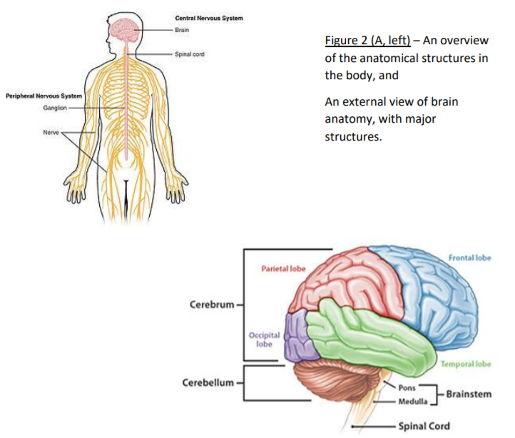
| CNS (Central Nervous System): the brain & spinal cord in vertebrates | PNS (Peripheral Nervous System): All other nervous tissue (nerves & ganglia). |
| Structures and Systems | Functions |
| White matter – myelinated axons in the CNS. Sometimes referred to as tracts, or fasciculi (or may have older, unique names). | Connections between CNS regions to coordinate functions. Allows integration of information. |
| Gray matter – neuron cell bodies in the CNS, includes cortical tissue (e.g. cerebral cortex) and nuclei (e.g. caudate nucleus assists with motor coordination). | Sites of information integration and decision-making, e.g. temporal cortex in the brain processes auditory signals and interprets speech. |
| Nerve – bundle of axons in the PNS | Can be classified as sensory (or afferent) for signals coming into the CNS from PNS, or motor (or efferent) for signals from CNS to PNS. Most nerves are mixed and carry both sensory and motor information. |
| Ganglia – clusters of neuron cell bodies in PNS | Various functions controlling and monitoring body functions, e.g. dorsal root ganglia are sensory neurons clustered lateral to the spinal cord. |
| Somatic Nervous System (or voluntary) | Nervous tissues related to conscious perception and voluntary body control. |
| Autonomic Nervous System (or visceral) – subdivided into the Sympathetic and Parasympathetic subdivisions. Nervous tissues related to unconscious perception and involuntary body control. |
Sympathetic – regulates ‘fight-or-flight’, short term stress responses (e.g., increase heart rate, pupil dilation); Parasympathetic – regulates ‘rest and digest’ functions (e.g., decrease in skeletal muscle activity, increase in digestive function – liver, pancreas function) |
Exercises:
Exercise 1:
Examine the microscope slides for neuron anatomy. For each of the listed slides, note the listed structures:
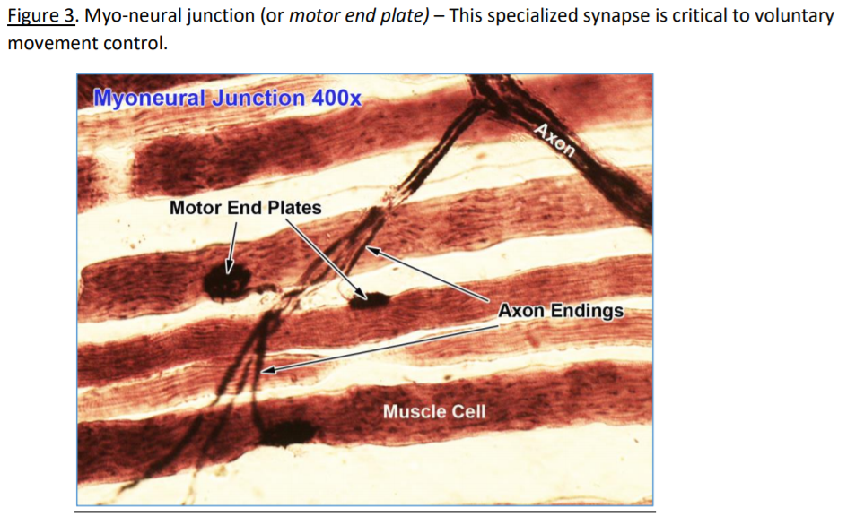
a. Motor end plate (or myo-neural junction) – the synaptic connection between a motor neuron and a skeletal muscle cell. Structures include multiple synapses (or motor end plates), axons, skeletal muscle cells with sarcomeres, and myelin sheath of Schwann cells on axons.
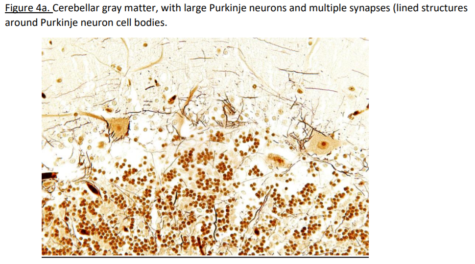
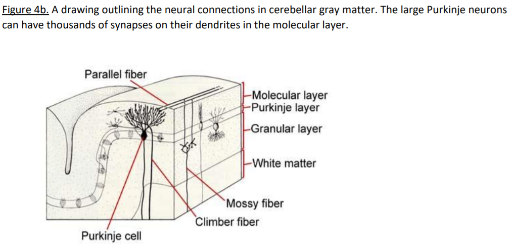
b. Cerebellum – the ‘little brain’ posterior to the brain stem has folded white and gray matter that is distinguishable with the naked eye. Within the gray matter, 3 distinct neuron layers (granular, Purkinje, molecular) reveal organizational structures.
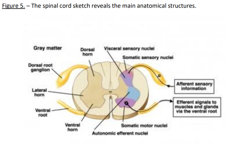
c. Spinal cord – a cross-section of the spinal cord reveals peripheral white matter and central gray matter. The small central canal contains ependymal glia. Large motor neuron cell bodies are located in the ventral gray matter – their axons form the ventral motor root of a spinal nerve. Sensory neuron cell bodies are in the dorsal root ganglia, and their axons form the dorsal sensory root of the spinal nerves.
Images:
Exercise 2:
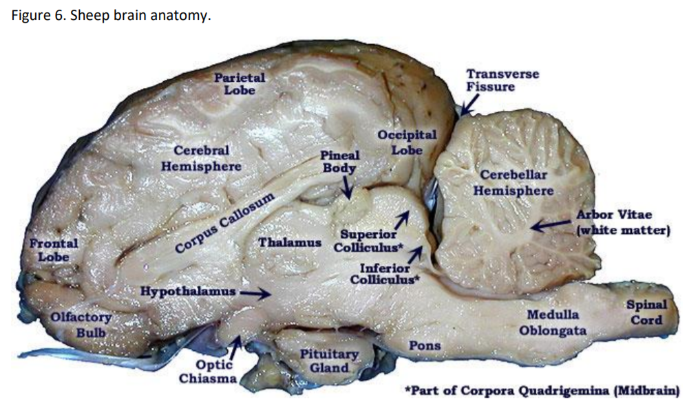
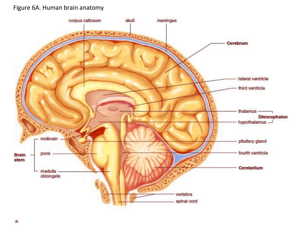
Gross anatomy. Use the models and/or brain specimens to examine, dissect, and identify the major anatomical regions of the brain. Not all structures may be present on the same model or specimen.
Cerebrum – cortex (or cortical gray matter – divided into lobes: frontal, temporal, parietal, occipital), sulci & gyri (folds of cortex), cerebral hemispheres, white matter (includes corpus callosum), cerebral nuclei (including basal nuclei, thalamus, hypothalamus), olfactory bulbs & tracts, optic nerves, optic tracts, optic chiasma, ventricles, and meninges.
Brain stem (includes medulla oblongata, pons, midbrain {includes sup. & inf. colliculi), cerebellum (arbor vitae is the white matter), cranial nerves (12 pairs total), and spinal cord.
Exercise 3:
Physiological functions of the nervous can be studied at a basic level by examining reflexes. Reflexes are the result of interconnected networks of neurons controlling specific functions.
3a. Reflex Physiology:
Using a meter stick, measure the speed of your neural network connecting your visual system and your somatic motor system. Work with a classmate, and perform the task 3 times each.
- Hold a meter stick vertically, with one end over the open hand of the first subject. The subject will hold his/her hand open, with the thumb two inches away from the rest of their fingers.
- Hold the stick so that the scale starts at ‘0’, even with the top of their hand, and the markings count upwards on the stick.
- Without warning, release the stick. When your classmate closes their hand to capture the stick, measure the distance the stick dropped by the markings at the top of your classmate’s hands holding the stick.
Results:
Student A: Trial 1 _______ cm; Trial 2 _______ cm; Trial 3 _______ cm = ________ (average)
Student B: Trial 1 _______ cm; Trial 2 _______ cm; Trial 3 _______ cm = ________ (average)
- Based on acceleration due to gravity (\(9.8\frac{m.}{sec^2}\) ), you can calculate the speed of the subject’s reflexes by \[time\;(sec)=\sqrt{\frac{2*distance\;(cm.)}{980\;(\frac{cm.}{sec^2})}}\]
- Convert your average distance to time:
- Student A = _____sec.
- Student B = _____sec.
- Compare your results with the class. Class range (maximum, minimum): ________ - ________ sec.
- There are at least 10 synapses involved in this response. Based on your results, what is the minimum amount of time that a neuron requires to receive and send a signal? ____________ seconds.
3b. Autonomic Reflexes:
Using a flashlight, test the pupil reflex for light responses.
- Have the subject look away from any strong light sources.
- While watching their eyes, bring a light toward their eye from the left side.
- Observe changes in the left pupil, as well as the right pupil.
- Compare the responses in the two eyes. Repeat on the right side.
- Explain why the pupils reacted as they did (did both react in the same manner?).
______________________________________________________________________________________________________________________________________________
______________________________________________________________________________________________________________________________________________
______________________________________________________________________________________________________________________________________________
______________________________________________________________________________________________________________________________________________
Exercise 4:
The somatosensory system is the largest sensing system in your body. This system produces sensory feedback whenever you come in physical contact with your environment. This sensory feedback includes body position (proprioception), sensing the movement of your body and limbs (kinesthesis), pain (nociception), temperature, and finally touch. For this last sense, you can map out touch receptor density in your skin using a simple technique. Most of the tactile feel we receive is gathered by four types of mechanoreceptors which are found in both layers of skin. The two receptors located near the top of the dermis, are called Merkel receptors and Meissner corpuscles. The other two mechanoreceptors located deep in the dermis and hypodermis layer and are the Ruffini and Pacinian corpuscles.
Working with a friend you are going to test each other's two-point threshold. A two-point threshold test seeks to find at what distance apart does a person perceive one point as two separate points. To test this, two points start together touching the skin. Incrementally, they are pulled further apart and reapplied to the skin until the subject can clearly tell there are two different points.
You will take measurements from four different body part locations: Fingertip, Cheek, Forearm, and Calf. You will perform this experiment twice (ascending and descending with the drafting compass). Finally, the test subject must always keep their eyes closed!
Experiment Steps:
- Before you start this experiment, be sure that you know how to use the compass. Practice touching your arm with it with the points at different distances (millimeter (mm.)) apart. Use the ruler to precisely position the points apart from each other. It is important during the experiment that you touch your partner's skin with both tips at the same time; otherwise, they will easily be able to tell there are two points. Once you have the hang of it you are ready to begin!
- Once your partner has closed their eyes, have them place their arm on the table with their palm facing up. Now take the compass, make sure the points are together (0 mm. apart), and start with your partner's fingertip.
- Touch the compass to their finger tip and ask them if they feel one point or two points. They should say one point because there is no distance between the points (0 mm). Remove the compass and increase the distance to 2 mm. Reapply the calipers, be sure to touch both tips at the same time, and ask again. If they still only feel one point, increase by another 2 mm. and reapply.
- Once your partner detects two points, move on to the next area of skin and repeat the process. Once you have completed the ascending or increasing distances now it’s time to repeat the process using the descending (decreasing distances) measurements. You can start at a distance of 7 mm. and repeat the process by decreasing the distance by 2 mm. each time. You can enter the data below. Remember to write down the distance that you feel only one point.
| Ascending | Descending | |||||||
| Subject | Fingertip | Cheek | Forearm | Calf | Fingertip | Cheek | Forearm | Calf |
| #1 | ||||||||
| #2 | ||||||||
| Average | ||||||||
Questions:
1. Why can your fingertip detect such small distances between points while your arms and legs cannot?
2. Would you expect to see a difference in males vs. females for the four recorded areas? What about children vs adults?
3. Why doesn't your brain have the sensitivity of your fingertips all over your body?