
Lab 6: Circulatory Systems
- Page ID
- 24395

\( \newcommand{\vecs}[1]{\overset { \scriptstyle \rightharpoonup} {\mathbf{#1}} } \)
\( \newcommand{\vecd}[1]{\overset{-\!-\!\rightharpoonup}{\vphantom{a}\smash {#1}}} \)
\( \newcommand{\dsum}{\displaystyle\sum\limits} \)
\( \newcommand{\dint}{\displaystyle\int\limits} \)
\( \newcommand{\dlim}{\displaystyle\lim\limits} \)
\( \newcommand{\id}{\mathrm{id}}\) \( \newcommand{\Span}{\mathrm{span}}\)
( \newcommand{\kernel}{\mathrm{null}\,}\) \( \newcommand{\range}{\mathrm{range}\,}\)
\( \newcommand{\RealPart}{\mathrm{Re}}\) \( \newcommand{\ImaginaryPart}{\mathrm{Im}}\)
\( \newcommand{\Argument}{\mathrm{Arg}}\) \( \newcommand{\norm}[1]{\| #1 \|}\)
\( \newcommand{\inner}[2]{\langle #1, #2 \rangle}\)
\( \newcommand{\Span}{\mathrm{span}}\)
\( \newcommand{\id}{\mathrm{id}}\)
\( \newcommand{\Span}{\mathrm{span}}\)
\( \newcommand{\kernel}{\mathrm{null}\,}\)
\( \newcommand{\range}{\mathrm{range}\,}\)
\( \newcommand{\RealPart}{\mathrm{Re}}\)
\( \newcommand{\ImaginaryPart}{\mathrm{Im}}\)
\( \newcommand{\Argument}{\mathrm{Arg}}\)
\( \newcommand{\norm}[1]{\| #1 \|}\)
\( \newcommand{\inner}[2]{\langle #1, #2 \rangle}\)
\( \newcommand{\Span}{\mathrm{span}}\) \( \newcommand{\AA}{\unicode[.8,0]{x212B}}\)
\( \newcommand{\vectorA}[1]{\vec{#1}} % arrow\)
\( \newcommand{\vectorAt}[1]{\vec{\text{#1}}} % arrow\)
\( \newcommand{\vectorB}[1]{\overset { \scriptstyle \rightharpoonup} {\mathbf{#1}} } \)
\( \newcommand{\vectorC}[1]{\textbf{#1}} \)
\( \newcommand{\vectorD}[1]{\overrightarrow{#1}} \)
\( \newcommand{\vectorDt}[1]{\overrightarrow{\text{#1}}} \)
\( \newcommand{\vectE}[1]{\overset{-\!-\!\rightharpoonup}{\vphantom{a}\smash{\mathbf {#1}}}} \)
\( \newcommand{\vecs}[1]{\overset { \scriptstyle \rightharpoonup} {\mathbf{#1}} } \)
\(\newcommand{\longvect}{\overrightarrow}\)
\( \newcommand{\vecd}[1]{\overset{-\!-\!\rightharpoonup}{\vphantom{a}\smash {#1}}} \)
\(\newcommand{\avec}{\mathbf a}\) \(\newcommand{\bvec}{\mathbf b}\) \(\newcommand{\cvec}{\mathbf c}\) \(\newcommand{\dvec}{\mathbf d}\) \(\newcommand{\dtil}{\widetilde{\mathbf d}}\) \(\newcommand{\evec}{\mathbf e}\) \(\newcommand{\fvec}{\mathbf f}\) \(\newcommand{\nvec}{\mathbf n}\) \(\newcommand{\pvec}{\mathbf p}\) \(\newcommand{\qvec}{\mathbf q}\) \(\newcommand{\svec}{\mathbf s}\) \(\newcommand{\tvec}{\mathbf t}\) \(\newcommand{\uvec}{\mathbf u}\) \(\newcommand{\vvec}{\mathbf v}\) \(\newcommand{\wvec}{\mathbf w}\) \(\newcommand{\xvec}{\mathbf x}\) \(\newcommand{\yvec}{\mathbf y}\) \(\newcommand{\zvec}{\mathbf z}\) \(\newcommand{\rvec}{\mathbf r}\) \(\newcommand{\mvec}{\mathbf m}\) \(\newcommand{\zerovec}{\mathbf 0}\) \(\newcommand{\onevec}{\mathbf 1}\) \(\newcommand{\real}{\mathbb R}\) \(\newcommand{\twovec}[2]{\left[\begin{array}{r}#1 \\ #2 \end{array}\right]}\) \(\newcommand{\ctwovec}[2]{\left[\begin{array}{c}#1 \\ #2 \end{array}\right]}\) \(\newcommand{\threevec}[3]{\left[\begin{array}{r}#1 \\ #2 \\ #3 \end{array}\right]}\) \(\newcommand{\cthreevec}[3]{\left[\begin{array}{c}#1 \\ #2 \\ #3 \end{array}\right]}\) \(\newcommand{\fourvec}[4]{\left[\begin{array}{r}#1 \\ #2 \\ #3 \\ #4 \end{array}\right]}\) \(\newcommand{\cfourvec}[4]{\left[\begin{array}{c}#1 \\ #2 \\ #3 \\ #4 \end{array}\right]}\) \(\newcommand{\fivevec}[5]{\left[\begin{array}{r}#1 \\ #2 \\ #3 \\ #4 \\ #5 \\ \end{array}\right]}\) \(\newcommand{\cfivevec}[5]{\left[\begin{array}{c}#1 \\ #2 \\ #3 \\ #4 \\ #5 \\ \end{array}\right]}\) \(\newcommand{\mattwo}[4]{\left[\begin{array}{rr}#1 \amp #2 \\ #3 \amp #4 \\ \end{array}\right]}\) \(\newcommand{\laspan}[1]{\text{Span}\{#1\}}\) \(\newcommand{\bcal}{\cal B}\) \(\newcommand{\ccal}{\cal C}\) \(\newcommand{\scal}{\cal S}\) \(\newcommand{\wcal}{\cal W}\) \(\newcommand{\ecal}{\cal E}\) \(\newcommand{\coords}[2]{\left\{#1\right\}_{#2}}\) \(\newcommand{\gray}[1]{\color{gray}{#1}}\) \(\newcommand{\lgray}[1]{\color{lightgray}{#1}}\) \(\newcommand{\rank}{\operatorname{rank}}\) \(\newcommand{\row}{\text{Row}}\) \(\newcommand{\col}{\text{Col}}\) \(\renewcommand{\row}{\text{Row}}\) \(\newcommand{\nul}{\text{Nul}}\) \(\newcommand{\var}{\text{Var}}\) \(\newcommand{\corr}{\text{corr}}\) \(\newcommand{\len}[1]{\left|#1\right|}\) \(\newcommand{\bbar}{\overline{\bvec}}\) \(\newcommand{\bhat}{\widehat{\bvec}}\) \(\newcommand{\bperp}{\bvec^\perp}\) \(\newcommand{\xhat}{\widehat{\xvec}}\) \(\newcommand{\vhat}{\widehat{\vvec}}\) \(\newcommand{\uhat}{\widehat{\uvec}}\) \(\newcommand{\what}{\widehat{\wvec}}\) \(\newcommand{\Sighat}{\widehat{\Sigma}}\) \(\newcommand{\lt}{<}\) \(\newcommand{\gt}{>}\) \(\newcommand{\amp}{&}\) \(\definecolor{fillinmathshade}{gray}{0.9}\)Most animals are complex multicellular organisms that require a mechanism for transporting nutrients throughout their bodies and removing waste products. The circulatory system has evolved over time from simple diffusion through cells in the early evolution of animals to a complex network of blood vessels that reach all parts of the human body. This extensive network supplies the cells, tissues, and organs with oxygen and nutrients, and removes carbon dioxide and waste, which are byproducts of respiration and other cellular activities.
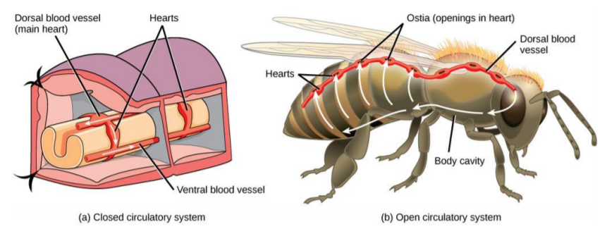
Circulatory systems differ significantly throughout the Animal Kingdom. In all vertebrate organisms, as well as some invertebrates, this is a closed-loop system, in which the blood is not free in a cavity. In a closed circulatory system, blood is contained inside blood vessels and circulates unidirectionally from the heart around the systemic circulatory route, then returns to the heart again. As opposed to a closed system, arthropods—including insects, crustaceans, and most mollusks—have an open circulatory system. In an open circulatory system, the blood is not enclosed in the blood vessels but is pumped into a cavity called a hemocoel and is called hemolymph because the blood mixes with the interstitial fluid.
Mammalian Heart and Blood Vessels:
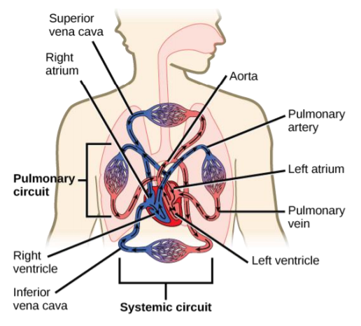
The mammalian circulatory system is divided into three circuits: the systemic circuit, the pulmonary circuit, and the coronary circuit. Blood is pumped from veins of the systemic circuit into the right atrium of the heart, then into the right ventricle. Blood then enters the pulmonary circuit and is oxygenated by the lungs. From the pulmonary circuit, blood re-enters the heart through the left atrium. From the left ventricle, blood re-enters the systemic circuit through the aorta and is distributed to the rest of the body.
Exercise 1: Structure of the Heart
Materials:
- Human Heart Model
Procedure:
- Using the model of the human heart and the chart provided, locate the different structures of the heart. Write the flow of the blood and the function of each structure in the space provided:
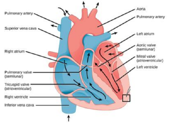
Right atrium: __________________________________________
Tricuspid valve: __________________________________________
Right ventricle: __________________________________________
Left atrium: __________________________________________
Bicuspid/Mitral valve: __________________________________________
Left ventricle: __________________________________________
Exercise 2: Blood Type and RH factor.
Blood is important for regulation of the body’s systems and homeostasis. Blood helps maintain homeostasis by stabilizing pH, temperature, osmotic pressure, and by eliminating excess heat. Blood supports growth by distributing nutrients and hormones, and by removing waste. Blood plays a protective role by transporting clotting factors and platelets to prevent blood loss and transporting the disease-fighting agents or white blood cells to sites of infection.
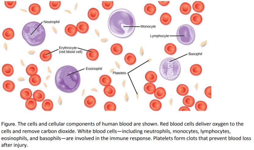
Microscope work: Use one of the light microscopes to explore a slide of a human blood smear. The leukocytes (white blood cells) are stained purple (nuclear staining) and far less common than the erythrocytes (red blood cells). Count the number of leukocytes in your view at the medium magnification. How many are there?
______________________________________________________________________________________________________________________________________________
Erythrocytes outnumber leukocytes 600:1. Therefore, estimate the number of erythrocytes in your view: _________________________________________________________
Why do you think the erythrocytes outnumber the leukocytes?
__________________________________________________________________________________________________________________________________________________________________________________________________________________________________________________________________________________________________________________________________________________________________________________________________________________________________________
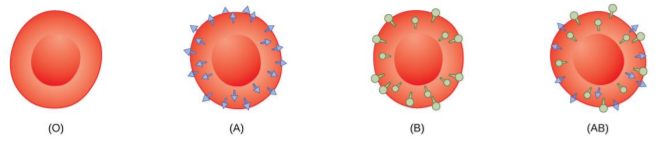
Red blood cells are coated in antigens made of glycolipids and glycoproteins. The composition of these molecules is determined by genetics, which have evolved over time. The two most well-known blood groups are the ABO and Rh systems. The surface antigens in the ABO blood group are glycolipids, called antigen A and antigen B. People with blood type A have antigen A, those with blood type B have antigen B, those with blood type AB have both antigens, and people with blood type O have neither antigen. Antibodies called agglutinogens are found in the blood plasma and react with the A or B antigens, if the two are mixed. When type A and type B blood are combined, agglutination (clumping) of the blood occurs because of antibodies in the plasma that bind with the opposing antigen; this causes clots that coagulate in the kidney causing kidney failure. Type O blood has neither A or B antigens, and therefore, type O blood can be given to all blood types. Type O negative blood is the universal donor. Type AB positive blood is the universal acceptor because it has both A and B antigen.
The Rh blood group was first discovered in Rhesus monkeys. Most people have the Rh antigen (Rh+) and do not have anti-Rh antibodies in their blood. The few people who do not have the Rh antigen and are Rh– can develop anti-Rh antibodies if exposed to Rh+ blood. This can happen after a blood transfusion or after an Rh– woman has an Rh+ baby. The first exposure does not usually cause a reaction; however, at the second exposure, enough antibodies have built up in the blood to produce a reaction that causes agglutination and breakdown of red blood cells. An injection can prevent this reaction.
Based on the information above, fill out the following chart regarding each blood type:
| Blood Type | Possible Genotype | Antigens on RBCs | Antibodies in Plasma |
| A | |||
| B | |||
| AB | |||
| O |
Materials:
- Blood typing kit (1/each group)
- 4 unknown simulated blood dropper bottles
- Anti –A, Anti-B, Anti-D (RH) sera dropper bottles
- Toothpicks
Procedure:
- Place one drop of simulated blood in the middle of each depression in the blood typing tray.
- Add one drop of Anti-A serum next to the A blood cells, one drop of Anti-B serum next to the B blood cells, and one drop of Anti-D serum next to box D blood cells.
- Mix the simulated blood with the Anti-serum using the toothpick for about two minutes
- Depend on the type of the antigen in the blood, the blood mixture will clump or the mixture will not change.
- Record your observation on the following table.
| Simulated Blood | Blood Type | RH (+,-) |
| Unknown Sample 1 | ||
| Unknown Sample 2 | ||
| Unknown Sample 3 | ||
| Unknown Sample 4 |
Exercise 3: Measuring Blood Pressure and Pulse Rate
Blood pressure (BP) is the pressure exerted by blood on the walls of a blood vessel that helps to push blood through the body. Blood moving through the blood vessels exerts pressure against the vessel walls. This blood pressure is highest in the aorta. It decreases as the blood moves through the arterioles, capillaries, venules, and veins. Many factors can affect blood pressure, such as hormones, stress, exercise, eating, sitting, and standing. Blood flow through the body is regulated by the size of blood vessels, by the action of smooth muscle, by one-way valves, and by the fluid pressure of the blood itself.
There are two components to blood pressure:
- Systolic blood pressure: measures the amount of pressure that blood exerts on vessels while the heart is beating (contracting). The pressure in the vessel is highest at this time. The optimal systolic blood pressure is 120 mmHg.
- Diastolic blood pressure: measures the pressure in the vessels between heartbeats (resting). The pressure is at its lowest point. The optimal diastolic blood pressure is 80 mmHg.
When we measure blood pressure, we are actually measuring the systolic pressure and the diastolic pressure separately. This is why you always see blood pressure reported as two numbers, one "over" the other. For example in a blood pressure reading of 130/85, this means that the systolic pressure is 130 and the diastolic pressure is 85.
Materials:
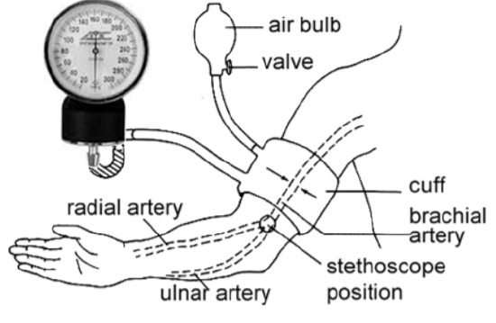
- Sphygmomanometer (pronounced sfig-mo-muh-NAM-eh-ter) consists of an inflatable cuff, an inflation pump, a gauge to register pressure, and a controlled exhaust valve.
- Stethoscope: to listen to the different sounds that occur
Procedure:
You will work in pairs to measure the effect of exercise on pulse rate and blood pressure.
- Have your partner sit down and relax for 2 minutes. Place your index and middle fingers in the groove on the inside of his/her wrist. Just slide your fingers across the tendons until they slip into soft tissue.
- Wait until you clearly feel beats coming with a regular rhythm.
- Count the number of pulses in 15 seconds.
- Multiply the number of pulses by 4 to get the number of beats per minute.
- Record this number in the table below.
- Attach the inflatable cuff around your partner’s arm above the elbow as shown in the figure above. Tuck the flap of the bag under the fold.
- Place the stethoscope over the brachial artery (underneath the cuff as shown in the figure above.
- You begin by inflating the cuff to about 200 mm. The blood flow below stops and you will hear no sound in the brachial artery when you listen with the stethoscope.
- If the pressure has gone below 200 mmHg, inflate the cuff again.
- Slowly begin releasing the pressure in the cuff. The blood begins to flow through the artery causing vibrations and turbulence that are audible through the stethoscope
- The first tapping sound you hear indicates that blood has entered the artery. Record this reading as the systolic pressure.
- You continue to deflate the cuff until you stop hearing any sound. The last tapping sound you hear indicates the diastolic pressure.
- Now go outside and exercise for exactly 5 minutes. You can do jumping jacks, run up and down the stairs or do push-ups.
- Immediately after sitting down measure the pulse rate/min (Steps 1–3) and the blood pressure (steps 4–10) and record your data.
- Measure the beat rate and the blood pressure of your partner. Record the results in the following table.
| Student 1 | Student 2 | |||||
| Beats/Minute | Systolic | Diastolic | Beats/Minute | Systolic | Diastolic | |
| Resting | ||||||
| After Exercise | ||||||
Questions:
1. How does exercise affect pulse rate and blood pressure?
2. Explain what happens to the circulatory system during exercise. Include the major organs involved in your explanation.
3. Why does increased physical activity increases heart rate?
4. Why is heart rate lower in an individual who does aerobic exercise regularly?
5. How and why does heart rate change with body position?