
3.4.3: Protists
- Page ID
- 108074
\( \newcommand{\vecs}[1]{\overset { \scriptstyle \rightharpoonup} {\mathbf{#1}} } \)
\( \newcommand{\vecd}[1]{\overset{-\!-\!\rightharpoonup}{\vphantom{a}\smash {#1}}} \)
\( \newcommand{\dsum}{\displaystyle\sum\limits} \)
\( \newcommand{\dint}{\displaystyle\int\limits} \)
\( \newcommand{\dlim}{\displaystyle\lim\limits} \)
\( \newcommand{\id}{\mathrm{id}}\) \( \newcommand{\Span}{\mathrm{span}}\)
( \newcommand{\kernel}{\mathrm{null}\,}\) \( \newcommand{\range}{\mathrm{range}\,}\)
\( \newcommand{\RealPart}{\mathrm{Re}}\) \( \newcommand{\ImaginaryPart}{\mathrm{Im}}\)
\( \newcommand{\Argument}{\mathrm{Arg}}\) \( \newcommand{\norm}[1]{\| #1 \|}\)
\( \newcommand{\inner}[2]{\langle #1, #2 \rangle}\)
\( \newcommand{\Span}{\mathrm{span}}\)
\( \newcommand{\id}{\mathrm{id}}\)
\( \newcommand{\Span}{\mathrm{span}}\)
\( \newcommand{\kernel}{\mathrm{null}\,}\)
\( \newcommand{\range}{\mathrm{range}\,}\)
\( \newcommand{\RealPart}{\mathrm{Re}}\)
\( \newcommand{\ImaginaryPart}{\mathrm{Im}}\)
\( \newcommand{\Argument}{\mathrm{Arg}}\)
\( \newcommand{\norm}[1]{\| #1 \|}\)
\( \newcommand{\inner}[2]{\langle #1, #2 \rangle}\)
\( \newcommand{\Span}{\mathrm{span}}\) \( \newcommand{\AA}{\unicode[.8,0]{x212B}}\)
\( \newcommand{\vectorA}[1]{\vec{#1}} % arrow\)
\( \newcommand{\vectorAt}[1]{\vec{\text{#1}}} % arrow\)
\( \newcommand{\vectorB}[1]{\overset { \scriptstyle \rightharpoonup} {\mathbf{#1}} } \)
\( \newcommand{\vectorC}[1]{\textbf{#1}} \)
\( \newcommand{\vectorD}[1]{\overrightarrow{#1}} \)
\( \newcommand{\vectorDt}[1]{\overrightarrow{\text{#1}}} \)
\( \newcommand{\vectE}[1]{\overset{-\!-\!\rightharpoonup}{\vphantom{a}\smash{\mathbf {#1}}}} \)
\( \newcommand{\vecs}[1]{\overset { \scriptstyle \rightharpoonup} {\mathbf{#1}} } \)
\(\newcommand{\longvect}{\overrightarrow}\)
\( \newcommand{\vecd}[1]{\overset{-\!-\!\rightharpoonup}{\vphantom{a}\smash {#1}}} \)
\(\newcommand{\avec}{\mathbf a}\) \(\newcommand{\bvec}{\mathbf b}\) \(\newcommand{\cvec}{\mathbf c}\) \(\newcommand{\dvec}{\mathbf d}\) \(\newcommand{\dtil}{\widetilde{\mathbf d}}\) \(\newcommand{\evec}{\mathbf e}\) \(\newcommand{\fvec}{\mathbf f}\) \(\newcommand{\nvec}{\mathbf n}\) \(\newcommand{\pvec}{\mathbf p}\) \(\newcommand{\qvec}{\mathbf q}\) \(\newcommand{\svec}{\mathbf s}\) \(\newcommand{\tvec}{\mathbf t}\) \(\newcommand{\uvec}{\mathbf u}\) \(\newcommand{\vvec}{\mathbf v}\) \(\newcommand{\wvec}{\mathbf w}\) \(\newcommand{\xvec}{\mathbf x}\) \(\newcommand{\yvec}{\mathbf y}\) \(\newcommand{\zvec}{\mathbf z}\) \(\newcommand{\rvec}{\mathbf r}\) \(\newcommand{\mvec}{\mathbf m}\) \(\newcommand{\zerovec}{\mathbf 0}\) \(\newcommand{\onevec}{\mathbf 1}\) \(\newcommand{\real}{\mathbb R}\) \(\newcommand{\twovec}[2]{\left[\begin{array}{r}#1 \\ #2 \end{array}\right]}\) \(\newcommand{\ctwovec}[2]{\left[\begin{array}{c}#1 \\ #2 \end{array}\right]}\) \(\newcommand{\threevec}[3]{\left[\begin{array}{r}#1 \\ #2 \\ #3 \end{array}\right]}\) \(\newcommand{\cthreevec}[3]{\left[\begin{array}{c}#1 \\ #2 \\ #3 \end{array}\right]}\) \(\newcommand{\fourvec}[4]{\left[\begin{array}{r}#1 \\ #2 \\ #3 \\ #4 \end{array}\right]}\) \(\newcommand{\cfourvec}[4]{\left[\begin{array}{c}#1 \\ #2 \\ #3 \\ #4 \end{array}\right]}\) \(\newcommand{\fivevec}[5]{\left[\begin{array}{r}#1 \\ #2 \\ #3 \\ #4 \\ #5 \\ \end{array}\right]}\) \(\newcommand{\cfivevec}[5]{\left[\begin{array}{c}#1 \\ #2 \\ #3 \\ #4 \\ #5 \\ \end{array}\right]}\) \(\newcommand{\mattwo}[4]{\left[\begin{array}{rr}#1 \amp #2 \\ #3 \amp #4 \\ \end{array}\right]}\) \(\newcommand{\laspan}[1]{\text{Span}\{#1\}}\) \(\newcommand{\bcal}{\cal B}\) \(\newcommand{\ccal}{\cal C}\) \(\newcommand{\scal}{\cal S}\) \(\newcommand{\wcal}{\cal W}\) \(\newcommand{\ecal}{\cal E}\) \(\newcommand{\coords}[2]{\left\{#1\right\}_{#2}}\) \(\newcommand{\gray}[1]{\color{gray}{#1}}\) \(\newcommand{\lgray}[1]{\color{lightgray}{#1}}\) \(\newcommand{\rank}{\operatorname{rank}}\) \(\newcommand{\row}{\text{Row}}\) \(\newcommand{\col}{\text{Col}}\) \(\renewcommand{\row}{\text{Row}}\) \(\newcommand{\nul}{\text{Nul}}\) \(\newcommand{\var}{\text{Var}}\) \(\newcommand{\corr}{\text{corr}}\) \(\newcommand{\len}[1]{\left|#1\right|}\) \(\newcommand{\bbar}{\overline{\bvec}}\) \(\newcommand{\bhat}{\widehat{\bvec}}\) \(\newcommand{\bperp}{\bvec^\perp}\) \(\newcommand{\xhat}{\widehat{\xvec}}\) \(\newcommand{\vhat}{\widehat{\vvec}}\) \(\newcommand{\uhat}{\widehat{\uvec}}\) \(\newcommand{\what}{\widehat{\wvec}}\) \(\newcommand{\Sighat}{\widehat{\Sigma}}\) \(\newcommand{\lt}{<}\) \(\newcommand{\gt}{>}\) \(\newcommand{\amp}{&}\) \(\definecolor{fillinmathshade}{gray}{0.9}\)Unit 3.4.3 - Protists
- Please read and watch the following Learning Resources
- Reading the material for understanding, and taking notes during videos, will take approximately 1 hour.
- Optional Activities are embedded.
- Bolded terms are located at the end of the unit in the Glossary. There is also a Unit Summary at the end of the Unit.
- To navigate to Unit 3.4.4, use the Contents menu at the top of the page OR the right arrow on the side of the page.
- If on a mobile device, use the Contents menu at the top of the page OR the links at the bottom of the page.
- Describe the cell structure characteristics of protists
- Describe the metabolic diversity of protists
- Describe the life cycle diversity of protists
- Describe representative protist organisms from each of the six presently recognized supergroups of eukaryotes
- Identify the evolutionary relationships of plants, animals, and fungi within the six presently recognized supergroups of eukaryotes
Introduction
Humans have been familiar with macroscopic organisms (organisms big enough to see with the unaided eye) since before there was written history, and it is likely that most cultures distinguished between animals and land plants, and most probably included the macroscopic fungi as plants. Therefore, it became an interesting challenge to deal with the world of microorganisms once microscopes were developed a few centuries ago. Many different naming schemes were used over the last couple of centuries, but it has become the most common practice to refer to eukaryotes that are not land plants, animals, or fungi as protists.
The name protist was first suggested by Ernst Haeckel in the late nineteenth century. It has been applied in many contexts and has been formally used to represent a kingdom-level taxon called Protista. Many modern systematists (biologists who study the relationships among organisms) are naming taxa as groups of organisms thought to include all the descendants of a LCA (monophyletic group). During the past two decades, the field of molecular genetics has demonstrated that some protists are more related to animals, plants, or fungi than they are to other protists. Therefore, not including animals, plants and fungi make the kingdom Protista a paraphyletic group, or one that does not include all descendants of its common ancestor Figure \(\PageIndex{1}\). For this reason, protist lineages originally classified into the kingdom Protista continue to be examined and debated. In the meantime, the term “protist” is still used informally to describe this diverse group of eukaryotes.
This 4-minute video, explore the many varied types of protists and their evolutionary history.
Question after watching: Why are all three types of algae placed in Protista rather than in Kingdom Plantae?
Classifying Protists
In the span of several decades, the Kingdom Protista has been disassembled because sequence analyses have revealed new genetic (and therefore evolutionary) relationships among these eukaryotes. Moreover, protists that exhibit similar morphological features may have evolved analogous structures because of similar selective pressures—rather than because of recent common ancestry. This convergent evolution is one reason why protist classification is so challenging. The emerging classification scheme groups the entire domain Eukaryota into six “supergroups” that contain all the protists as well as animals, plants ("land plants" in the figure), and fungi that evolved from a common ancestor (Figure \(\PageIndex{2}\)). The supergroups are believed to be monophyletic, meaning that all organisms within each supergroup are believed to have evolved from a single common ancestor, and thus all members are most closely related to each other than to organisms outside that group. There is still evidence lacking for the monophyly of some groups.
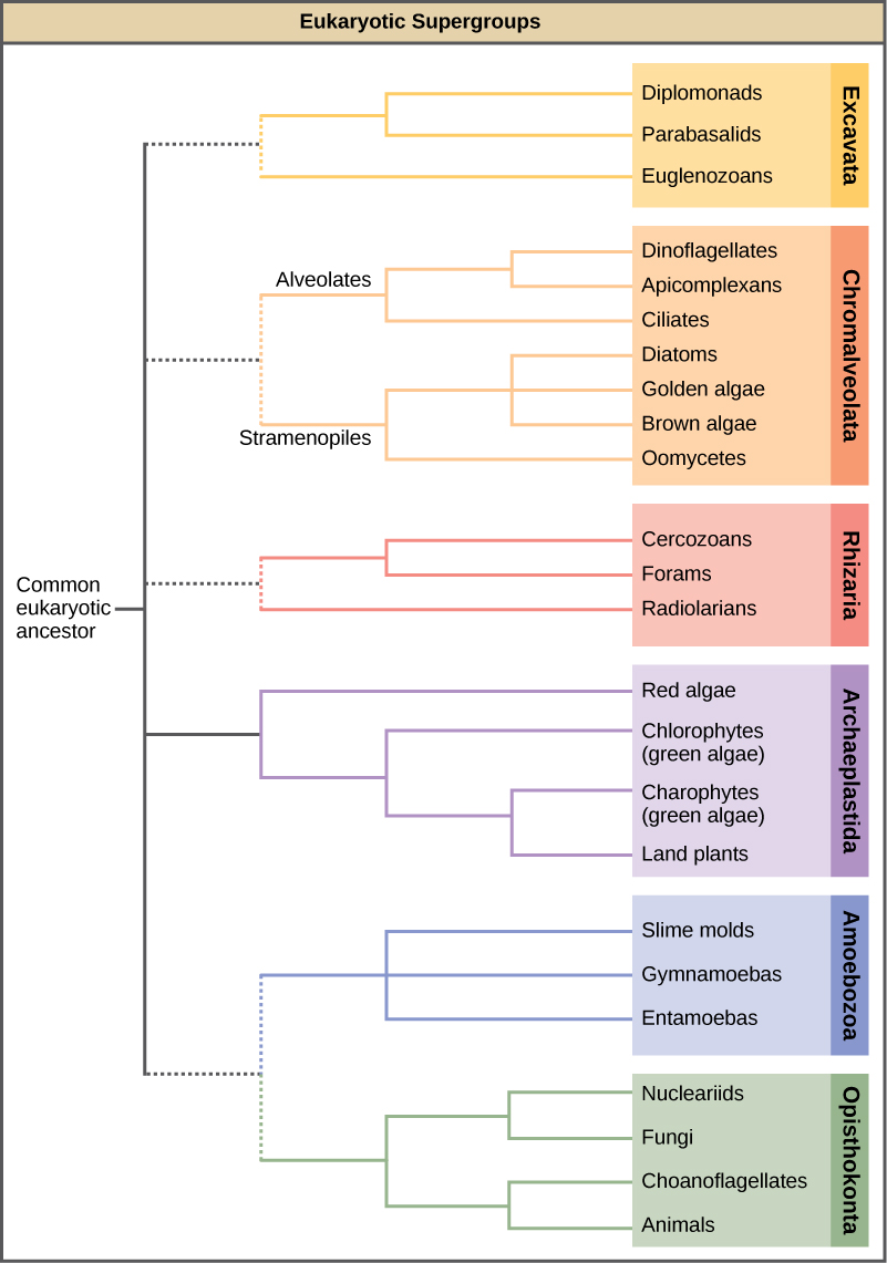
The classification of eukaryotes is still in flux, and the six supergroups may be modified or replaced by a more appropriate hierarchy as genetic, morphological, and ecological data accumulate. Keep in mind that the classification scheme presented here is just one of several hypotheses, and the true evolutionary relationships are still to be determined.
Cell Structure
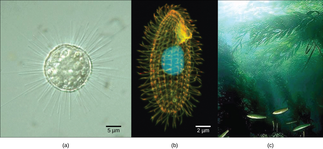
The cells of protists are among the most elaborate of all cells. Most protists are microscopic and unicellular, but some multicellular forms exist. A few protists live as colonies that behave in some ways as a group of independent free-living cells and in other ways as a multicellular organism. Not all protists are microscopic. Some have huge, macroscopic cells, such as the plasmodia (giant amoebae) of myxomycete slime molds or the marine green alga Caulerpa. Some protists are multicellular, such as the red, green, and brown seaweeds. It is among the protists that one finds the wealth of ways that organisms can grow. In fact, many protist cells are multinucleated; in some species, the nuclei are different sizes and have distinct roles in protist cell function.
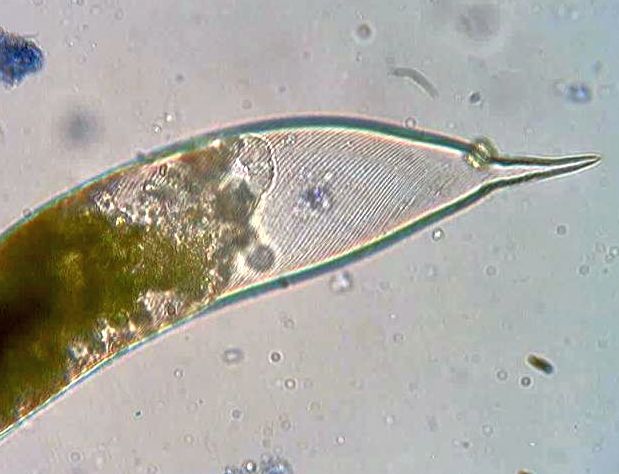
Single protist cells range in size from less than a micrometer to three meters in length, to hectares in area. Protist cells may be enveloped by animal-like cell membranes or plant-like cell walls. Others are encased in glassy silica-based shells or wound with pellicles of interlocking protein strips. The pellicle functions like a flexible coat of armor, preventing the protist from being torn or pierced without compromising its range of motion.
Metabolism
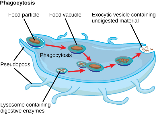
Protists exhibit many forms of nutrition and may be aerobic or anaerobic. Protists that store energy by photosynthesis belong to a group of photoautotrophs and are characterized by the presence of chloroplasts. Other protists are heterotrophic and consume organic materials (such as other organisms) to obtain nutrition. Amoebas and some other heterotrophic protist species ingest particles by a process called phagocytosis, in which the cell membrane engulfs a food particle and brings it inward, pinching off an intracellular membranous sac, or vesicle, called a food vacuole (Figure \(\PageIndex{4}\)). The food particle is broken down into small molecules that can diffuse into the cytoplasm and be used in cellular metabolism. Undigested materials are expelled from the cell via exocytosis.
Subtypes of heterotrophs, called saprobes, absorb nutrients from dead organisms or their organic wastes. Some protists can function as mixotrophs, obtaining nutrition by photoautotrophic or heterotrophic routes, depending on whether sunlight or organic nutrients are available.
In this 11-minute video, the diverse ways that protist extract energy from their environment are explored.
Question after watching: What is the advantage of being a mixotroph in aquatic environments?
Motility
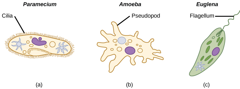
Most protists are motile, but different types of protists have evolved different modes of movement (Figure \(\PageIndex{5}\)). Some protists have one or more flagella, which they rotate or whip. Others are covered in rows or tufts of tiny cilia that they coordinately beat to swim. Still others form cytoplasmic extensions called pseudopodia anywhere on the cell, anchor the pseudopodia to a substrate, and pull themselves forward. Some protists can move toward or away from a stimulus, a movement referred to as taxis. Movement toward light, termed phototaxis, is accomplished by coupling their locomotion strategy with a light-sensing organ.
In this 11-minute video, find out how these single-celled eukaryotes move through their viscous environment.
Question after watching: After making a table with the three main types of possible movements, which representative organisms would you include to help you remember each one?
Life Cycles
Protists reproduce by many mechanisms. Most undergo some form of asexual reproduction, such as binary fission, to produce two daughter cells. In protists, binary fission can occur along the transverse or longitudinal axis of the cell, depending on the axis of orientation. Sometimes Paramecium uses binary fission to reproduce. Some protists such as the true slime molds exhibit multiple fission and simultaneously divide into many daughter cells. Others produce tiny buds that go on to divide and grow to the size of the parental protist.
Sexual reproduction, involving meiosis and fertilization, is also found among protists, and many protist species can switch from asexual to sexual reproduction when necessary. Sexual reproduction is often associated with periods of stress such as when nutrients are rare or environmental changes occur. Sexual reproduction may allow the protist to recombine genes and produce new variations in its progeny that may be better suited to surviving in the new environment.
In addition, sexual reproduction is often associated with the creation of resistant cysts that are a protective, resting stage of the life cycle of the protist. Depending on their habitat, the cysts may be particularly resistant to temperature extremes, desiccation, or low pH. This strategy also allows certain protists to “wait out” stressors until their environment becomes more favorable for survival or until they are carried (such as by wind, water, or transport on a larger organism) to a different environment because cysts exhibit little cellular metabolism and leave the organism in a sort of suspended animation.
Protist life cycles range from simple to complex. Certain parasitic protists have complicated life cycles and must infect different host species at different developmental stages to complete their life cycle. Some protists are unicellular in the haploid form and multicellular in the diploid form, a strategy employed by animals (gametes are unicellular, but fertilized eggs rapidly develop and divide to form multicellular animals). Other protists have multicellular stages in both haploid and diploid forms, a strategy called alternation of generations that is also used by plants.
In this 8.5-minute video, explore the ways that protists reproduce.
Question after watching: Explain in your own words why sexual reproduction can be useful if a protist’s environment changes.
Habitats
Nearly all protists exist in aquatic environments, including freshwater and marine environments, damp soil, and even snow. They are also common in the digestive tracts of animals and in the vascular tissues of plants. Others invade the cells of other protists, animals, and plants. Protists function in diverse ecological niches. Whereas some protist species are essential components of the food chain and generators of biomass, others function in the decomposition of organic materials. Still other protists are dangerous human pathogens or causative agents of devastating plant diseases. Several protist species are parasites that infect animals or plants. A few protist species live on dead organisms or their wastes and contribute to their decay.
Protists with the capabilities to perform photosynthesis and to absorb nutrients from dead organisms are called ______________.
- photoautotrophs
- mixotrophs
- saprobes
- heterotrophs
- Answer
-
B. mixotrophs
Which of these locomotor organs would likely be the shortest?
- a flagellum
- a cilium
- an extended pseudopod
- a pellicle
- Answer
-
B. a cilium
Thumbnail: This scanning electron micrograph (SEM) revealed some of the external ultrastructural details displayed by a flagellated Giardia lamblia protozoan parasite. G. lamblia is the organism responsible for causing the diarrheal disease "giardiasis". (Public Domain; CDC / Janice Haney Carr).