
2.E: How We See the Invisible World (Exercises)
- Page ID
- 10399

\( \newcommand{\vecs}[1]{\overset { \scriptstyle \rightharpoonup} {\mathbf{#1}} } \)
\( \newcommand{\vecd}[1]{\overset{-\!-\!\rightharpoonup}{\vphantom{a}\smash {#1}}} \)
\( \newcommand{\id}{\mathrm{id}}\) \( \newcommand{\Span}{\mathrm{span}}\)
( \newcommand{\kernel}{\mathrm{null}\,}\) \( \newcommand{\range}{\mathrm{range}\,}\)
\( \newcommand{\RealPart}{\mathrm{Re}}\) \( \newcommand{\ImaginaryPart}{\mathrm{Im}}\)
\( \newcommand{\Argument}{\mathrm{Arg}}\) \( \newcommand{\norm}[1]{\| #1 \|}\)
\( \newcommand{\inner}[2]{\langle #1, #2 \rangle}\)
\( \newcommand{\Span}{\mathrm{span}}\)
\( \newcommand{\id}{\mathrm{id}}\)
\( \newcommand{\Span}{\mathrm{span}}\)
\( \newcommand{\kernel}{\mathrm{null}\,}\)
\( \newcommand{\range}{\mathrm{range}\,}\)
\( \newcommand{\RealPart}{\mathrm{Re}}\)
\( \newcommand{\ImaginaryPart}{\mathrm{Im}}\)
\( \newcommand{\Argument}{\mathrm{Arg}}\)
\( \newcommand{\norm}[1]{\| #1 \|}\)
\( \newcommand{\inner}[2]{\langle #1, #2 \rangle}\)
\( \newcommand{\Span}{\mathrm{span}}\) \( \newcommand{\AA}{\unicode[.8,0]{x212B}}\)
\( \newcommand{\vectorA}[1]{\vec{#1}} % arrow\)
\( \newcommand{\vectorAt}[1]{\vec{\text{#1}}} % arrow\)
\( \newcommand{\vectorB}[1]{\overset { \scriptstyle \rightharpoonup} {\mathbf{#1}} } \)
\( \newcommand{\vectorC}[1]{\textbf{#1}} \)
\( \newcommand{\vectorD}[1]{\overrightarrow{#1}} \)
\( \newcommand{\vectorDt}[1]{\overrightarrow{\text{#1}}} \)
\( \newcommand{\vectE}[1]{\overset{-\!-\!\rightharpoonup}{\vphantom{a}\smash{\mathbf {#1}}}} \)
\( \newcommand{\vecs}[1]{\overset { \scriptstyle \rightharpoonup} {\mathbf{#1}} } \)
\( \newcommand{\vecd}[1]{\overset{-\!-\!\rightharpoonup}{\vphantom{a}\smash {#1}}} \)
2.1: The Properties of Light
Visible light consists of electromagnetic waves that behave like other waves. Hence, many of the properties of light that are relevant to microscopy can be understood in terms of light’s behavior as a wave. An important property of light waves is the wavelength, or the distance between one peak of a wave and the next peak. The height of each peak (or depth of each trough) is called the amplitude.
Multiple Choice
Which of the following has the highest energy?
- light with a long wavelength
- light with an intermediate wavelength
- light with a short wavelength
- It is impossible to tell from the information given.
- Answer
-
C
You place a specimen under the microscope and notice that parts of the specimen begin to emit light immediately. These materials can be described as _____________.
- fluorescent
- phosphorescent
- transparent
- opaque
- Answer
-
A
Fill in the Blank
When you see light bend as it moves from air into water, you are observing _________.
- Answer
-
refraction
Short Answer
Explain how a prism separates white light into different colors.
Critical Thinking
In Figure 2.1.6, which of the following has the lowest energy?
- visible light
- X-rays
- ultraviolet rays
- infrared rays
2.2: Peering into the Invisible World
Italian scholar Girolamo Fracastoro is regarded as the first person to formally postulate that disease was spread by tiny invisible seminaria. He proposed that these seeds could attach themselves to certain objects that supported their transfer from person to person. However, since the technology for seeing such tiny objects did not yet exist, the existence of the seminaria remained hypothetical for a little over a century—an invisible world waiting to be revealed.
Short Answer
Why is Antonie van Leeuwenhoek’s work much better known than that of Zaccharias Janssen?
Why did the cork cells observed by Robert Hooke appear to be empty, as opposed to being full of other structures?
Multiple Choice
Who was the first to describe “cells” in dead cork tissue?
- Hans Janssen
- Zaccharias Janssen
- Antonie van Leeuwenhoek
- Robert Hooke
- Answer
-
D
Who is the probable inventor of the compound microscope?
- Girolamo Fracastoro
- Zaccharias Janssen
- Antonie van Leeuwenhoek
- Robert Hooke
- Answer
-
B
Fill in the Blank
A microscope that uses multiple lenses is called a _________ microscope.
- Answer
-
compound
2.3: Instruments of Microscopy
The 20th century saw the development of microscopes that leveraged nonvisible light, such as fluorescence microscopy, which uses an ultraviolet light source, and electron microscopy, which uses short-wavelength electron beams. These advances led to major improvements in magnification, resolution, and contrast. In this section, we survey the broad range of modern microscopic technology and common applications for each type of microscope.
Multiple Choice
Which would be the best choice for viewing internal structures of a living protist such as a Paramecium?
- a brightfield microscope with a stain
- a brightfield microscope without a stain
- a darkfield microscope
- a transmission electron microscope
- Answer
-
C
Which type of microscope is especially useful for viewing thick structures such as biofilms?
- a transmission electron microscope
- a scanning electron microscopes
- a phase-contrast microscope
- a confocal scanning laser microscope
- an atomic force microscope
- Answer
-
D
Which type of microscope would be the best choice for viewing very small surface structures of a cell?
- a transmission electron microscope
- a scanning electron microscope
- a brightfield microscope
- a darkfield microscope
- a phase-contrast microscope
- Answer
-
B
What type of microscope uses an annular stop?
- a transmission electron microscope
- a scanning electron microscope
- a brightfield microscope
- a darkfield microscope
- a phase-contrast microscope
- Answer
-
E
What type of microscope uses a cone of light so that light only hits the specimen indirectly, producing a darker image on a brighter background?
- a transmission electron microscope
- a scanning electron microscope
- a brightfield microscope
- a darkfield microscope
- a phase-contrast microscope
- Answer
-
D
Fill in the Blank
Chromophores that absorb and then emit light are called __________.
- Answer
-
fluorochromes
In a(n) _______ microscope, a probe located just above the specimen moves up and down in response to forces between the atoms and the tip of the probe.
- Answer
-
atomic force microscope
What is the total magnification of a specimen that is being viewed with a standard ocular lens and a 40⨯ objective lens?
- Answer
-
400⨯
Short Answer
What is the function of the condenser in a brightfield microscope?
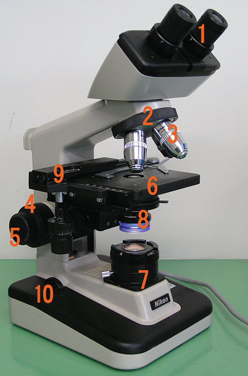
Label each component of the brightfield microscope.
Critical Thinking
When focusing a light microscope, why is it best to adjust the focus using the coarse focusing knob before using the fine focusing knob?
You need to identify structures within a cell using a microscope. However, the image appears very blurry even though you have a high magnification. What are some things that you could try to improve the resolution of the image? Describe the most basic factors that affect resolution when you first put the slide onto the stage; then consider more specific factors that could affect resolution for 40⨯ and 100⨯ lenses.
2.4: Staining Microscopic Specimens
In their natural state, most of the cells and microorganisms that we observe under the microscope lack color and contrast. This makes it difficult, if not impossible, to detect important cellular structures and their distinguishing characteristics without artificially treating specimens. We focus on the most clinically relevant techniques developed to identify specific microbes, cellular structures, DNA sequences, or indicators of infection in tissue samples, under the microscope.
Multiple Choice
What mordant is used in Gram staining?
- crystal violet
- safranin
- acid-alcohol
- iodine
- Answer
-
D
What is one difference between specimen preparation for a transmission electron microscope (TEM) and preparation for a scanning electron microscope (SEM)?
- Only the TEM specimen requires sputter coating.
- Only the SEM specimen requires sputter-coating.
- Only the TEM specimen must be dehydrated.
- Only the SEM specimen must be dehydrated.
- Answer
-
B
Fill in the Blank
Ziehl-Neelsen staining, a type of _______ staining, is diagnostic for Mycobacterium tuberculosis.
- Answer
-
acid-fast
The _______ is used to differentiate bacterial cells based on the components of their cell walls.
- Answer
-
Gram stain
Short Answer
How could you identify whether a particular bacterial sample contained specimens with mycolic acid-rich cell walls?
Critical Thinking
You use the Gram staining procedure to stain an L-form bacterium (a bacterium that lacks a cell wall). What color will the bacterium be after the staining procedure is finished?