
1.2: Cellular Organization - Prokaryotic and Eukaryotic Cells
- Page ID
- 2698

\( \newcommand{\vecs}[1]{\overset { \scriptstyle \rightharpoonup} {\mathbf{#1}} } \)
\( \newcommand{\vecd}[1]{\overset{-\!-\!\rightharpoonup}{\vphantom{a}\smash {#1}}} \)
\( \newcommand{\id}{\mathrm{id}}\) \( \newcommand{\Span}{\mathrm{span}}\)
( \newcommand{\kernel}{\mathrm{null}\,}\) \( \newcommand{\range}{\mathrm{range}\,}\)
\( \newcommand{\RealPart}{\mathrm{Re}}\) \( \newcommand{\ImaginaryPart}{\mathrm{Im}}\)
\( \newcommand{\Argument}{\mathrm{Arg}}\) \( \newcommand{\norm}[1]{\| #1 \|}\)
\( \newcommand{\inner}[2]{\langle #1, #2 \rangle}\)
\( \newcommand{\Span}{\mathrm{span}}\)
\( \newcommand{\id}{\mathrm{id}}\)
\( \newcommand{\Span}{\mathrm{span}}\)
\( \newcommand{\kernel}{\mathrm{null}\,}\)
\( \newcommand{\range}{\mathrm{range}\,}\)
\( \newcommand{\RealPart}{\mathrm{Re}}\)
\( \newcommand{\ImaginaryPart}{\mathrm{Im}}\)
\( \newcommand{\Argument}{\mathrm{Arg}}\)
\( \newcommand{\norm}[1]{\| #1 \|}\)
\( \newcommand{\inner}[2]{\langle #1, #2 \rangle}\)
\( \newcommand{\Span}{\mathrm{span}}\) \( \newcommand{\AA}{\unicode[.8,0]{x212B}}\)
\( \newcommand{\vectorA}[1]{\vec{#1}} % arrow\)
\( \newcommand{\vectorAt}[1]{\vec{\text{#1}}} % arrow\)
\( \newcommand{\vectorB}[1]{\overset { \scriptstyle \rightharpoonup} {\mathbf{#1}} } \)
\( \newcommand{\vectorC}[1]{\textbf{#1}} \)
\( \newcommand{\vectorD}[1]{\overrightarrow{#1}} \)
\( \newcommand{\vectorDt}[1]{\overrightarrow{\text{#1}}} \)
\( \newcommand{\vectE}[1]{\overset{-\!-\!\rightharpoonup}{\vphantom{a}\smash{\mathbf {#1}}}} \)
\( \newcommand{\vecs}[1]{\overset { \scriptstyle \rightharpoonup} {\mathbf{#1}} } \)
\( \newcommand{\vecd}[1]{\overset{-\!-\!\rightharpoonup}{\vphantom{a}\smash {#1}}} \)
\(\newcommand{\avec}{\mathbf a}\) \(\newcommand{\bvec}{\mathbf b}\) \(\newcommand{\cvec}{\mathbf c}\) \(\newcommand{\dvec}{\mathbf d}\) \(\newcommand{\dtil}{\widetilde{\mathbf d}}\) \(\newcommand{\evec}{\mathbf e}\) \(\newcommand{\fvec}{\mathbf f}\) \(\newcommand{\nvec}{\mathbf n}\) \(\newcommand{\pvec}{\mathbf p}\) \(\newcommand{\qvec}{\mathbf q}\) \(\newcommand{\svec}{\mathbf s}\) \(\newcommand{\tvec}{\mathbf t}\) \(\newcommand{\uvec}{\mathbf u}\) \(\newcommand{\vvec}{\mathbf v}\) \(\newcommand{\wvec}{\mathbf w}\) \(\newcommand{\xvec}{\mathbf x}\) \(\newcommand{\yvec}{\mathbf y}\) \(\newcommand{\zvec}{\mathbf z}\) \(\newcommand{\rvec}{\mathbf r}\) \(\newcommand{\mvec}{\mathbf m}\) \(\newcommand{\zerovec}{\mathbf 0}\) \(\newcommand{\onevec}{\mathbf 1}\) \(\newcommand{\real}{\mathbb R}\) \(\newcommand{\twovec}[2]{\left[\begin{array}{r}#1 \\ #2 \end{array}\right]}\) \(\newcommand{\ctwovec}[2]{\left[\begin{array}{c}#1 \\ #2 \end{array}\right]}\) \(\newcommand{\threevec}[3]{\left[\begin{array}{r}#1 \\ #2 \\ #3 \end{array}\right]}\) \(\newcommand{\cthreevec}[3]{\left[\begin{array}{c}#1 \\ #2 \\ #3 \end{array}\right]}\) \(\newcommand{\fourvec}[4]{\left[\begin{array}{r}#1 \\ #2 \\ #3 \\ #4 \end{array}\right]}\) \(\newcommand{\cfourvec}[4]{\left[\begin{array}{c}#1 \\ #2 \\ #3 \\ #4 \end{array}\right]}\) \(\newcommand{\fivevec}[5]{\left[\begin{array}{r}#1 \\ #2 \\ #3 \\ #4 \\ #5 \\ \end{array}\right]}\) \(\newcommand{\cfivevec}[5]{\left[\begin{array}{c}#1 \\ #2 \\ #3 \\ #4 \\ #5 \\ \end{array}\right]}\) \(\newcommand{\mattwo}[4]{\left[\begin{array}{rr}#1 \amp #2 \\ #3 \amp #4 \\ \end{array}\right]}\) \(\newcommand{\laspan}[1]{\text{Span}\{#1\}}\) \(\newcommand{\bcal}{\cal B}\) \(\newcommand{\ccal}{\cal C}\) \(\newcommand{\scal}{\cal S}\) \(\newcommand{\wcal}{\cal W}\) \(\newcommand{\ecal}{\cal E}\) \(\newcommand{\coords}[2]{\left\{#1\right\}_{#2}}\) \(\newcommand{\gray}[1]{\color{gray}{#1}}\) \(\newcommand{\lgray}[1]{\color{lightgray}{#1}}\) \(\newcommand{\rank}{\operatorname{rank}}\) \(\newcommand{\row}{\text{Row}}\) \(\newcommand{\col}{\text{Col}}\) \(\renewcommand{\row}{\text{Row}}\) \(\newcommand{\nul}{\text{Nul}}\) \(\newcommand{\var}{\text{Var}}\) \(\newcommand{\corr}{\text{corr}}\) \(\newcommand{\len}[1]{\left|#1\right|}\) \(\newcommand{\bbar}{\overline{\bvec}}\) \(\newcommand{\bhat}{\widehat{\bvec}}\) \(\newcommand{\bperp}{\bvec^\perp}\) \(\newcommand{\xhat}{\widehat{\xvec}}\) \(\newcommand{\vhat}{\widehat{\vvec}}\) \(\newcommand{\uhat}{\widehat{\uvec}}\) \(\newcommand{\what}{\widehat{\wvec}}\) \(\newcommand{\Sighat}{\widehat{\Sigma}}\) \(\newcommand{\lt}{<}\) \(\newcommand{\gt}{>}\) \(\newcommand{\amp}{&}\) \(\definecolor{fillinmathshade}{gray}{0.9}\)- Briefly describe why, in terms of differences in cell size, a eukaryotic cell is structurally more complex and compartmentalized than a cell that is prokaryotic.
- When given a description, determine whether a cell is prokaryotic or eukaryotic and explain why.
- Briefly state why viruses are not considered as prokaryotic nor eukaryotic.
According to the cell theory, the cell is the basic unit of life. All living organisms are composed of one or more cells. Based on the organization of their cellular structures, all living cells can be divided into two groups: prokaryotic and eukaryotic (also spelled procaryotic and eucaryotic). Animals, plants, fungi, protozoans, and algae all possess eukaryotic cell types. Only bacteria have prokaryotic cell types.
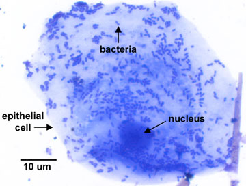
Prokaryotic cells are generally much smaller and more simple than eukaryotic (Figure \(\PageIndex{1}\)). Prokaryotic cells are, in fact, able to be structurally more simple because of their small size. The smaller a cell, the greater is its surface-to-volume ratio (the surface area of a cell compared to its volume).
The surface area of a spherical object can be calculated using the following formula:
\[S = 4\, \pi\, r^2\]
The volume of a spherical object can be calculated using the formula:
\[V = \dfrac{4}{3}\, \pi\, r^3 \]
For example, a spherical cell 1 micrometer (µm) in diameter - the average size of a coccus-shaped bacterium - has a surface-to-volume ratio of approximately 6:1, while a spherical cell having a diameter of 20 µm has a surface-to-volume ratio of approximately 0.3:1.
A large surface-to-volume ratio, as seen in smaller prokaryotic cells, means that nutrients can easily and rapidly reach any part of the cells interior. However, in the larger eukaryotic cell, the limited surface area when compared to its volume means nutrients cannot rapidly diffuse to all interior parts of the cell. That is why eukaryotic cells require a variety of specialized internal organelles to carry out metabolism, provide energy, and transport chemicals throughout the cell. Both, however, must carry out the same life processes. Some features distinguishing prokaryotic and eukaryotic cells are shown in Table \(\PageIndex{1}\). All of these features will be discussed in detail later in Unit 1.
Table \(\PageIndex{1}\): Eukaryotic Versus Prokaryotic CellsNuclear Body
eukaryotic cell
a. The nuclear body is bounded by a nuclear membrane having pores connecting it with the endoplasmic reticulum (see Figure \(\PageIndex{2}\) and Figure \(\PageIndex{3}\)).
b. It contains one or more paired, linear chromosomes composed of deoxyribonucleic acid (DNA) associated with histone proteins ).
c. A nucleolus is present. Ribosomal RNA (rRNA) is transcribed and assembled in the nucleolus.
d. The nuclear body is called a nucleus.
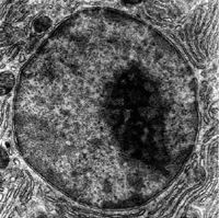
An electron micrograph of a cell nucleus, showing the darkly stained nucleolus. (Public Domain; US National Institute of General Medical Sciences/National Institutes of Health)
prokaryotic cell
a. The nuclear body is not bounded by a nuclear membrane (see Figure \(\PageIndex{4}\)).
b. It usually contains one circular chromosome composed of deoxyribonucleic acid (DNA) associated with histone-like proteins.
c. There is no nucleolus.
d. The nuclear body is called a nucleoid .
Cell Division
eukaryotic cell
a. The nucleus divides by mitosis .
b. Haploid (1N) sex cells in diploid or 2N organisms are produced through meiosis .
prokaryotic cell
a. The cell usually divides by binary fission . There is no mitosis.
b. Prokaryotic cells are haploid. Meiosis is not needed.
Cytoplasmic Membrane - also known as a cell membrane or plasma membrane
eukaryotic cell
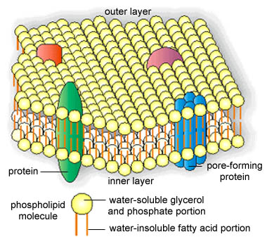
a. The cytoplasmic membrane (see Figure \(\PageIndex{2}\) and Figure \(\PageIndex{3}\)) is a fluid phospholipid bilayer (see Figure \(\PageIndex{5}\)) containing sterols (see Figure \(\PageIndex{6}\)) .
b. The membrane is capable of endocytosis (phagocytosis and pinocytosis) and exocytosis .
prokaryotic cell
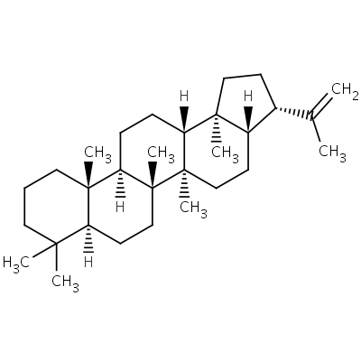
a. The cytoplasmic membrane (Figure \(\PageIndex{4}\)) is a fluid phospholipid bilayer (Figure \(\PageIndex{5}\)) that usually lacking sterols. Bacteria generally contain sterol-like molecules called hopanoids (Figure \(\PageIndex{7}\)).

b.The membrane is incapable of endocytosis and exocytosis.
Cytoplasmic Structures
eukaryotic cell
a. The ribosomes are composed of a 60S and a 40S subunit that come together during protein synthesis to form an 80S ribosome .
- Ribosomal subunit densities: 60S and 40S
b. Internal membrane-bound organelles such as mitochondria , endoplasmic reticulum , Golgi apparatus , vacuoles, and lysosomes are present (see Figure \(\PageIndex{2}\) and Figure \(\PageIndex{3}\)).
c. Chloroplasts serve as organelles for photosynthesis.
d. A mitotic spindle involved in mitosis is present during cell division.
e. A cytoskeleton is present. It contains microtubules, actin micofilaments, and intermediate filaments. These collectively play a role in giving shape to cells, allowing for cell movement, movement of organelles within the cell and endocytosis, and cell division.
- Electron micrograph of a cytoplasmic membrane courtesy of Dennis Kunkel's Microscopy
- Electron micrograph of mitochondria courtesy of Dennis Kunkel's Microscopy
- Electron micrograph of rough endoplasmic reticulum courtesy of Dennis Kunkel's Microscopy
- Electron micrograph of a Golgi apparatus courtesy of Dennis Kunkel's Microscopy
prokaryotic cell
a. The ribosomes are composed of a 50S and a 30S subunit that come together during protein synthesis to form a 70S ribosome . See Figure \(\PageIndex{8}\).
- Ribosomal subunit densities: 50S and 30S
b. Internal membrane-bound organelles such as mitochondria, endoplasmic reticulum, Golgi apparatus, vacuoles, and lysosomes are absent (see Figure \(\PageIndex{4}\))
c. There are no chloroplasts. Photosynthesis usually takes place in infoldings or extensions derived from the cytoplasmic membrane.
d. There is no mitosis and no mitotic spindle.
e. The various structural filaments in the cytoplasm collectively make up the prokaryotic cytoskeleton. Cytoskeletal filaments play essential roles in determining the shape of a bacterium (coccus, bacillus, or spiral) and are also critical in the process of cell division by binary fission and in determining bacterial polarity.
Respiratory Enzymes and Electron Transport Chains
eukaryotic cell
- The electron transport system is located in the inner membrane of the mitochondria. It contributes to the production of ATP molecules via chemiosmosis.
-Electron micrograph of a mitochondrion from the Biology Department at the University of New Mexico.
prokaryotic cell
- The electron transport system is located in the cytoplasmic membrane. It contributes to the production of ATP molecules via chemiosmosis.
Cell Wall
eukaryotic cell
a. Plant cells, algae, and fungi have cell walls, usually composed of cellulose or chitin. Eukaryotic cell walls are never composed of peptidoglycan (see Figure \(\PageIndex{3}\)).
b. Animal cells and protozoans lack cell walls (see Figure \(\PageIndex{2}\)).
prokaryotic cell
a. With few exceptions, members of the domain Bacteria have cell walls composed of peptidoglycan (see Figure \(\PageIndex{4}\)).
b. Members of the domain Archae have cell walls composed of protein, a complex carbohydrate, or unique molecules resembling but not the same as peptidoglycan.
Locomotor Organelles
eukaryotic cell
- Eukaryotic cells may have flagella or cilia. Flagella and cilia are organelles involved in locomotion and in eukaryotic cells consist of a distinct arrangement of sliding microtubules surrounded by a membrane. The microtubule arrangement is referred to as a 2X9+2 arrangement (see Figure \(\PageIndex{9}\)).
- Electron micrograph of cilia showing microtubules courtesy of Dennis Kunkel's Microscopy
- YouTube movie of motile sperm.
prokaryotic cell
- Many prokaryotes have flagella, each composed of a single, rotating fibril and usually not surrounded by a membrane (see Figure \(\PageIndex{10}\)). There are no cilia.
- Movie of motile Rhodobacter spheroides with fluorescent labelled-flagella. Courtesy of Dr. Howard C. Berg from the Roland Institute at Harvard.
Representative Organisms
- eukaryotic cell: The domain Eukarya: animals, plants, algae, protozoans, and fungi (yeasts, molds, mushrooms).
- prokaryotic cell: The domain Bacteria and the domain Archae.
Since viruses are acellular- they contain no cellular organelles, cannot grow and divide, and carry out no independent metabolism - they are considered neither prokaryotic nor eukaryotic. Because viruses are not cells and have no cellular organelles, they can only replicate and assemble inside a living host cell. They turn the host cell into a factory for manufacturing viral parts and viral enzymes and assembling the viral components.
Viruses, which possess both living and nonliving characteristics, will be discussed in Unit 4. Recently, viruses have been declared as living entities based on the large number of protein folds encoded by viral genomes that are shared with the genomes of cells. This indicates that viruses likely arose from multiple ancient cells.
Summary
- There are two basic types of cells in nature: prokaryotic and eukaryotic.
- Prokaryotic cells are structurally simpler than eukaryotic cells.
- The smaller a cell, the greater its surface to volume ratio.
- The smaller the surface to volume ratio, the more structurally complex (compartmentalized) a cell needs to be in order to carry out life functions.
- There are fundamental differences between prokaryotic and eukaryotic cells.
- Bacteria are prokaryotic cells; fungi, protozoa, algae, plants, and animals are composed of eukaryotic cells.
- Viruses are not cells so they are neither prokaryotic nor eukaryotic. They can replicate only inside a living cell.