Biology LibreTexts

5.2: Comparing Prokaryotic and Eukaryotic Cells

  • Page ID
    68560
  • \( \newcommand{\vecs}[1]{\overset { \scriptstyle \rightharpoonup} {\mathbf{#1}} } \) \( \newcommand{\vecd}[1]{\overset{-\!-\!\rightharpoonup}{\vphantom{a}\smash {#1}}} \)\(\newcommand{\id}{\mathrm{id}}\) \( \newcommand{\Span}{\mathrm{span}}\) \( \newcommand{\kernel}{\mathrm{null}\,}\) \( \newcommand{\range}{\mathrm{range}\,}\) \( \newcommand{\RealPart}{\mathrm{Re}}\) \( \newcommand{\ImaginaryPart}{\mathrm{Im}}\) \( \newcommand{\Argument}{\mathrm{Arg}}\) \( \newcommand{\norm}[1]{\| #1 \|}\) \( \newcommand{\inner}[2]{\langle #1, #2 \rangle}\) \( \newcommand{\Span}{\mathrm{span}}\) \(\newcommand{\id}{\mathrm{id}}\) \( \newcommand{\Span}{\mathrm{span}}\) \( \newcommand{\kernel}{\mathrm{null}\,}\) \( \newcommand{\range}{\mathrm{range}\,}\) \( \newcommand{\RealPart}{\mathrm{Re}}\) \( \newcommand{\ImaginaryPart}{\mathrm{Im}}\) \( \newcommand{\Argument}{\mathrm{Arg}}\) \( \newcommand{\norm}[1]{\| #1 \|}\) \( \newcommand{\inner}[2]{\langle #1, #2 \rangle}\) \( \newcommand{\Span}{\mathrm{span}}\)\(\newcommand{\AA}{\unicode[.8,0]{x212B}}\)

    Cells fall into one of two broad categories: prokaryotic and eukaryotic. The predominantly single-celled organisms of the domains Bacteria and Archaea are classified as prokaryotes (pro– = before; –karyon– = nucleus). Animal cells, plant cells, fungi, and protists are eukaryotes (eu– = true).

    All cells share four common components: 1) a plasma membrane, an outer covering that separates the cell’s interior from its surrounding environment; 2) cytoplasm, consisting of a gel-like region within the cell in which other cellular components are found; 3) DNA, the genetic material of the cell; and 4) ribosomes, particles that synthesize proteins.

    Components of Prokaryotic Cells

    Prokaryotes differ from eukaryotic cells in several important ways. A prokaryotic cell is a simple, single-celled (unicellular) organism that lacks a nucleus, or any other membrane-bound organelle. We will shortly come to see that this is significantly different in eukaryotes. Prokaryotic DNA is found in the central part of the cell: a darkened region called the nucleoid (Figure \(\PageIndex{1}\)).

    In this illustration, the prokaryotic cell has an oval shape. The circular chromosome is concentrated in a region called the nucleoid. The fluid inside the cell is called the cytoplasm. Ribosomes, depicted as small circles, float in the cytoplasm. The cytoplasm is encased by a plasma membrane, which in turn is encased by a cell wall. A capsule surrounds the cell wall. The bacterium depicted has a flagellum protruding from one narrow end. Pili are small protrusions that project from the capsule in all directions.
    Figure \(\PageIndex{1}\): This figure shows the generalized structure of a prokaryotic cell.

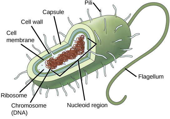
    Unlike Archaea and eukaryotes, bacteria have a cell wall made of peptidoglycan, comprised of sugars and amino acids, and many have a polysaccharide (carbohydrate) capsule (Figure \(\PageIndex{1}\)). The cell wall acts as an extra layer of protection, helps the cell maintain its shape, and prevents dehydration. The capsule enables the cell to attach to surfaces in its environment. Some prokaryotes have flagella, pili, or fimbriae. Flagella are used for locomotion, while most pili are used to exchange genetic material during a type of reproduction called conjugation.

    Components of Eukaryotic Cells

    In nature, the relationship between form and function is apparent at all levels, including the level of the cell, and this will become clear as we explore eukaryotic cells. The principle “form follows function” is found in many contexts. For example, birds and fish have streamlined bodies that allow them to move quickly through the medium in which they live, be it air or water. It means that, in general, one can deduce the function of a structure by looking at its form, because the two are matched.

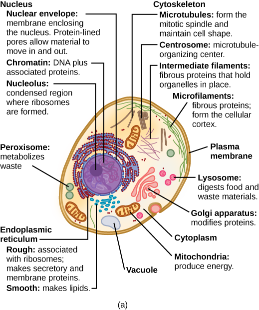
    A eukaryotic cell is a cell that has a membrane-bound nucleus and other membrane-bound compartments or sacs, called organelles, which have specialized functions. The rest of this chapter will discuss functions of the various organelles. The word eukaryotic means “true kernel” or “true nucleus,” alluding to the presence of the membrane-bound nucleus in these cells. The word “organelle” means “little organ,” and, as already mentioned, organelles have specialized cellular functions, just as the organs of your body have specialized functions. 

    drawing of a cell
    Figure \(\PageIndex{2}\): A generalized eukaryotic cell showing some of the organelles.  (Photo credit: MediranWikimedia. 14 Aug 2002)

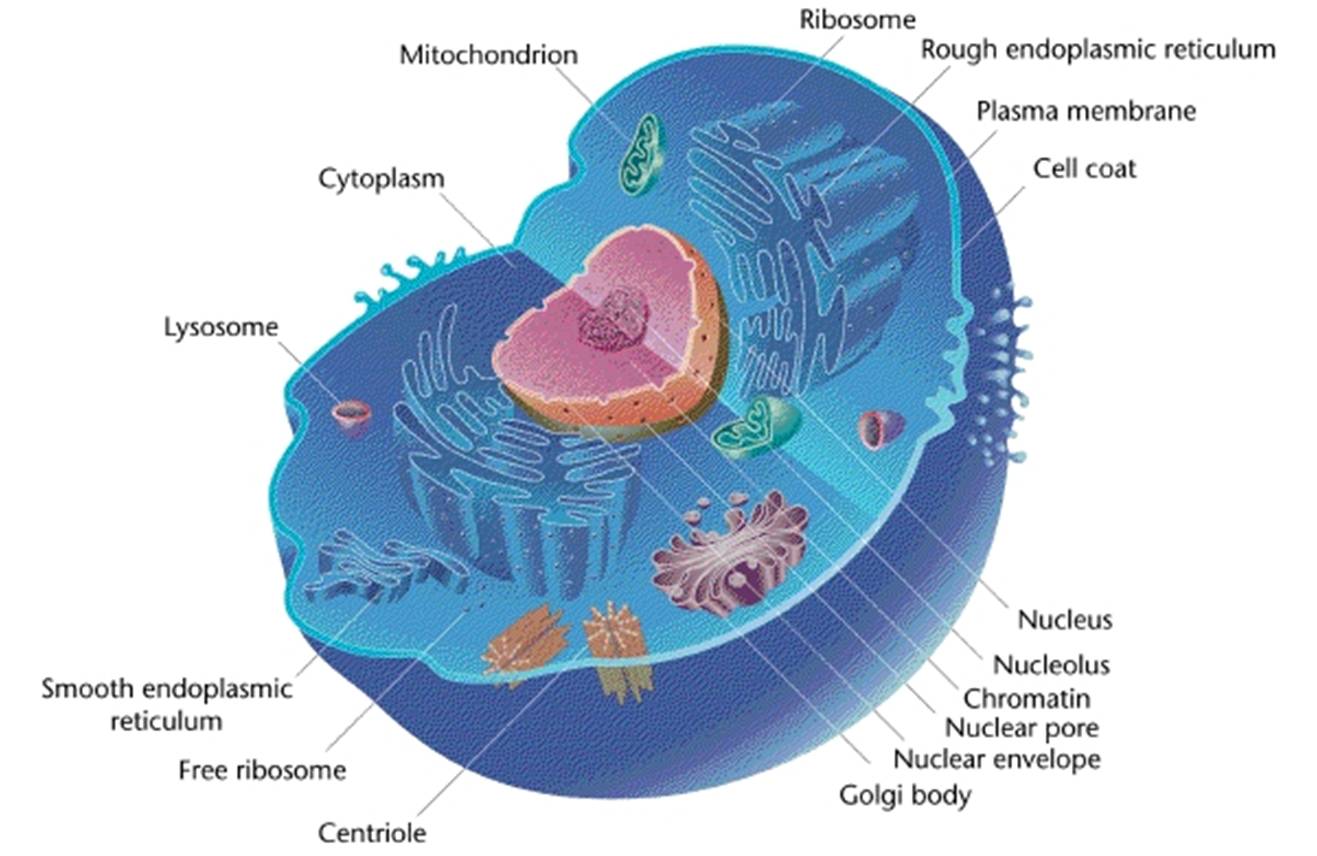
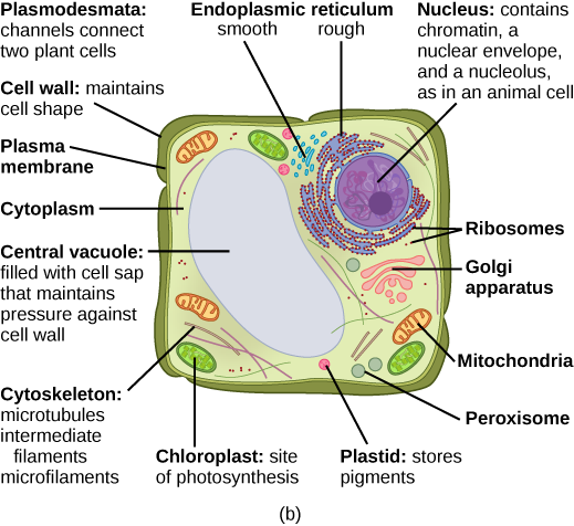
    Both animals and plants are eukaryotes. Despite their fundamental similarities, there are some striking differences between animal and plant cells. Animal cells have centrioles, centrosomes (discussed under the cytoskeleton), and lysosomes, whereas plant cells do not. Plant cells have a cell wall, chloroplasts, plasmodesmata, and plastids used for storage, and a large central vacuole, whereas animal cells do not.

    Part a: This illustration shows a typical eukaryotic cell, which is egg shaped. The fluid inside the cell is called the cytoplasm, and the cell is surrounded by a cell membrane. The nucleus takes up about one-half of the width of the cell. Inside the nucleus is the chromatin, which is comprised of DNA and associated proteins. A region of the chromatin is condensed into the nucleolus, a structure in which ribosomes are synthesized. The nucleus is encased in a nuclear envelope, which is perforated by protein-lined pores that allow entry of material into the nucleus.  The nucleus is surrounded by the rough and smooth endoplasmic reticulum, or ER. The smooth ER is the site of lipid synthesis. The rough ER has embedded ribosomes that give it a bumpy appearance. It synthesizes membrane and secretory proteins. Besides the ER, many other organelles float inside the cytoplasm. These include the Golgi apparatus, which modifies proteins and lipids synthesized in the ER. The Golgi apparatus is made of layers of flat membranes. Mitochondria, which produce energy for the cell, have an outer membrane and a highly folded inner membrane. Other, smaller organelles include peroxisomes that metabolize waste, lysosomes that digest food, and vacuoles. Ribosomes, responsible for protein synthesis, also float freely in the cytoplasm and are depicted as small dots. The last cellular component shown is the cytoskeleton, which has four different types of components: microfilaments, intermediate filaments, microtubules, and centrosomes. Microfilaments are fibrous proteins that line the cell membrane and make up the cellular cortex. Intermediate filaments are fibrous proteins that hold organelles in place. Microtubules form the mitotic spindle and maintain cell shape. Centrosomes are made of two tubular structures at right angles to one another. They form the microtubule-organizing center.
    Part b: This illustration depicts a typical eukaryotic plant cell. The nucleus of a plant cell contains chromatin and a nucleolus, the same as in an animal cell. Other structures that a plant cell has in common with an animal cell include rough and smooth ER, the Golgi apparatus, mitochondria, peroxisomes, and ribosomes. The fluid inside the plant cell is called the cytoplasm, just as in an animal cell. The plant cell has three of the four cytoskeletal components found in animal cells: microtubules, intermediate filaments, and microfilaments. Plant cells do not have centrosomes. Plants have five structures not found in animals cells: plasmodesmata, chloroplasts, plastids, a central vacuole, and a cell wall. Plasmodesmata form channels between adjacent plant cells. Chloroplasts are responsible for photosynthesis; they have an outer membrane, an inner membrane, and stack of membranes inside the inner membrane. The central vacuole is a very large, fluid-filled structure that maintains pressure against the cell wall. Plastids store pigments. The cell wall is localized outside the cell membrane.

    Query \(\PageIndex{1}\)

    Query \(\PageIndex{2}\)

    Cell Size

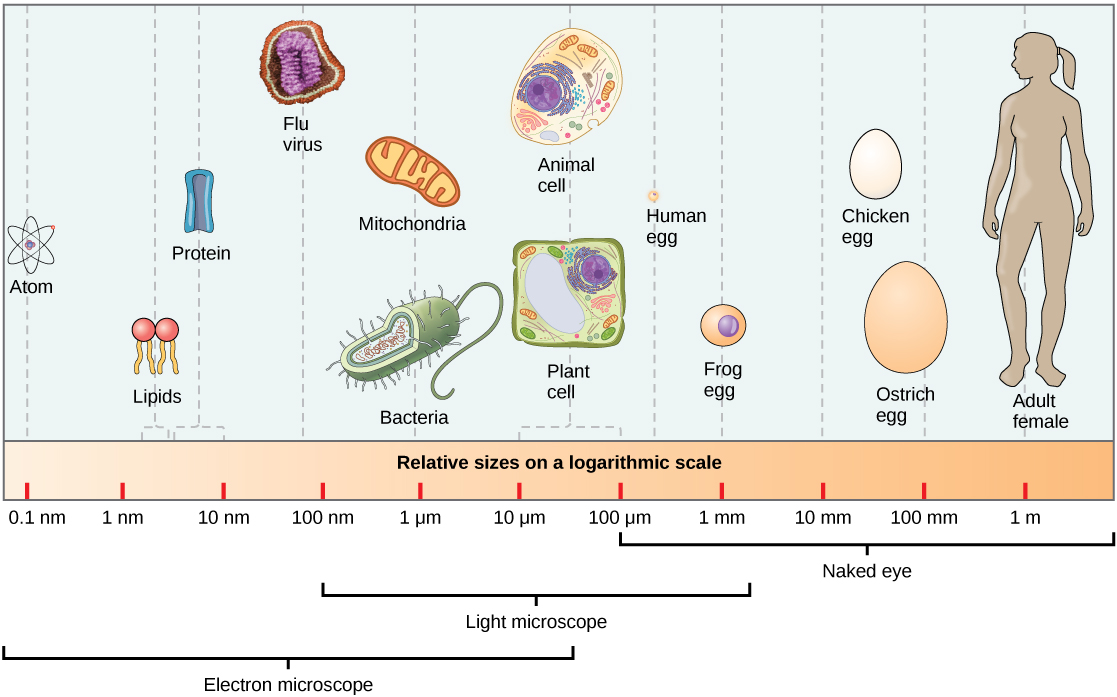
    At 0.1–5.0 μm in diameter, prokaryotic cells are significantly smaller than eukaryotic cells, which have diameters ranging from 10–100 μm (Figure \(\PageIndex{3}\)). The small size of prokaryotes allows ions and organic molecules that enter them to quickly spread to other parts of the cell. Similarly, any wastes produced within a prokaryotic cell can quickly move out. However, larger eukaryotic cells have evolved different structural adaptations to enhance cellular transport. Indeed, the large size of these cells would not be possible without these adaptations. In general, cell size is limited because volume increases much more quickly than does cell surface area. As a cell becomes larger, it becomes more and more difficult for the cell to acquire sufficient materials to support the processes inside the cell, because the relative size of the surface area across which materials must be transported declines.

    Relative sizes on a logarithmic scale, from 0.1 nm to 1 m, are shown. Objects are shown from smallest to largest. The smallest object shown, an atom, is about 1 nm in size. The next largest objects shown are lipids and proteins; these molecules are between 1 and 10 nm. Bacteria are about 100 nm, and mitochondria are about 1 µm. Plant and animal cells are both between 10 and 100 µm. A human egg is between 100 µm and 1 mm. A frog egg is about 1 mm, a chicken egg and an ostrich egg are both between 10 and 100 mm, but an ostrich egg is larger. For comparison, a human is approximately 1 m tall.
    Figure \(\PageIndex{3}\): This figure shows the relative sizes of different kinds of cells and cellular components. An adult human is shown for comparison.

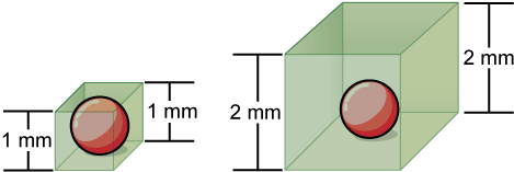
    Small size, in general, is necessary for all cells, whether prokaryotic or eukaryotic. Let’s examine why that is so. First, we’ll consider the area and volume of a typical cell. Not all cells are spherical in shape, but most tend to approximate a sphere. You may remember from your geometry course that the formula for the surface area of a sphere is 4πr2, while the formula for its volume is 4πr3/3. Thus, as the radius of a cell increases, its surface area increases as the square of its radius, but its volume increases as the cube of its radius (much more rapidly). Therefore, as a cell increases in size, its surface area-to-volume ratio decreases. This same principle would apply if the cell had the shape of a cube (Figure \(\PageIndex{4}\)). If the cell grows too large, the plasma membrane will not have sufficient surface area to support the rate of diffusion required for the increased volume. In other words, as a cell grows, it becomes less efficient. One way to become more efficient is to divide; another way is to develop organelles that perform specific tasks. These adaptations lead to the development of more sophisticated cells called eukaryotic cells.

    cube cells
    Figure \(\PageIndex{4}\): Volume increases faster than surface area. The surface area of the small cell is 1mm x 1mm x 6 sides = 6mm2. The volume of the small cell is 1mm x 1mm x 1mm = 1mm3. This gives a surface area to volume ratio of 6:1. The surface area of the larger cell is 2mm x 2mm x 6 sides = 24mm2. The volume of the large cell is 2mm x 2mm x 2mm = 8mm3. This gives a surface area to volume ratio of 3:1 (24:8 reduces to 3:1).

    Query \(\PageIndex{3}\)

    References

    Unless otherwise noted, images on this page are licensed under CC-BY 4.0 by OpenStax.

    Text adapted from: OpenStax, Concepts of Biology. OpenStax CNX. May 18, 2016 http://cnx.org/contents/b3c1e1d2-839...9a8aafbdd@9.10


    5.2: Comparing Prokaryotic and Eukaryotic Cells is shared under a CC BY license and was authored, remixed, and/or curated by LibreTexts.