
6.5: The Lower Limbs
- Page ID
- 59393
\( \newcommand{\vecs}[1]{\overset { \scriptstyle \rightharpoonup} {\mathbf{#1}} } \)
\( \newcommand{\vecd}[1]{\overset{-\!-\!\rightharpoonup}{\vphantom{a}\smash {#1}}} \)
\( \newcommand{\dsum}{\displaystyle\sum\limits} \)
\( \newcommand{\dint}{\displaystyle\int\limits} \)
\( \newcommand{\dlim}{\displaystyle\lim\limits} \)
\( \newcommand{\id}{\mathrm{id}}\) \( \newcommand{\Span}{\mathrm{span}}\)
( \newcommand{\kernel}{\mathrm{null}\,}\) \( \newcommand{\range}{\mathrm{range}\,}\)
\( \newcommand{\RealPart}{\mathrm{Re}}\) \( \newcommand{\ImaginaryPart}{\mathrm{Im}}\)
\( \newcommand{\Argument}{\mathrm{Arg}}\) \( \newcommand{\norm}[1]{\| #1 \|}\)
\( \newcommand{\inner}[2]{\langle #1, #2 \rangle}\)
\( \newcommand{\Span}{\mathrm{span}}\)
\( \newcommand{\id}{\mathrm{id}}\)
\( \newcommand{\Span}{\mathrm{span}}\)
\( \newcommand{\kernel}{\mathrm{null}\,}\)
\( \newcommand{\range}{\mathrm{range}\,}\)
\( \newcommand{\RealPart}{\mathrm{Re}}\)
\( \newcommand{\ImaginaryPart}{\mathrm{Im}}\)
\( \newcommand{\Argument}{\mathrm{Arg}}\)
\( \newcommand{\norm}[1]{\| #1 \|}\)
\( \newcommand{\inner}[2]{\langle #1, #2 \rangle}\)
\( \newcommand{\Span}{\mathrm{span}}\) \( \newcommand{\AA}{\unicode[.8,0]{x212B}}\)
\( \newcommand{\vectorA}[1]{\vec{#1}} % arrow\)
\( \newcommand{\vectorAt}[1]{\vec{\text{#1}}} % arrow\)
\( \newcommand{\vectorB}[1]{\overset { \scriptstyle \rightharpoonup} {\mathbf{#1}} } \)
\( \newcommand{\vectorC}[1]{\textbf{#1}} \)
\( \newcommand{\vectorD}[1]{\overrightarrow{#1}} \)
\( \newcommand{\vectorDt}[1]{\overrightarrow{\text{#1}}} \)
\( \newcommand{\vectE}[1]{\overset{-\!-\!\rightharpoonup}{\vphantom{a}\smash{\mathbf {#1}}}} \)
\( \newcommand{\vecs}[1]{\overset { \scriptstyle \rightharpoonup} {\mathbf{#1}} } \)
\(\newcommand{\longvect}{\overrightarrow}\)
\( \newcommand{\vecd}[1]{\overset{-\!-\!\rightharpoonup}{\vphantom{a}\smash {#1}}} \)
\(\newcommand{\avec}{\mathbf a}\) \(\newcommand{\bvec}{\mathbf b}\) \(\newcommand{\cvec}{\mathbf c}\) \(\newcommand{\dvec}{\mathbf d}\) \(\newcommand{\dtil}{\widetilde{\mathbf d}}\) \(\newcommand{\evec}{\mathbf e}\) \(\newcommand{\fvec}{\mathbf f}\) \(\newcommand{\nvec}{\mathbf n}\) \(\newcommand{\pvec}{\mathbf p}\) \(\newcommand{\qvec}{\mathbf q}\) \(\newcommand{\svec}{\mathbf s}\) \(\newcommand{\tvec}{\mathbf t}\) \(\newcommand{\uvec}{\mathbf u}\) \(\newcommand{\vvec}{\mathbf v}\) \(\newcommand{\wvec}{\mathbf w}\) \(\newcommand{\xvec}{\mathbf x}\) \(\newcommand{\yvec}{\mathbf y}\) \(\newcommand{\zvec}{\mathbf z}\) \(\newcommand{\rvec}{\mathbf r}\) \(\newcommand{\mvec}{\mathbf m}\) \(\newcommand{\zerovec}{\mathbf 0}\) \(\newcommand{\onevec}{\mathbf 1}\) \(\newcommand{\real}{\mathbb R}\) \(\newcommand{\twovec}[2]{\left[\begin{array}{r}#1 \\ #2 \end{array}\right]}\) \(\newcommand{\ctwovec}[2]{\left[\begin{array}{c}#1 \\ #2 \end{array}\right]}\) \(\newcommand{\threevec}[3]{\left[\begin{array}{r}#1 \\ #2 \\ #3 \end{array}\right]}\) \(\newcommand{\cthreevec}[3]{\left[\begin{array}{c}#1 \\ #2 \\ #3 \end{array}\right]}\) \(\newcommand{\fourvec}[4]{\left[\begin{array}{r}#1 \\ #2 \\ #3 \\ #4 \end{array}\right]}\) \(\newcommand{\cfourvec}[4]{\left[\begin{array}{c}#1 \\ #2 \\ #3 \\ #4 \end{array}\right]}\) \(\newcommand{\fivevec}[5]{\left[\begin{array}{r}#1 \\ #2 \\ #3 \\ #4 \\ #5 \\ \end{array}\right]}\) \(\newcommand{\cfivevec}[5]{\left[\begin{array}{c}#1 \\ #2 \\ #3 \\ #4 \\ #5 \\ \end{array}\right]}\) \(\newcommand{\mattwo}[4]{\left[\begin{array}{rr}#1 \amp #2 \\ #3 \amp #4 \\ \end{array}\right]}\) \(\newcommand{\laspan}[1]{\text{Span}\{#1\}}\) \(\newcommand{\bcal}{\cal B}\) \(\newcommand{\ccal}{\cal C}\) \(\newcommand{\scal}{\cal S}\) \(\newcommand{\wcal}{\cal W}\) \(\newcommand{\ecal}{\cal E}\) \(\newcommand{\coords}[2]{\left\{#1\right\}_{#2}}\) \(\newcommand{\gray}[1]{\color{gray}{#1}}\) \(\newcommand{\lgray}[1]{\color{lightgray}{#1}}\) \(\newcommand{\rank}{\operatorname{rank}}\) \(\newcommand{\row}{\text{Row}}\) \(\newcommand{\col}{\text{Col}}\) \(\renewcommand{\row}{\text{Row}}\) \(\newcommand{\nul}{\text{Nul}}\) \(\newcommand{\var}{\text{Var}}\) \(\newcommand{\corr}{\text{corr}}\) \(\newcommand{\len}[1]{\left|#1\right|}\) \(\newcommand{\bbar}{\overline{\bvec}}\) \(\newcommand{\bhat}{\widehat{\bvec}}\) \(\newcommand{\bperp}{\bvec^\perp}\) \(\newcommand{\xhat}{\widehat{\xvec}}\) \(\newcommand{\vhat}{\widehat{\vvec}}\) \(\newcommand{\uhat}{\widehat{\uvec}}\) \(\newcommand{\what}{\widehat{\wvec}}\) \(\newcommand{\Sighat}{\widehat{\Sigma}}\) \(\newcommand{\lt}{<}\) \(\newcommand{\gt}{>}\) \(\newcommand{\amp}{&}\) \(\definecolor{fillinmathshade}{gray}{0.9}\)Information
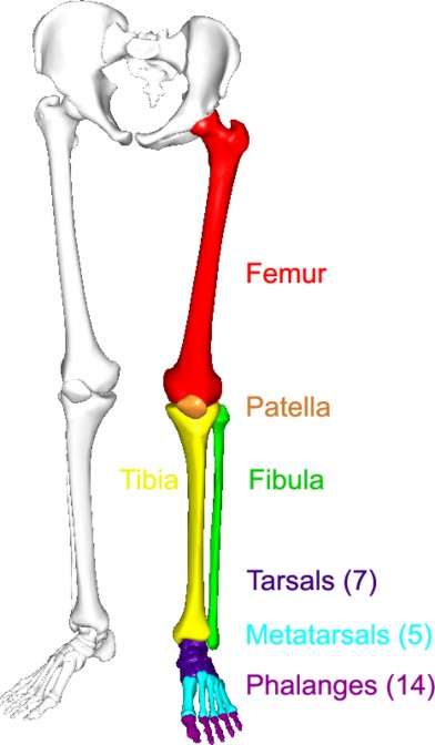
For anatomists, the lower limb consists of the thigh (the upper leg), the leg (the lower leg), and the foot. The thigh consists of a single bone, the femur. The leg consists of two long bones, the tibia and fibula, and the sesamoid bone, the patella, that serves as the knee cap. The foot consists of 26 bones, which are grouped into the tarsals, metatarsals, and phalanges.
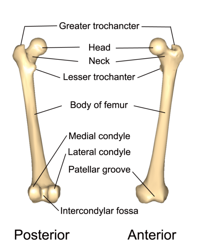
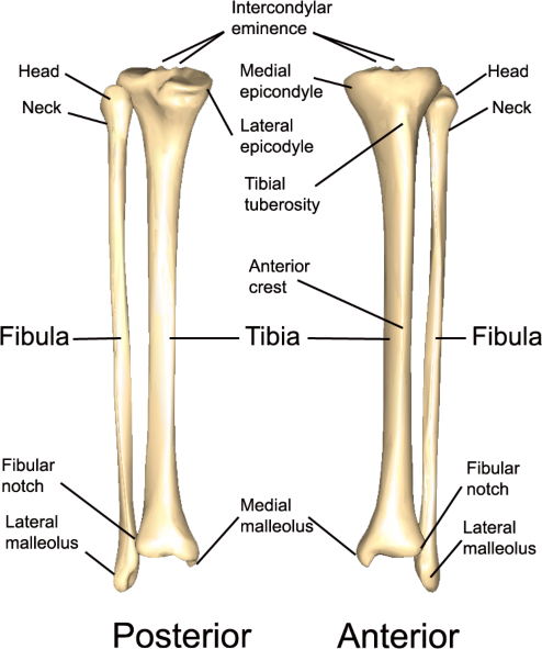
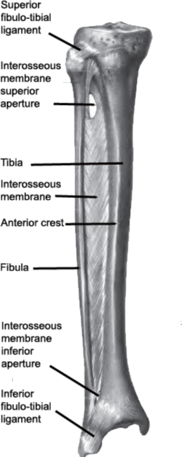
The major processes and markings of the femur, patella, and tibia & fibula bones are shown in Figures \(\PageIndex{2}\), \(\PageIndex{3}\), and \(\PageIndex{4}\), respectively. The interosseous membrane connecting the tibia and fibula bones is shown in Figure \(\PageIndex{5}\)
Lateral condyle
Medial condyle
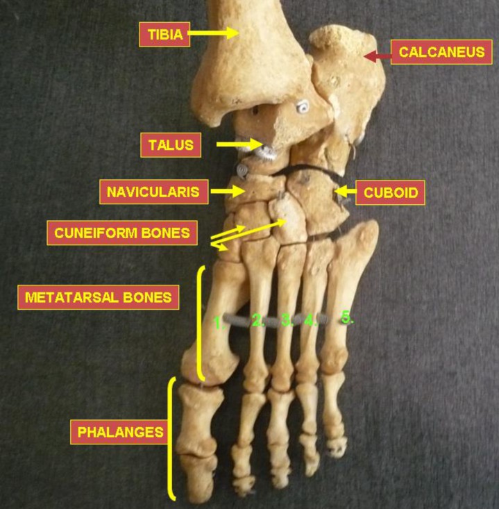
The bones of the foot are shown in Figure 6.20 below. The calcaneus is the heel bone, and the talus bone forms the ankle joint with the tibia and fibula. The calcaneus and tarsus are two of the seven tarsal bones that are posterior to the first long bones of the foot, the metatarsal bones. The bones of the toes are phalanges, the same name used for finger bones.
LAB 6 EXERCISE \(\PageIndex{1}\)
- Using one of the full skeletons in the room, fill out the table below with three or four steps to determine whether an individual bone comes from the anatomical left or anatomical right. You must use any features that are already filled in.
- You can describe any features on that bone and which direction it has to face to allow you to determine whether that particular bone came from anatomical left or anatomical right.
- Sample instructions for the clavicle are provided as an example for all subsequent exercises. Use proper anatomical terminology. Use terms which will make sense to anyone schooled in anatomy if they read it. Use as many steps as you need, not necessarily four.
| Femur – Anatomical left from anatomical right. | |
| 1 | Head |
| 2 | |
| 3 | |
| 4 |
| Tibia – Anatomical left from anatomical right. | |
| 1 | Tibial tuberosity |
| 2 | |
| 3 | |
| 4 |
| Fibula – Anatomical left from anatomical right. | |
| 1 | Lateral malleolus |
| 2 | |
| 3 | |
| 4 |
- Using one of the full skeletons again, fill out the tables below with three or four steps to determine how to distinguish the calcaneus bone from the talus bone.
- Use proper anatomical terminology. Use terms which will make sense to anyone schooled in anatomy if they read it. Use as many steps as you need, not necessarily three.
| Calcaneus bone | |
| 1 | |
| 2 | |
| 3 |
| Talus bone | |
| 1 | |
| 2 | |
| 3 |
LICENSES AND ATTRIBUTIONS
CC LICENSED CONTENT, ORIGINALA&P Labs. Authored by: Ross Whitwam. Provided by: Mississippi University for Women. Located at: http://www.muw.edu. License: CC BY-SA: Attribution-ShareAlike
Figure \(\PageIndex{1}\). The bones of the left lower limb.. Authored by: Images in Figure 7-18 were made out of, or made from, content published in a BodyParts3D/Anatomography web site. The content of their website is published under the Creative Commons Attribution 2.1 Japan license. The author and licenser of the contents is http://lifesciencedb.jp/bp3d/?lng=en.. Located at: . License: CC BY-SA: Attribution-ShareAlike
Figure \(\PageIndex{2}\). The left femur and its various processes and markings.. Authored by: Images in Figure 7-19 were made out of, or made from, content published in a BodyParts3D/Anatomography web site. The content of their website is published under the Creative Commons Attribution 2.1 Japan license. The author and licenser of the contents is http://lifesciencedb.jp/bp3d/?lng=en.. Located at: . License: CC BY-SA: Attribution-ShareAlike
Figure \(\PageIndex{4}\). The left tibia and femur and their various processes and markings.. Authored by: Images in Figure 7-21 were made out of, or made from, content published in a BodyParts3D/Anatomography web site. The content of their website is published under the Creative Commons Attribution 2.1 Japan license. The author and licenser of the contents is http://lifesciencedb.jp/bp3d/?lng=en.. Located at: . License: CC BY-SA: Attribution-ShareAlike
CC LICENSED CONTENT, SHARED PREVIOUSLY
Figure \(\PageIndex{5}\). The interosseous membrane of the left leg.. Authored by: Berichard. Located at: https://commons.wikimedia.org/wiki/F...ia_Fibula1.png. License: CC BY-SA: Attribution-ShareAlike
Figure \(\PageIndex{6}\). The bones of the left foot.. Authored by: Anatomist90. Located at: upload.wikimedia.org/wikiped...7/Foot_bones_-_tarsus%2C_metatarsus_and_phalanges.jpg. License: CC BY-SA: Attribution-ShareAlike
PUBLIC DOMAIN CONTENT
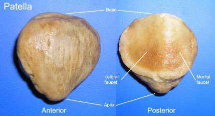
Figure \(\PageIndex3}\). The Right patella.. Authored by: Palica . Located at: commons.wikimedia.org/wiki/F...e:Patella_post.jpg. License: Public Domain: No Known Copyright