
6.2: The Upper Limbs
- Page ID
- 59390
\( \newcommand{\vecs}[1]{\overset { \scriptstyle \rightharpoonup} {\mathbf{#1}} } \)
\( \newcommand{\vecd}[1]{\overset{-\!-\!\rightharpoonup}{\vphantom{a}\smash {#1}}} \)
\( \newcommand{\dsum}{\displaystyle\sum\limits} \)
\( \newcommand{\dint}{\displaystyle\int\limits} \)
\( \newcommand{\dlim}{\displaystyle\lim\limits} \)
\( \newcommand{\id}{\mathrm{id}}\) \( \newcommand{\Span}{\mathrm{span}}\)
( \newcommand{\kernel}{\mathrm{null}\,}\) \( \newcommand{\range}{\mathrm{range}\,}\)
\( \newcommand{\RealPart}{\mathrm{Re}}\) \( \newcommand{\ImaginaryPart}{\mathrm{Im}}\)
\( \newcommand{\Argument}{\mathrm{Arg}}\) \( \newcommand{\norm}[1]{\| #1 \|}\)
\( \newcommand{\inner}[2]{\langle #1, #2 \rangle}\)
\( \newcommand{\Span}{\mathrm{span}}\)
\( \newcommand{\id}{\mathrm{id}}\)
\( \newcommand{\Span}{\mathrm{span}}\)
\( \newcommand{\kernel}{\mathrm{null}\,}\)
\( \newcommand{\range}{\mathrm{range}\,}\)
\( \newcommand{\RealPart}{\mathrm{Re}}\)
\( \newcommand{\ImaginaryPart}{\mathrm{Im}}\)
\( \newcommand{\Argument}{\mathrm{Arg}}\)
\( \newcommand{\norm}[1]{\| #1 \|}\)
\( \newcommand{\inner}[2]{\langle #1, #2 \rangle}\)
\( \newcommand{\Span}{\mathrm{span}}\) \( \newcommand{\AA}{\unicode[.8,0]{x212B}}\)
\( \newcommand{\vectorA}[1]{\vec{#1}} % arrow\)
\( \newcommand{\vectorAt}[1]{\vec{\text{#1}}} % arrow\)
\( \newcommand{\vectorB}[1]{\overset { \scriptstyle \rightharpoonup} {\mathbf{#1}} } \)
\( \newcommand{\vectorC}[1]{\textbf{#1}} \)
\( \newcommand{\vectorD}[1]{\overrightarrow{#1}} \)
\( \newcommand{\vectorDt}[1]{\overrightarrow{\text{#1}}} \)
\( \newcommand{\vectE}[1]{\overset{-\!-\!\rightharpoonup}{\vphantom{a}\smash{\mathbf {#1}}}} \)
\( \newcommand{\vecs}[1]{\overset { \scriptstyle \rightharpoonup} {\mathbf{#1}} } \)
\(\newcommand{\longvect}{\overrightarrow}\)
\( \newcommand{\vecd}[1]{\overset{-\!-\!\rightharpoonup}{\vphantom{a}\smash {#1}}} \)
\(\newcommand{\avec}{\mathbf a}\) \(\newcommand{\bvec}{\mathbf b}\) \(\newcommand{\cvec}{\mathbf c}\) \(\newcommand{\dvec}{\mathbf d}\) \(\newcommand{\dtil}{\widetilde{\mathbf d}}\) \(\newcommand{\evec}{\mathbf e}\) \(\newcommand{\fvec}{\mathbf f}\) \(\newcommand{\nvec}{\mathbf n}\) \(\newcommand{\pvec}{\mathbf p}\) \(\newcommand{\qvec}{\mathbf q}\) \(\newcommand{\svec}{\mathbf s}\) \(\newcommand{\tvec}{\mathbf t}\) \(\newcommand{\uvec}{\mathbf u}\) \(\newcommand{\vvec}{\mathbf v}\) \(\newcommand{\wvec}{\mathbf w}\) \(\newcommand{\xvec}{\mathbf x}\) \(\newcommand{\yvec}{\mathbf y}\) \(\newcommand{\zvec}{\mathbf z}\) \(\newcommand{\rvec}{\mathbf r}\) \(\newcommand{\mvec}{\mathbf m}\) \(\newcommand{\zerovec}{\mathbf 0}\) \(\newcommand{\onevec}{\mathbf 1}\) \(\newcommand{\real}{\mathbb R}\) \(\newcommand{\twovec}[2]{\left[\begin{array}{r}#1 \\ #2 \end{array}\right]}\) \(\newcommand{\ctwovec}[2]{\left[\begin{array}{c}#1 \\ #2 \end{array}\right]}\) \(\newcommand{\threevec}[3]{\left[\begin{array}{r}#1 \\ #2 \\ #3 \end{array}\right]}\) \(\newcommand{\cthreevec}[3]{\left[\begin{array}{c}#1 \\ #2 \\ #3 \end{array}\right]}\) \(\newcommand{\fourvec}[4]{\left[\begin{array}{r}#1 \\ #2 \\ #3 \\ #4 \end{array}\right]}\) \(\newcommand{\cfourvec}[4]{\left[\begin{array}{c}#1 \\ #2 \\ #3 \\ #4 \end{array}\right]}\) \(\newcommand{\fivevec}[5]{\left[\begin{array}{r}#1 \\ #2 \\ #3 \\ #4 \\ #5 \\ \end{array}\right]}\) \(\newcommand{\cfivevec}[5]{\left[\begin{array}{c}#1 \\ #2 \\ #3 \\ #4 \\ #5 \\ \end{array}\right]}\) \(\newcommand{\mattwo}[4]{\left[\begin{array}{rr}#1 \amp #2 \\ #3 \amp #4 \\ \end{array}\right]}\) \(\newcommand{\laspan}[1]{\text{Span}\{#1\}}\) \(\newcommand{\bcal}{\cal B}\) \(\newcommand{\ccal}{\cal C}\) \(\newcommand{\scal}{\cal S}\) \(\newcommand{\wcal}{\cal W}\) \(\newcommand{\ecal}{\cal E}\) \(\newcommand{\coords}[2]{\left\{#1\right\}_{#2}}\) \(\newcommand{\gray}[1]{\color{gray}{#1}}\) \(\newcommand{\lgray}[1]{\color{lightgray}{#1}}\) \(\newcommand{\rank}{\operatorname{rank}}\) \(\newcommand{\row}{\text{Row}}\) \(\newcommand{\col}{\text{Col}}\) \(\renewcommand{\row}{\text{Row}}\) \(\newcommand{\nul}{\text{Nul}}\) \(\newcommand{\var}{\text{Var}}\) \(\newcommand{\corr}{\text{corr}}\) \(\newcommand{\len}[1]{\left|#1\right|}\) \(\newcommand{\bbar}{\overline{\bvec}}\) \(\newcommand{\bhat}{\widehat{\bvec}}\) \(\newcommand{\bperp}{\bvec^\perp}\) \(\newcommand{\xhat}{\widehat{\xvec}}\) \(\newcommand{\vhat}{\widehat{\vvec}}\) \(\newcommand{\uhat}{\widehat{\uvec}}\) \(\newcommand{\what}{\widehat{\wvec}}\) \(\newcommand{\Sighat}{\widehat{\Sigma}}\) \(\newcommand{\lt}{<}\) \(\newcommand{\gt}{>}\) \(\newcommand{\amp}{&}\) \(\definecolor{fillinmathshade}{gray}{0.9}\)Information
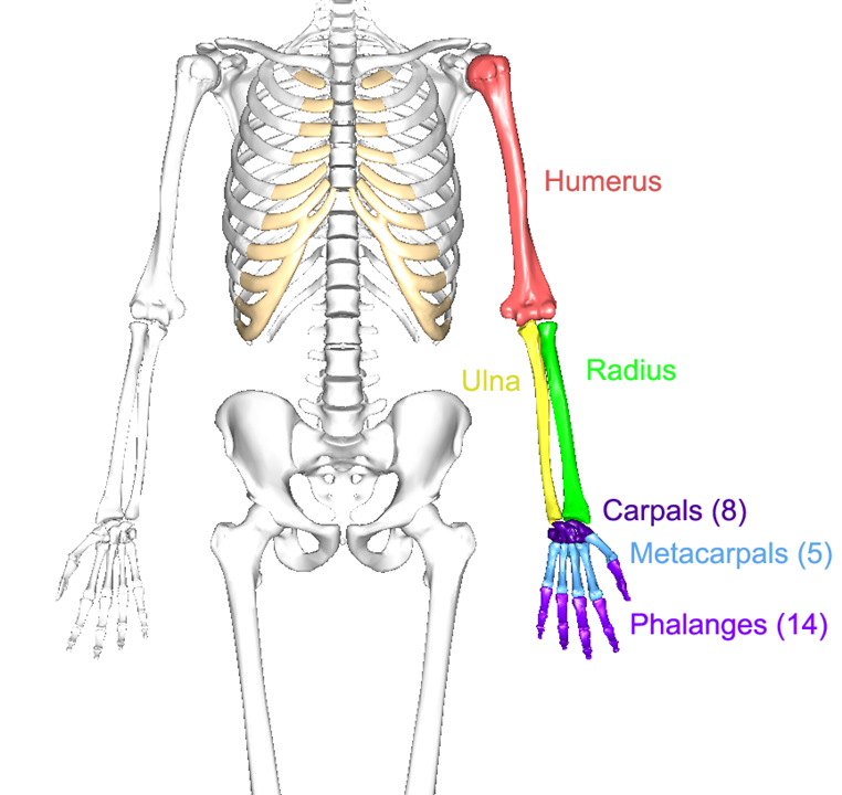
For anatomists, the upper limb consists of the arm (the upper arm), the forearm (the lower arm), and the hand. The arm consists of a single bone, the humerus. The forearm consists of two bones, the ulna and radius. And the hand consists of 27 bones, which are grouped into the phalanges, metacarpals, and carpals.
The major processes and markings of the humerus, ulna, and radius bones are shown in Figures \(\PageIndex{2}\), \(\PageIndex{3}\), and \(\PageIndex{4}\), respectively.
The bones of the hands are divided into three groups. The carpals articulate with the ulna and radius bones of the forearm and are named after the carpus, or wrist. There are 8 carpal bones and each has its own name. In Figure \(\PageIndex{5}\) they are numbered so that 1-trapezium, 2-trapezoid, 3-capitate, 4-hamate, 5-pisiform, 6-triquetrum, 7-lunate, 8- scaphoid.

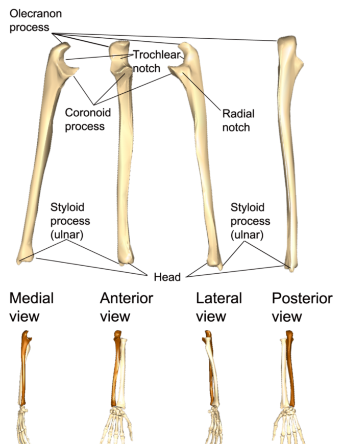
Figure \(\PageIndex{3}\): The left ulna (in brown) and its major markings and processes. (CC-BY-SA, BodyParts3D/Anatomography)

Figure \(\PageIndex{4}\): he left radius (in brown) and its major markings and processes. (CC-BY-SA, BodyParts3D/Anatomography)
The metacarpals are five individual bones that are wrapped in muscle and collective tissue to create a single solid mass that serves as the palm of the hand. They are numbered I – V, starting with the metacarpal under the thumb and moving sequentially to the little finger.
Metacarpals are individually named according to the hand they come from and their number. So the metacarpal under the little finger in Figure 6.9 is named left metacarpal V.
There are 14 bones that make up the fingers. All are called phalanges (singular is phalanx). Each finger is made up of three phalanges, labelled the proximal, middle and distal phalanges as you move farther out from the metacarpals. Each thumb only has two phalanges, labelled proximal and distal. The phalanges are numbered I through V, like the metacarpals. Each phalanx then is named according to which hand it comes from, which number it is, and whether it is proximal, middle, or distal. So the second phalanx on the pointing finger in Figure is the left medial phalanx II.
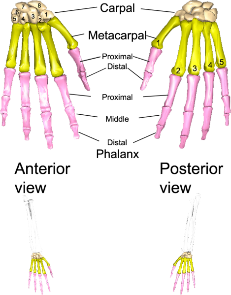 Figure \(\PageIndex{5}\): The bones of the left hand. (CC-BY-SA, BodyParts3D/Anatomography)
LAB 6 EXERCISE \(\PageIndex{1}\):
- Using one of the full skeletons in the room, fill out the table below with three or four steps to determine whether an individual bone comes from the anatomical left or anatomical right. You must use any features that are already filled in.
- You can describe any features on that bone and which direction it has to face to allow you to determine whether that particular bone came from anatomical left or anatomical right.
- Sample instructions for the clavicle are provided as an example for all subsequent exercises. Use proper anatomical terminology. Use terms which will make sense to anyone schooled in anatomy if they read it. Use as many steps as you need, not necessarily four.
Humerus – Anatomical left from anatomical right.
1. Olecranon fossa -
2.
3.
4.
Ulna – Anatomical left from anatomical right.
1. Radial notch -
2.
3.
4.
Radius – Anatomical left from anatomical right.
1. Styloid process -
2.
3.
4.
LICENSES AND ATTRIBUTIONS
CC LICENSED CONTENT, ORIGINAL
A&P Labs. Authored by: Ross Whitwam. Provided by: Mississippi University for Women. Located at: http://www.muw.edu. License: CC BY-SA: Attribution-ShareAlike
Fig \(\PageIndex{1}\):. The bones of the left upper limb.. Authored by: Images in Figure 7-8 were made out of, or made from, content published in a BodyParts3D/Anatomography web site. The content of their website is published under the Creative Commons Attribution 2.1 Japan license. The author and licenser of the contents is http://lifesciencedb.jp/bp3d/?lng=en. Located at: http://lifesciencedb.jp/bp3d/?lng=en. License: CC BY-SA: Attribution-ShareAlike
Figure \(\PageIndex{2}\):. The left humerus and its various processes and markings.. Authored by: Images in Figure 7-9 were made out of, or made from, content published in a BodyParts3D/Anatomography web site. The content of their website is published under the Creative Commons Attribution 2.1 Japan license. The author and licenser of the contents is http://lifesciencedb.jp/bp3d/?lng=en. Located at: http://lifesciencedb.jp/bp3d/?lng=en. License: CC BY-SA: Attribution-ShareAlike
Figure \(\PageIndex{3}\):. The left ulna (in brown) and its major markings and processes.. Authored by: Images in Figure 7-10 were made out of, or made from, content published in a BodyParts3D/Anatomography web site. The content of their website is published under the Creative Commons Attribution 2.1 Japan license. The author and licenser of the contents is http://lifesciencedb.jp/bp3d/?lng=en. Located at: http://lifesciencedb.jp/bp3d/?lng=en. License: CC BY-SA: Attribution- ShareAlike
Figure \(\PageIndex{4}\):. The left radius (in brown) and its major markings and processes.. Authored by: Images in Figure 7-11 were made out of, or made from, content published in a BodyParts3D/Anatomography web site. The content of their website is published under the Creative Commons Attribution 2.1 Japan license. The author and licenser of the contents is http://lifesciencedb.jp/bp3d/?lng=en. Located at: http://lifesciencedb.jp/bp3d/?lng=en. License: CC BY-SA: Attribution- ShareAlike
Figure \(\PageIndex{5}\):. The bones of the left hand.. Authored by: Images in Figure 7-12 were made out of, or made from, content published in a BodyParts3D/Anatomography web site. The content of their website is published under the Creative Commons Attribution 2.1 Japan license. The author and licenser of the contents is http://lifesciencedb.jp/bp3d/?lng=en. Located at: http://lifesciencedb.jp/bp3d/?lng=en. License: CC BY-SA: Attribution-ShareAlike