
5.2: The Vertebral Column
- Page ID
- 59385
\( \newcommand{\vecs}[1]{\overset { \scriptstyle \rightharpoonup} {\mathbf{#1}} } \)
\( \newcommand{\vecd}[1]{\overset{-\!-\!\rightharpoonup}{\vphantom{a}\smash {#1}}} \)
\( \newcommand{\dsum}{\displaystyle\sum\limits} \)
\( \newcommand{\dint}{\displaystyle\int\limits} \)
\( \newcommand{\dlim}{\displaystyle\lim\limits} \)
\( \newcommand{\id}{\mathrm{id}}\) \( \newcommand{\Span}{\mathrm{span}}\)
( \newcommand{\kernel}{\mathrm{null}\,}\) \( \newcommand{\range}{\mathrm{range}\,}\)
\( \newcommand{\RealPart}{\mathrm{Re}}\) \( \newcommand{\ImaginaryPart}{\mathrm{Im}}\)
\( \newcommand{\Argument}{\mathrm{Arg}}\) \( \newcommand{\norm}[1]{\| #1 \|}\)
\( \newcommand{\inner}[2]{\langle #1, #2 \rangle}\)
\( \newcommand{\Span}{\mathrm{span}}\)
\( \newcommand{\id}{\mathrm{id}}\)
\( \newcommand{\Span}{\mathrm{span}}\)
\( \newcommand{\kernel}{\mathrm{null}\,}\)
\( \newcommand{\range}{\mathrm{range}\,}\)
\( \newcommand{\RealPart}{\mathrm{Re}}\)
\( \newcommand{\ImaginaryPart}{\mathrm{Im}}\)
\( \newcommand{\Argument}{\mathrm{Arg}}\)
\( \newcommand{\norm}[1]{\| #1 \|}\)
\( \newcommand{\inner}[2]{\langle #1, #2 \rangle}\)
\( \newcommand{\Span}{\mathrm{span}}\) \( \newcommand{\AA}{\unicode[.8,0]{x212B}}\)
\( \newcommand{\vectorA}[1]{\vec{#1}} % arrow\)
\( \newcommand{\vectorAt}[1]{\vec{\text{#1}}} % arrow\)
\( \newcommand{\vectorB}[1]{\overset { \scriptstyle \rightharpoonup} {\mathbf{#1}} } \)
\( \newcommand{\vectorC}[1]{\textbf{#1}} \)
\( \newcommand{\vectorD}[1]{\overrightarrow{#1}} \)
\( \newcommand{\vectorDt}[1]{\overrightarrow{\text{#1}}} \)
\( \newcommand{\vectE}[1]{\overset{-\!-\!\rightharpoonup}{\vphantom{a}\smash{\mathbf {#1}}}} \)
\( \newcommand{\vecs}[1]{\overset { \scriptstyle \rightharpoonup} {\mathbf{#1}} } \)
\(\newcommand{\longvect}{\overrightarrow}\)
\( \newcommand{\vecd}[1]{\overset{-\!-\!\rightharpoonup}{\vphantom{a}\smash {#1}}} \)
\(\newcommand{\avec}{\mathbf a}\) \(\newcommand{\bvec}{\mathbf b}\) \(\newcommand{\cvec}{\mathbf c}\) \(\newcommand{\dvec}{\mathbf d}\) \(\newcommand{\dtil}{\widetilde{\mathbf d}}\) \(\newcommand{\evec}{\mathbf e}\) \(\newcommand{\fvec}{\mathbf f}\) \(\newcommand{\nvec}{\mathbf n}\) \(\newcommand{\pvec}{\mathbf p}\) \(\newcommand{\qvec}{\mathbf q}\) \(\newcommand{\svec}{\mathbf s}\) \(\newcommand{\tvec}{\mathbf t}\) \(\newcommand{\uvec}{\mathbf u}\) \(\newcommand{\vvec}{\mathbf v}\) \(\newcommand{\wvec}{\mathbf w}\) \(\newcommand{\xvec}{\mathbf x}\) \(\newcommand{\yvec}{\mathbf y}\) \(\newcommand{\zvec}{\mathbf z}\) \(\newcommand{\rvec}{\mathbf r}\) \(\newcommand{\mvec}{\mathbf m}\) \(\newcommand{\zerovec}{\mathbf 0}\) \(\newcommand{\onevec}{\mathbf 1}\) \(\newcommand{\real}{\mathbb R}\) \(\newcommand{\twovec}[2]{\left[\begin{array}{r}#1 \\ #2 \end{array}\right]}\) \(\newcommand{\ctwovec}[2]{\left[\begin{array}{c}#1 \\ #2 \end{array}\right]}\) \(\newcommand{\threevec}[3]{\left[\begin{array}{r}#1 \\ #2 \\ #3 \end{array}\right]}\) \(\newcommand{\cthreevec}[3]{\left[\begin{array}{c}#1 \\ #2 \\ #3 \end{array}\right]}\) \(\newcommand{\fourvec}[4]{\left[\begin{array}{r}#1 \\ #2 \\ #3 \\ #4 \end{array}\right]}\) \(\newcommand{\cfourvec}[4]{\left[\begin{array}{c}#1 \\ #2 \\ #3 \\ #4 \end{array}\right]}\) \(\newcommand{\fivevec}[5]{\left[\begin{array}{r}#1 \\ #2 \\ #3 \\ #4 \\ #5 \\ \end{array}\right]}\) \(\newcommand{\cfivevec}[5]{\left[\begin{array}{c}#1 \\ #2 \\ #3 \\ #4 \\ #5 \\ \end{array}\right]}\) \(\newcommand{\mattwo}[4]{\left[\begin{array}{rr}#1 \amp #2 \\ #3 \amp #4 \\ \end{array}\right]}\) \(\newcommand{\laspan}[1]{\text{Span}\{#1\}}\) \(\newcommand{\bcal}{\cal B}\) \(\newcommand{\ccal}{\cal C}\) \(\newcommand{\scal}{\cal S}\) \(\newcommand{\wcal}{\cal W}\) \(\newcommand{\ecal}{\cal E}\) \(\newcommand{\coords}[2]{\left\{#1\right\}_{#2}}\) \(\newcommand{\gray}[1]{\color{gray}{#1}}\) \(\newcommand{\lgray}[1]{\color{lightgray}{#1}}\) \(\newcommand{\rank}{\operatorname{rank}}\) \(\newcommand{\row}{\text{Row}}\) \(\newcommand{\col}{\text{Col}}\) \(\renewcommand{\row}{\text{Row}}\) \(\newcommand{\nul}{\text{Nul}}\) \(\newcommand{\var}{\text{Var}}\) \(\newcommand{\corr}{\text{corr}}\) \(\newcommand{\len}[1]{\left|#1\right|}\) \(\newcommand{\bbar}{\overline{\bvec}}\) \(\newcommand{\bhat}{\widehat{\bvec}}\) \(\newcommand{\bperp}{\bvec^\perp}\) \(\newcommand{\xhat}{\widehat{\xvec}}\) \(\newcommand{\vhat}{\widehat{\vvec}}\) \(\newcommand{\uhat}{\widehat{\uvec}}\) \(\newcommand{\what}{\widehat{\wvec}}\) \(\newcommand{\Sighat}{\widehat{\Sigma}}\) \(\newcommand{\lt}{<}\) \(\newcommand{\gt}{>}\) \(\newcommand{\amp}{&}\) \(\definecolor{fillinmathshade}{gray}{0.9}\)The Hyoid Bone
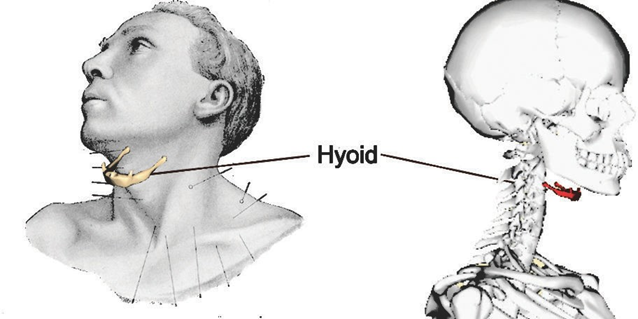
The hyoid bone in the neck is the only bone in the body that does not articulate directly with at least one other bone. It is U-shaped and is held in place by, and helps anchor, muscles that connect to the floor of the mouth and the tongue. It helps provide greater movement of the tongue and larynx, and so is crucial to human speech.
In about 50% of strangulations, the hyoid bone is fractured.
LICENSES AND ATTRIBUTIONS
CC LICENSED CONTENT, ORIGINAL
A&P Labs. Authored by: Ross Whitwam. Provided by: Mississippi University for Women. Located at: http://www.muw.edu/. License: CC BY-SA: Attribution-ShareAlike
CC LICENSED CONTENT, SHARED PREVIOUSLY
Figure \(\PageIndex{1}\). The hyoid bone. Authored by: Was a bee. Located at: https://commons.wikimedia.org/wiki/File:Hyoid_bone_animation.gif. License: CC BY-SA: Attribution-ShareAlike
PUBLIC DOMAIN CONTENT
Figure \(\PageIndex{1}\). The hyoid bone. Authored by: Henry Grey, Henry Vandyke Carter. Provided by: Anatomy of the Human Body, 20th Edition (1918). Located at: https://commons.wikimedia.org/wiki/File:Gray1194.png. License: Public Domain: No Known Copyright
The Vertebral Column
The vertebral column is more colloquially called the backbone or the spine. It consists of 24 vertebrae bones, and two bones from the axial section of the pelvic girdle, the sacrum and the coccyx.
The vertebrae are divided into three groups. There are seven cervical vertebrae (names C1 through C7), twelve thoracic vertebrae (named T1 through T12), and five lumbar vertebrae (named L1 through L5).
You can use a meal-related mnemonic to remember them – imagine a crunchy breakfast at 7 am (7 cervical vertebrae), a tasty lunch at 12 noon (12 thoracic vertebrae), and a light dinner at 5 pm (5 lumbar vertebrae).
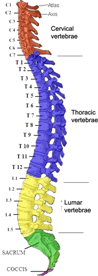
The first two cervical vertebrae have alternate names to C1 and C2. C1 is also called the atlas. In Greek mythology, Atlas was a titan who held the entire world on his shoulders. As the first vertebra in the column, Atlas in a sense holds up the skull. C2 is also called the axis. The axis allows both the skull and the atlas to rotate, so the head can be turned from side to side by neck muscles.
All three types of vertebrae have some structural features in common and some features that should allow you to readily distinguish vertebrae in one category from another.
All vertebrae, except C1 and C2, the atlas and axis, have a solid round portion on their anterior side called the body of the vertebra. The body is what allows the vertebrae in the vertebral column to be stacked upon one another, separated by pads of fibrocartilage called the intervertebral discs. The lower you go in the vertebral column, the larger the vertebrae’s bodies become.
The axis, or C2 vertebra, also has a bulbous vertical process not found in any of the other vertebrae. This is called the dens and it is what allows the axis vertebra above it to rotate.
Posterior to the vertebral body is a large opening in each vertebra called the vertebral foramen. This is the hole through which the spinal cord passes.
Posterior to the vertebral foramen there is a central process jutting out of each vertebra. This is the spinous process. It points more or less downwards when the vertebrae are correctly stacked into their column.
Surrounding the central spinous processes, there are other processes whose position and number vary depending on whether a vertebra is cervical, thoracic, or lumbar. Some of these processes allow a vertebra to articulate with the vertebrae superior and inferior to it, others, found only on thoracic vertebrae, allow a pair of ribs to articulate with the vertebra.
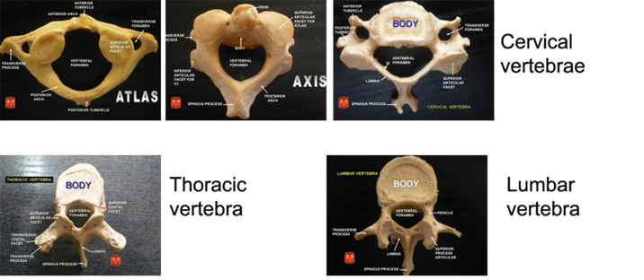
Figure \(\PageIndex{3}\): The three types of vertebrae. All are being viewed from behind. Note that C1 and C2 vertebrae, the atlas and axis, do not have vertebral bodies. (CC-BY-SA, Anatomist90, Wikimedia)
Figure \(\PageIndex{2}\) The three types of vertebrae. All are being viewed from behind. Note that C1 and C2 vertebrae, the atlas and axis, do not have vertebral bodies.
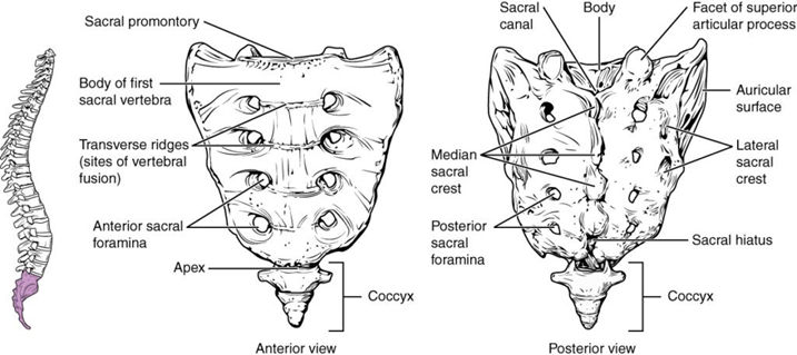
The sacrum is part of both the vertebral column and the pelvic girdle. It articulates with the intervertebral disc under the L5 vertebra above it, and with two coxal bones lateral to it.
The sacrum starts out as five vertebrae that fuse to form the one structure. This fusion is not complete until somewhere between the 18th and 30th year.
The coccyx is a vestigial tailbone. It is the evolutionary remnant of an ancestral species to humans that did have tails. It no longer serves as a functional tail, but some muscles, tendons, and ligaments do attach to it, making it useful. It forms from the fusion of usually three vertebrae, but a small proportion of the population have four or even five vertebrae in their coccyx.
Lab 5 Exercise \(\PageIndex{1}\)
1. Real or replica vertebrae bones may be found in the cabinets and on the back counters. Identify which in your group are the atlas, axis, cervical vertebrae, thoracic vertebrae, and lumbar vertebrae.
2. Stack your vertebrae in the correct order and in the correct orientation.
LICENSES AND ATTRIBUTIONS
CC LICENSED CONTENT, ORIGINAL
A&P Labs. Authored by: Ross Whitwam. Provided by: Mississippi University for Women. Located at: http://www.muw.edu/. License: CC BY-SA: Attribution-ShareAlike CC LICENSED CONTENT, SHARED PREVIOUSLY
Figure \(\PageIndex{3}\). The three types of vertebrae. All are being viewed from behind. Note that C1 and C2 vertebrae, the atlas and axis, do not have vertebral bodies. . Authored by: Anatomist90. Located at: https://commons.wikimedia.org/wiki/F..._vertebrae.jpg. License: CC BY- SA: Attribution-ShareAlike
Figure \(\PageIndex{4}\). The sacrum and coccyx. Authored by: OpenStax College. Located at: http://cnx.org/resources/e394fc66a09810ea99d0165de1078c8af2d4dc46/720_Sacrum_and_Coccyx.jpg. License: CC BY-SA: Attribution-ShareAlike
PUBLIC DOMAIN CONTENT
Figure \(\PageIndex{2}\). The bones of the vertebral column. Authored by: Henry Grey, Henry Vandyke Carter. Located at: https://upload.wikimedia.org/wikiped...red_Ro_mod.JPG. Project: Anatomy of the Human Body, 20th Edition (1918). License: Public Domain: No Known Copyright