
5.4: Exercises
- Page ID
- 59387

\( \newcommand{\vecs}[1]{\overset { \scriptstyle \rightharpoonup} {\mathbf{#1}} } \)
\( \newcommand{\vecd}[1]{\overset{-\!-\!\rightharpoonup}{\vphantom{a}\smash {#1}}} \)
\( \newcommand{\dsum}{\displaystyle\sum\limits} \)
\( \newcommand{\dint}{\displaystyle\int\limits} \)
\( \newcommand{\dlim}{\displaystyle\lim\limits} \)
\( \newcommand{\id}{\mathrm{id}}\) \( \newcommand{\Span}{\mathrm{span}}\)
( \newcommand{\kernel}{\mathrm{null}\,}\) \( \newcommand{\range}{\mathrm{range}\,}\)
\( \newcommand{\RealPart}{\mathrm{Re}}\) \( \newcommand{\ImaginaryPart}{\mathrm{Im}}\)
\( \newcommand{\Argument}{\mathrm{Arg}}\) \( \newcommand{\norm}[1]{\| #1 \|}\)
\( \newcommand{\inner}[2]{\langle #1, #2 \rangle}\)
\( \newcommand{\Span}{\mathrm{span}}\)
\( \newcommand{\id}{\mathrm{id}}\)
\( \newcommand{\Span}{\mathrm{span}}\)
\( \newcommand{\kernel}{\mathrm{null}\,}\)
\( \newcommand{\range}{\mathrm{range}\,}\)
\( \newcommand{\RealPart}{\mathrm{Re}}\)
\( \newcommand{\ImaginaryPart}{\mathrm{Im}}\)
\( \newcommand{\Argument}{\mathrm{Arg}}\)
\( \newcommand{\norm}[1]{\| #1 \|}\)
\( \newcommand{\inner}[2]{\langle #1, #2 \rangle}\)
\( \newcommand{\Span}{\mathrm{span}}\) \( \newcommand{\AA}{\unicode[.8,0]{x212B}}\)
\( \newcommand{\vectorA}[1]{\vec{#1}} % arrow\)
\( \newcommand{\vectorAt}[1]{\vec{\text{#1}}} % arrow\)
\( \newcommand{\vectorB}[1]{\overset { \scriptstyle \rightharpoonup} {\mathbf{#1}} } \)
\( \newcommand{\vectorC}[1]{\textbf{#1}} \)
\( \newcommand{\vectorD}[1]{\overrightarrow{#1}} \)
\( \newcommand{\vectorDt}[1]{\overrightarrow{\text{#1}}} \)
\( \newcommand{\vectE}[1]{\overset{-\!-\!\rightharpoonup}{\vphantom{a}\smash{\mathbf {#1}}}} \)
\( \newcommand{\vecs}[1]{\overset { \scriptstyle \rightharpoonup} {\mathbf{#1}} } \)
\(\newcommand{\longvect}{\overrightarrow}\)
\( \newcommand{\vecd}[1]{\overset{-\!-\!\rightharpoonup}{\vphantom{a}\smash {#1}}} \)
\(\newcommand{\avec}{\mathbf a}\) \(\newcommand{\bvec}{\mathbf b}\) \(\newcommand{\cvec}{\mathbf c}\) \(\newcommand{\dvec}{\mathbf d}\) \(\newcommand{\dtil}{\widetilde{\mathbf d}}\) \(\newcommand{\evec}{\mathbf e}\) \(\newcommand{\fvec}{\mathbf f}\) \(\newcommand{\nvec}{\mathbf n}\) \(\newcommand{\pvec}{\mathbf p}\) \(\newcommand{\qvec}{\mathbf q}\) \(\newcommand{\svec}{\mathbf s}\) \(\newcommand{\tvec}{\mathbf t}\) \(\newcommand{\uvec}{\mathbf u}\) \(\newcommand{\vvec}{\mathbf v}\) \(\newcommand{\wvec}{\mathbf w}\) \(\newcommand{\xvec}{\mathbf x}\) \(\newcommand{\yvec}{\mathbf y}\) \(\newcommand{\zvec}{\mathbf z}\) \(\newcommand{\rvec}{\mathbf r}\) \(\newcommand{\mvec}{\mathbf m}\) \(\newcommand{\zerovec}{\mathbf 0}\) \(\newcommand{\onevec}{\mathbf 1}\) \(\newcommand{\real}{\mathbb R}\) \(\newcommand{\twovec}[2]{\left[\begin{array}{r}#1 \\ #2 \end{array}\right]}\) \(\newcommand{\ctwovec}[2]{\left[\begin{array}{c}#1 \\ #2 \end{array}\right]}\) \(\newcommand{\threevec}[3]{\left[\begin{array}{r}#1 \\ #2 \\ #3 \end{array}\right]}\) \(\newcommand{\cthreevec}[3]{\left[\begin{array}{c}#1 \\ #2 \\ #3 \end{array}\right]}\) \(\newcommand{\fourvec}[4]{\left[\begin{array}{r}#1 \\ #2 \\ #3 \\ #4 \end{array}\right]}\) \(\newcommand{\cfourvec}[4]{\left[\begin{array}{c}#1 \\ #2 \\ #3 \\ #4 \end{array}\right]}\) \(\newcommand{\fivevec}[5]{\left[\begin{array}{r}#1 \\ #2 \\ #3 \\ #4 \\ #5 \\ \end{array}\right]}\) \(\newcommand{\cfivevec}[5]{\left[\begin{array}{c}#1 \\ #2 \\ #3 \\ #4 \\ #5 \\ \end{array}\right]}\) \(\newcommand{\mattwo}[4]{\left[\begin{array}{rr}#1 \amp #2 \\ #3 \amp #4 \\ \end{array}\right]}\) \(\newcommand{\laspan}[1]{\text{Span}\{#1\}}\) \(\newcommand{\bcal}{\cal B}\) \(\newcommand{\ccal}{\cal C}\) \(\newcommand{\scal}{\cal S}\) \(\newcommand{\wcal}{\cal W}\) \(\newcommand{\ecal}{\cal E}\) \(\newcommand{\coords}[2]{\left\{#1\right\}_{#2}}\) \(\newcommand{\gray}[1]{\color{gray}{#1}}\) \(\newcommand{\lgray}[1]{\color{lightgray}{#1}}\) \(\newcommand{\rank}{\operatorname{rank}}\) \(\newcommand{\row}{\text{Row}}\) \(\newcommand{\col}{\text{Col}}\) \(\renewcommand{\row}{\text{Row}}\) \(\newcommand{\nul}{\text{Nul}}\) \(\newcommand{\var}{\text{Var}}\) \(\newcommand{\corr}{\text{corr}}\) \(\newcommand{\len}[1]{\left|#1\right|}\) \(\newcommand{\bbar}{\overline{\bvec}}\) \(\newcommand{\bhat}{\widehat{\bvec}}\) \(\newcommand{\bperp}{\bvec^\perp}\) \(\newcommand{\xhat}{\widehat{\xvec}}\) \(\newcommand{\vhat}{\widehat{\vvec}}\) \(\newcommand{\uhat}{\widehat{\uvec}}\) \(\newcommand{\what}{\widehat{\wvec}}\) \(\newcommand{\Sighat}{\widehat{\Sigma}}\) \(\newcommand{\lt}{<}\) \(\newcommand{\gt}{>}\) \(\newcommand{\amp}{&}\) \(\definecolor{fillinmathshade}{gray}{0.9}\)
Lab 5 Exercise \(\PageIndex{1}\)
1. The instructor will provide you with a single rib from the human body. On that individual rib, identify which end is the head, and which is the anterior end.
2. On one of the intact skeletons in the lab, identify all the following components of the thoracic cage:
| the true ribs | the false ribs | the floating ribs |
| costal cartilag | sternum | xiphoid process |
| manubrium | sternal body |
E
Lab 5 Exercise \(\PageIndex{1}\)
1. The instructor will provide you with a single rib from the human body. On that individual rib, identify which end is the head, and which is the anterior end.
2. On one of the intact skeletons in the lab, identify all the following components of the thoracic cage:
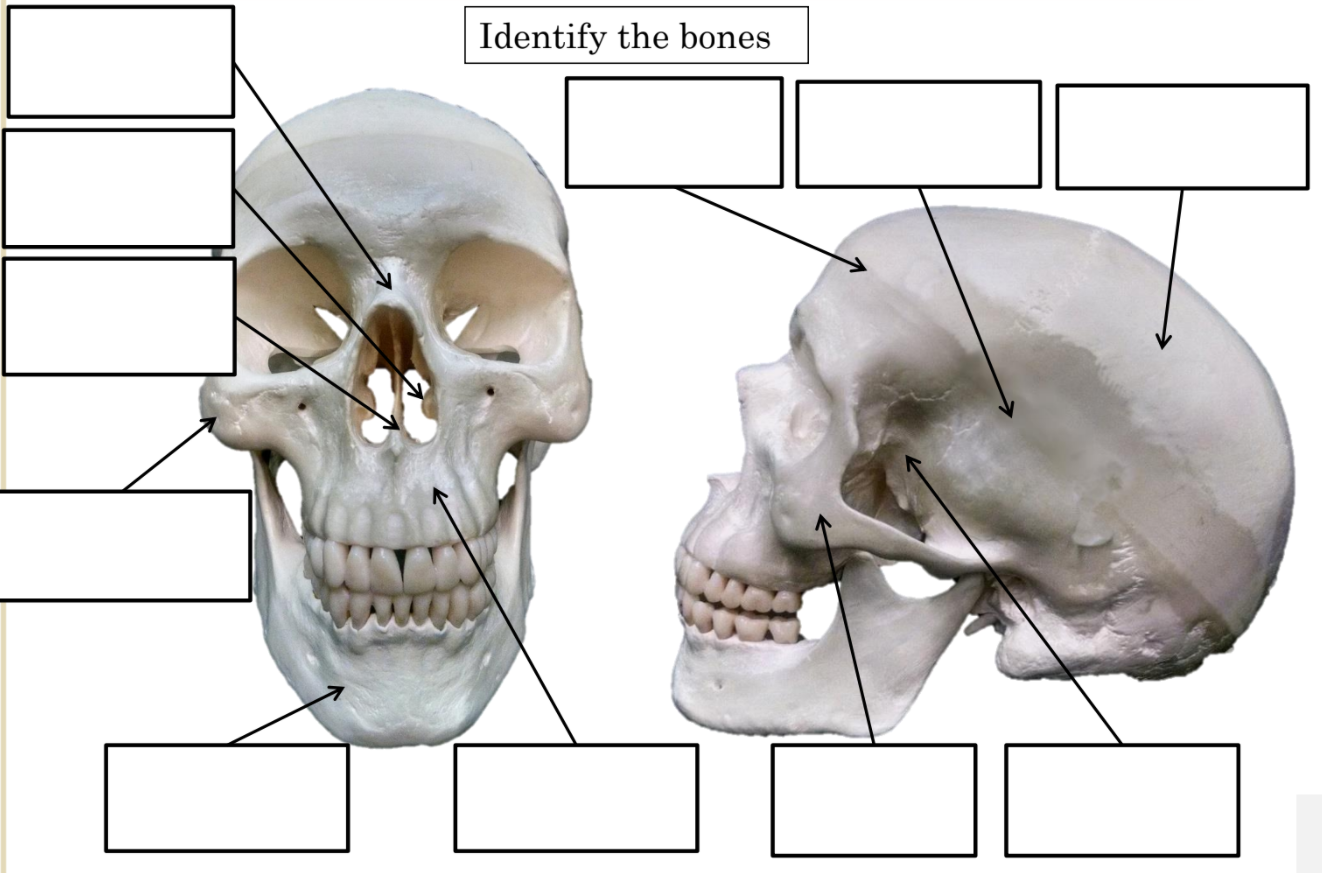
| 1 | Zygomatic bones | 6 | Inferior orbital foramen |
| 2 | Lacrimal bones | 7 | Glabella |
| 3 | Coronal suture | 8 | Superior orbital fissure |
| 4 | Sagittal suture | 9 | Inferior orbital fissure |
| 5 | Superior orbital notch/foramen |
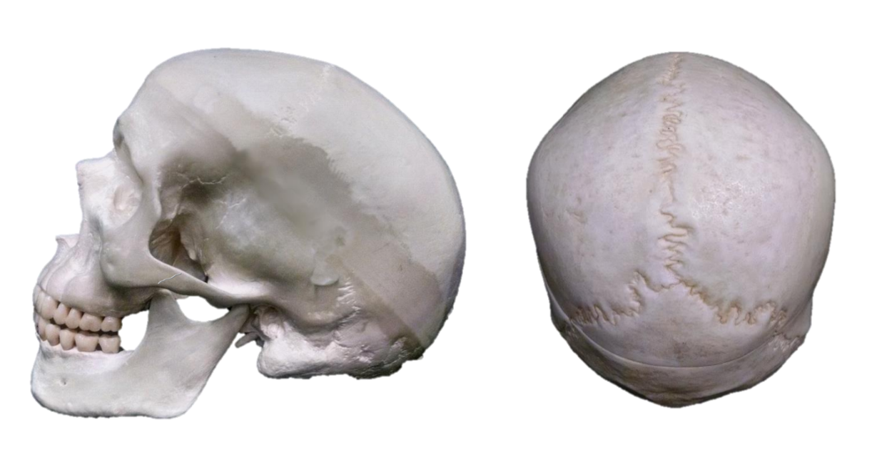
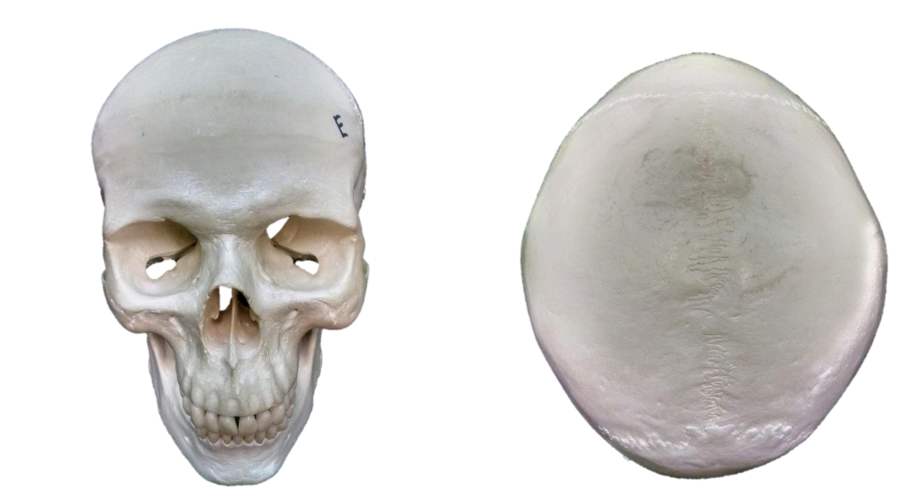
| 1 | Occipital bone | 7 | Mastoid process |
| 2 | Temporal bone | 8 | Glabella |
| 3 | Sphenoid bone | 9 | Styloid process |
| 4 | Maxilla | 10 | Lambdoid suture |
| 5 | Mandible | 11 | Zygomatic process of the temporal bone |
| 6 | External acoustic meatus | 12 | Temporal process of the zygomatic bone |
| 13 | Sutural bones |
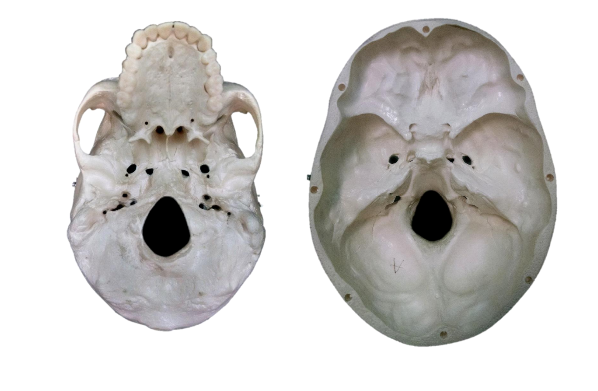
| 1 | Zygomatic arch8 | 8 | Internal acoustic meatus |
| 2 | Foramen ovale | 9 | Occipital condyle |
| 3 | Foramen spinosum | 10 | Mandibular fossa |
| 4 | Foramen lacerum | 11 | Ethmoid bone |
| 5 | Jugular foramen | 12 | Anterior cranial fossa |
| 6 | Carotid canal | 13 | Middle cranial fossa |
| 7 | Foramen magnum | 14 | Posterior cranial fossa. |
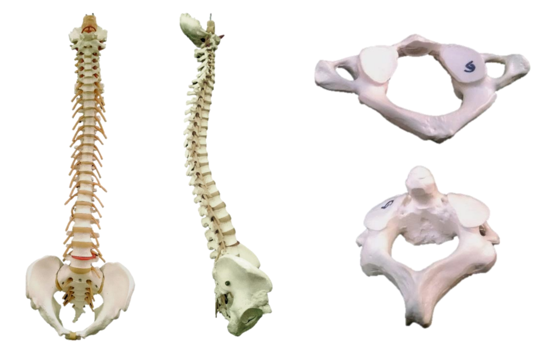
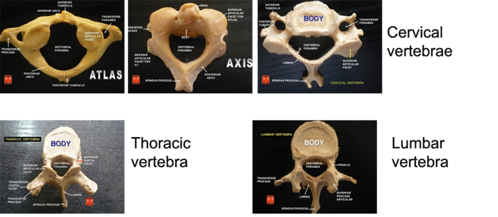
| 1 | Cervical vertebrae | 8 | Lumbar curvature |
| 2 | Cervical curvature | 9 | Atlas |
| 3 | Thoracic vertebrae | 10 | C1 |
| 4 | Thoracic curvature | 11 | C2 |
| 5 | 5 | 12 | 12 |
| 6 | Carotid canal | 13 | Axis |
| 7 | 7 | 14 | Dens |
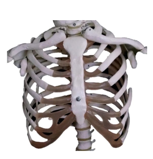
| 1 | True ribs | 3 | False ribs |
| 2 | Floating ribs | 4 | Costal cartilages. |
Lab 5 Exercise \(\PageIndex{3}\)
1. Using one of the full skeletons in the room, fill out the tables below with three or four steps to determine whether each individual upper limb bone comes from the anatomical left or anatomical right.
2. You can describe any features on that bone and which direction it has to face to allow you to determine whether that particular bone came from anatomical left or anatomical right.
3. You don't have to use anatomical jargon if you don't want. Use terms which will make sense to you when you read it again. Use as many steps as you need, not necessarily four.
| Humerus – Anatomical left from anatomical right |
| 1. |
| 2. |
| 3. |
| 4. |
| Ulna – Anatomical left from anatomical right |
| 1. |
| 2. |
| 3. |
| 4. |
| Radius – Anatomical left from anatomical right |
| 1. |
| 2. |
| 3. |
| 4. |
Lab 5 Exercise \(\PageIndex{4}\)
1. Using one of the full skeletons in the room, fill out the tables below with three or four steps to determine whether an individual coxal bone comes from the anatomical left or the anatomical right.
2. You can describe any features on the bone and which direction it has to face to allow you to determine whether that particular bone came from anatomical left or anatomical right.
3. You don't have to use anatomical jargon if you don't want. Use terms which will make sense to you when you read it again. Use as many steps as you need, not necessarily four.
| Coxal bone – Anatomical left from anatomical right |
| 1. |
| 2. |
| 3. |
| 4. |
e}\
e}
Exercise \(\PageIndex{5}\)
1. There will be an intact pelvis set up at the instructor’s station. Using the criteria in this image determine if the pelvis came from a male or female. Give three lines of evidence to support your conclusion.
| Male or female? | |
| Evidence 1 | |
| Evidence 2 | |
| Evidence 2 |
Exercise \(\PageIndex{1}\)
1. Using one of the full skeletons in the room, fill out the tables below with three or four steps to determine whether each individual lower limb bone comes from the anatomical left or anatomical right.
2. You can describe any features on that bone and which direction it has to face to allow you to determine whether that particular bone came from anatomical left or anatomical right.
3. You don't have to use anatomical jargon if you don't want. Use terms which will make sense to you when you read it again. Use as many steps as you need, not necessarily four.
| Femur – Anatomical left from anatomical right |
| 1. |
| 2. |
| 3. |
| 4. |
| Tibia – Anatomical left from anatomical right |
| 1. |
| 2. |
| 3. |
| 4. |
| Fibula – Anatomical left from anatomical right |
| 1. |
| 2. |
| 3. |
| 4. |

)