
5.1: Bones of the Skull


- Last updated
- Sep 14, 2021
- Save as PDF
( \newcommand{\kernel}{\mathrm{null}\,}\)
Information
The bones of the human body can be divided into two broad groups, the axial skeleton and the appendicular skeleton. The axial skeleton comprises the bones found along the central axis traveling down the center of the body. The appendicular skeleton comprises the bones appended to the central axis.
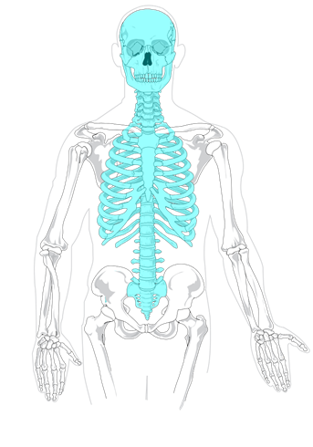
The axial skeleton consists of the bones of the skull, the bones of the inner ear (known as ossicles), the hyoid bone in the throat, and the bones of the vertebral column, including the sacrum and coccyx bones in the center of the pelvic girdle.
LICENSES AND ATTRIBUTIONS
CC LICENSED CONTENT, ORIGINAL
A&P Labs. Authored by: Ross Whitwam. Provided by: Mississippi University for Women. Located at: http://www.muw.edu. License: CC BY- SA: Attribution-ShareAlike
PUBLIC DOMAIN CONTENT
Figure 5.1.1 The axial skeleton highlighted in blue.. Authored by: Axial_skeleton_diagram.svg: LadyofHats Mariana Ruiz Villarreal. Located at: https://commons.wikimedia.org/wiki/F...gram_blank.svg. License: Public Domain: No Known Copyright
The Bones of the Skull
There is only one movable joint in the skull. That is the joint connecting the lower jaw, or mandible, to the rest of the skull. All the other bones in the skull are firmly attached to one another by sutures.
Sutures are rigid immovable connections holding bones tightly to one another. Some of the sutures in the skull take a few months-to-years after birth to completely form.
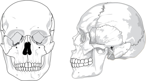
The brain is encased in the cranium of the skull. The bones that make up the cranium are called the cranial bones. The remainder of the bones in the skull are the facial bones.
Figure 5.1.2 and Figure 5.1.3 show all the bones of the skull, as they appear from the outside. In Figure 5.1.5, some of the bones of the hard palate forming the roof of the mouth are visible because the mandible is not present. Figure 5.1.9 also shows the foramen magnum, the large hole at the base of the skull that allows the spinal cord to attach to the brain


Figure 5.1.3: The bones of the skull, anterior view. (Public Domain, Wikimedia)

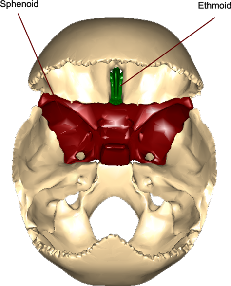
Figure 5.1.5: The interior of the cranial cavity, viewed from above and behind, with the parietal bones removed. (CC-BY-SA, BodyParts3D/Anatomography)
The sphenoid bone, from the outside, appears to contribute to only a small portion of the cranium, but when the parietal bones are removed and the interior of the cranial cavity (where the brain would be housed) is viewed, you can see the butterfly-like shape of the sphenoid bone makes a large contribution to the floor of the cranial cavity. The ethmoid bone, which from the outside is only visible in the eye sockets and as the upper conchae (internal bumps) of the nasal cavity, also contributes to the floor of the cranial cavity. The contributions of these two bones to the floor of the cranial cavity are shown in Figure 5.1.5.
What is commonly referred to as the “cheekbone” is really the processes of two bones connected together: the zygomatic process of the temporal bone is sutured to the temporal process of the zygomatic bone to produce the zygomatic arch.
There are three prominent bone markings on the temporal bones. The external acoustic meatus is the opening that leads to the organs of the inner ear. The styloid process is a thin, pen-like projection where muscles and ligaments of the neck are attached. The mastoid process is a wide and rough projection that serves as another attachment point for neck muscles.
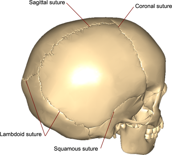
While all the bones of the skull, other than the mandible, are sutured to one another, the flat bones of the cranium are visibly sutured where they articulate to another. There are four different cranial sutures.
The coronal suture is the articulation point of the frontal bone with the two parietal bones. The sagittal suture is the articulation point between the two parietal bones.
The squamous sutures are the articulation points between the each temporal bone and the parietal bone superior to it.
The lambdoid suture is the articulation point between the occipital bone and the two parietal bones.
Lab 5 Exercise 5.1.1
Models of human skulls may be found in the cabinets. On the models identify all of the following and on the diagrams below be able to label and/or color in all the following bones, processes, and foramina:
(Public Domain, Pixabay)
|
Bones |
Sutures |
Foramina |
Processes |
|
B1 – frontal |
S1 – coronal |
F1 – supraorbital |
P1 – mastoid |
|
B2 – parietal |
S2 – squamous |
F2 – infraorbital |
P2 – styloid |
|
B3 – occipital |
S3 – lambdoid |
F3 – mental |
P3 – zygomatic |
|
B4 – temporal |
|
|
P4 temporal |
|
B5 – sphenoid |
|
||
|
B6 – ethmoid |
|||
|
B7 – lacrimal |
|||
|
B8 – nasal |
|||
|
B9 – maxilla |
|||
|
B10 – zygomatic |
|||
|
B11 – mandible |
|||
|
B12 – vomer |
|||
|
B13 – palatine (not visible here) |
|||
LICENSES AND ATTRIBUTIONS
CC LICENSED CONTENT, ORIGINAL
A&P Labs. Authored by: Ross Whitwam. Provided by: Mississippi University for Women. Located at: http://www.muw.edu. License: CC BY- SA: Attribution-ShareAlike
CC LICENSED CONTENT, SPECIFIC ATTRIBUTION
Figure 5.1.4. The bones of the skull, inferior view, looking up. Mandible removed.. Authored by: Made out of, or made from, content published in a BodyParts3D/Anatomography web site. The content of their website is published under the Creative Commons Attribution 2.1 Japan license. The author and licenser of the contents is http://lifesciencedb.jp/bp3d/?lng=en. Located at: http://lifesciencedb.jp/bp3d/?lng=en. License: CC BY-SA: Attribution-ShareAlike
Figure 5.1.5. The interior of the cranial cavity, viewed from above and behind, with the parietal bones removed.. Authored by: Made out of, or made from, content published in a BodyParts3D/Anatomography web site. The content of their website is published under the Creative Commons Attribution 2.1 Japan license. The author and licenser of the contents is http://lifesciencedb.jp/bp3d/?lng=en. Located at: http://lifesciencedb.jp/bp3d/?lng=en. License: CC BY-SA: Attribution-ShareAlike
Figure 5.1.6. The cranial sutures.. Authored by: Made out of, or made from, content published in a BodyParts3D/Anatomography web site. The content of their website is published under the Creative Commons Attribution 2.1 Japan license. The author and licenser of the contents is http://lifesciencedb.jp/bp3d/?lng=en. Located at: http://lifesciencedb.jp/bp3d/?lng=en. License: Public Domain: No Known Copyright
PUBLIC DOMAIN CONTENT
Figure 5.1.2. The bones of the skull, left lateral view.. Authored by: LadyofHats Mariana Ruiz Villarreal. Located at: https://en.wikipedia.org/wiki/File:H...ed_(bones).svg. License: Public Domain: No Known Copyright
Figure 5.1.3. The bones of the skull, anterior view.. Authored by: LadyofHats Mariana Ruiz Villarreal. Located at: https://commons.wikimedia.org/wiki/F...ront_bones.svg. License: Public Domain: No Known Copyright
Lab exercises 5.1.1. Located at: https://pixabay.com/en/skull-diagram...anatomy-41553/. License: Public Domain: No Known Copyright