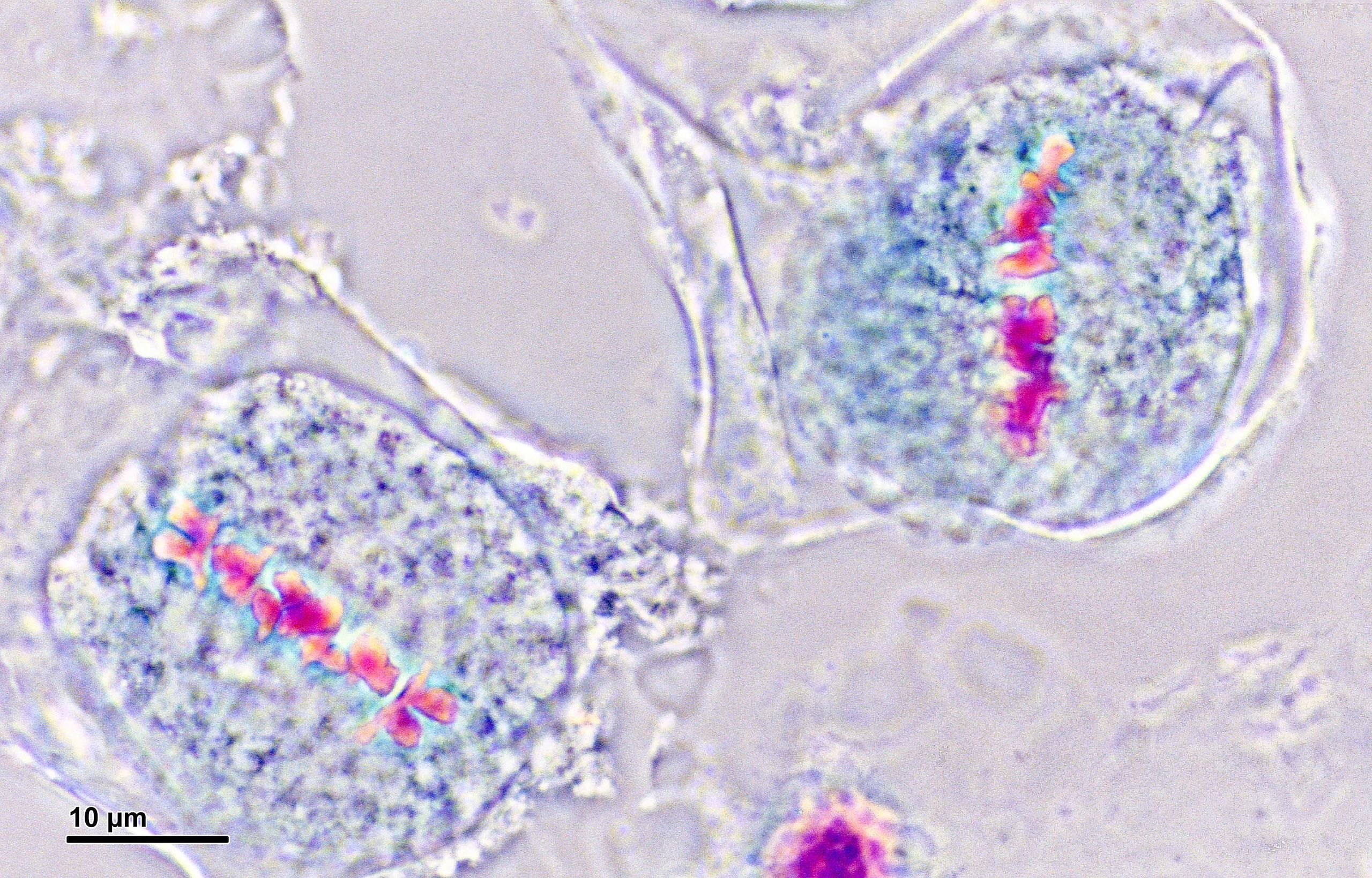
Figure 10.3.2.1: Dividing pollen mother cell, Prophase I. In the first image, the cell is in the early stages of prophase with the chromatin still condensing (appears neon pink). In the second image, twelve replicated chromosomes are visible, stained a deep blue. There is no visible nuclear envelope or nucleolus. Photos by Doc. RNDr. Josef Reischig, CSc., CC BY-SA 3.0, via Wikimedia Commons.Figure 10.3.2.2: Dividing pollen mother cell, Metaphase I. The chromosomes in these two cells (appearing neon pink) are lined up across the center of the cell where the new cell wall will form. Photo by Doc. RNDr. Josef Reischig, CSc., CC BY-SA 3.0, via Wikimedia Commons.Figure 10.3.2.3: Dividing pollen mother cell, Metaphase I and Anaphase I. Here chromosomes are shown as stained bright pink. In the two cells on the left, the chromosomes are in the middle of the cell. The lower of these two cells is clearly in Metaphase I, with the chromosomes forming a distinct line across the cell. The cell on the right is in Anaphase I, sister chromatids (now each a chromosome) have been separated and pulled to either side. Photo by Doc. RNDr. Josef Reischig, CSc., CC BY-SA 3.0, via Wikimedia Commons.Figure 10.3.2.4: Dividing pollen mother cells, Metaphase I and Telophase I. The cell on the top is in metaphase I, while the cell on the bottom is in telophase I. In the lower cell, there are two reforming nuclei. Distinct chromosomes are no longer distinguishable. Photo by Doc. RNDr. Josef Reischig, CSc., CC BY-SA 3.0, via Wikimedia Commons.
Meiosis II
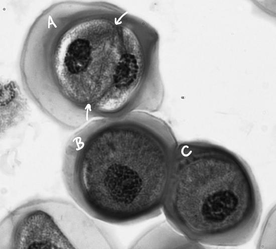
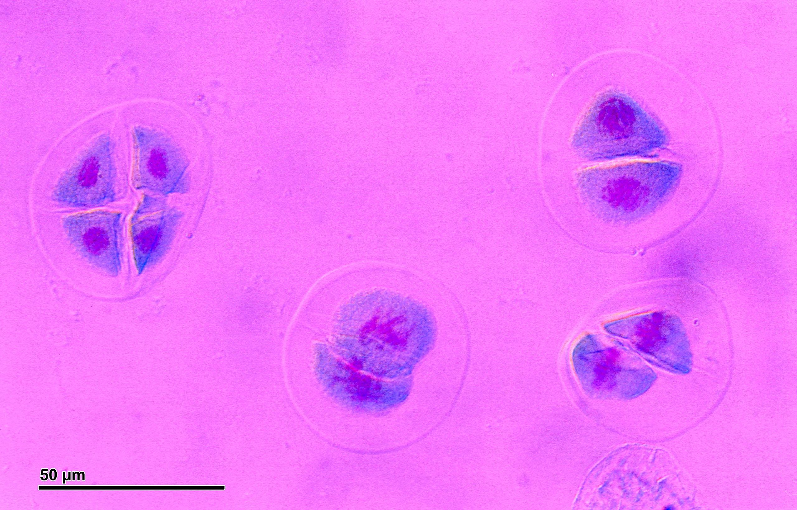
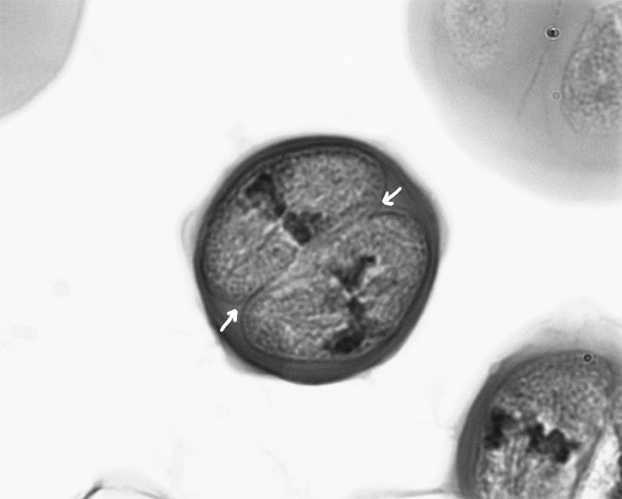
Figure 10.3.2.5: In the first image, cell A is clearly in prophase II. The white arrows point at the cell wall that formed during cytokinesis of meiosis I. There are two distinct nuclei with chromosomes condensing. Cells B and C are also in prophase II, but not clearly so (they appear to be in prophase I). This is because the second cell for each of these is under the first; the 3D nature of these structures makes it difficult to determine how many cells are present. In the second image, there are four dividing mother cells. The leftmost has four cells in it, while the other three have two. The three on the right are in prophase II, while the one on the left has completed meiosis. First photo by Maria Morrow, CC-BY 4.0 . Second photo by Doc. RNDr. Josef Reischig, CSc., CC BY-SA 3.0, via Wikimedia Commons.Figure \(\PageIndex{6\): A pollen mother cell in Metaphase II. There is a clearly formed cell wall (indicated by white arrows), formed during meiosis I. The chromosomes are lined up in the center of each newly formed cell. Photo by Maria Morrow, CC-BY 4.0.Figure 10.3.2.7: The three topmost cells are in telophase II, the lowest cell is in anaphase II. All four cells have a distinct cell wall. Photo by Doc. RNDr. Josef Reischig, CSc., CC BY-SA 3.0, via Wikimedia Commons.Figure 10.3.2.8: Four pollen grains. Each of these pollen grains is haploid and genetically distinct. Photo by Doc. RNDr. Josef Reischig, CSc., CC BY-SA 3.0, via Wikimedia Commons.


.jpg?revision=1&size=bestfit&height=330)
.jpg?revision=1&size=bestfit&width=510&height=325)
.jpg?revision=1&size=bestfit&width=575&height=368)
.jpg?revision=1&size=bestfit&width=660&height=422)
.jpg?revision=1&size=bestfit&width=671&height=428)
.jpg?revision=1&size=bestfit&width=612&height=391)
.jpg?revision=1&size=bestfit&width=856&height=548)
.jpg?revision=1&size=bestfit&width=702&height=450)

