Development
- Page ID
- 23999
\( \newcommand{\vecs}[1]{\overset { \scriptstyle \rightharpoonup} {\mathbf{#1}} } \)
\( \newcommand{\vecd}[1]{\overset{-\!-\!\rightharpoonup}{\vphantom{a}\smash {#1}}} \)
\( \newcommand{\dsum}{\displaystyle\sum\limits} \)
\( \newcommand{\dint}{\displaystyle\int\limits} \)
\( \newcommand{\dlim}{\displaystyle\lim\limits} \)
\( \newcommand{\id}{\mathrm{id}}\) \( \newcommand{\Span}{\mathrm{span}}\)
( \newcommand{\kernel}{\mathrm{null}\,}\) \( \newcommand{\range}{\mathrm{range}\,}\)
\( \newcommand{\RealPart}{\mathrm{Re}}\) \( \newcommand{\ImaginaryPart}{\mathrm{Im}}\)
\( \newcommand{\Argument}{\mathrm{Arg}}\) \( \newcommand{\norm}[1]{\| #1 \|}\)
\( \newcommand{\inner}[2]{\langle #1, #2 \rangle}\)
\( \newcommand{\Span}{\mathrm{span}}\)
\( \newcommand{\id}{\mathrm{id}}\)
\( \newcommand{\Span}{\mathrm{span}}\)
\( \newcommand{\kernel}{\mathrm{null}\,}\)
\( \newcommand{\range}{\mathrm{range}\,}\)
\( \newcommand{\RealPart}{\mathrm{Re}}\)
\( \newcommand{\ImaginaryPart}{\mathrm{Im}}\)
\( \newcommand{\Argument}{\mathrm{Arg}}\)
\( \newcommand{\norm}[1]{\| #1 \|}\)
\( \newcommand{\inner}[2]{\langle #1, #2 \rangle}\)
\( \newcommand{\Span}{\mathrm{span}}\) \( \newcommand{\AA}{\unicode[.8,0]{x212B}}\)
\( \newcommand{\vectorA}[1]{\vec{#1}} % arrow\)
\( \newcommand{\vectorAt}[1]{\vec{\text{#1}}} % arrow\)
\( \newcommand{\vectorB}[1]{\overset { \scriptstyle \rightharpoonup} {\mathbf{#1}} } \)
\( \newcommand{\vectorC}[1]{\textbf{#1}} \)
\( \newcommand{\vectorD}[1]{\overrightarrow{#1}} \)
\( \newcommand{\vectorDt}[1]{\overrightarrow{\text{#1}}} \)
\( \newcommand{\vectE}[1]{\overset{-\!-\!\rightharpoonup}{\vphantom{a}\smash{\mathbf {#1}}}} \)
\( \newcommand{\vecs}[1]{\overset { \scriptstyle \rightharpoonup} {\mathbf{#1}} } \)
\(\newcommand{\longvect}{\overrightarrow}\)
\( \newcommand{\vecd}[1]{\overset{-\!-\!\rightharpoonup}{\vphantom{a}\smash {#1}}} \)
\(\newcommand{\avec}{\mathbf a}\) \(\newcommand{\bvec}{\mathbf b}\) \(\newcommand{\cvec}{\mathbf c}\) \(\newcommand{\dvec}{\mathbf d}\) \(\newcommand{\dtil}{\widetilde{\mathbf d}}\) \(\newcommand{\evec}{\mathbf e}\) \(\newcommand{\fvec}{\mathbf f}\) \(\newcommand{\nvec}{\mathbf n}\) \(\newcommand{\pvec}{\mathbf p}\) \(\newcommand{\qvec}{\mathbf q}\) \(\newcommand{\svec}{\mathbf s}\) \(\newcommand{\tvec}{\mathbf t}\) \(\newcommand{\uvec}{\mathbf u}\) \(\newcommand{\vvec}{\mathbf v}\) \(\newcommand{\wvec}{\mathbf w}\) \(\newcommand{\xvec}{\mathbf x}\) \(\newcommand{\yvec}{\mathbf y}\) \(\newcommand{\zvec}{\mathbf z}\) \(\newcommand{\rvec}{\mathbf r}\) \(\newcommand{\mvec}{\mathbf m}\) \(\newcommand{\zerovec}{\mathbf 0}\) \(\newcommand{\onevec}{\mathbf 1}\) \(\newcommand{\real}{\mathbb R}\) \(\newcommand{\twovec}[2]{\left[\begin{array}{r}#1 \\ #2 \end{array}\right]}\) \(\newcommand{\ctwovec}[2]{\left[\begin{array}{c}#1 \\ #2 \end{array}\right]}\) \(\newcommand{\threevec}[3]{\left[\begin{array}{r}#1 \\ #2 \\ #3 \end{array}\right]}\) \(\newcommand{\cthreevec}[3]{\left[\begin{array}{c}#1 \\ #2 \\ #3 \end{array}\right]}\) \(\newcommand{\fourvec}[4]{\left[\begin{array}{r}#1 \\ #2 \\ #3 \\ #4 \end{array}\right]}\) \(\newcommand{\cfourvec}[4]{\left[\begin{array}{c}#1 \\ #2 \\ #3 \\ #4 \end{array}\right]}\) \(\newcommand{\fivevec}[5]{\left[\begin{array}{r}#1 \\ #2 \\ #3 \\ #4 \\ #5 \\ \end{array}\right]}\) \(\newcommand{\cfivevec}[5]{\left[\begin{array}{c}#1 \\ #2 \\ #3 \\ #4 \\ #5 \\ \end{array}\right]}\) \(\newcommand{\mattwo}[4]{\left[\begin{array}{rr}#1 \amp #2 \\ #3 \amp #4 \\ \end{array}\right]}\) \(\newcommand{\laspan}[1]{\text{Span}\{#1\}}\) \(\newcommand{\bcal}{\cal B}\) \(\newcommand{\ccal}{\cal C}\) \(\newcommand{\scal}{\cal S}\) \(\newcommand{\wcal}{\cal W}\) \(\newcommand{\ecal}{\cal E}\) \(\newcommand{\coords}[2]{\left\{#1\right\}_{#2}}\) \(\newcommand{\gray}[1]{\color{gray}{#1}}\) \(\newcommand{\lgray}[1]{\color{lightgray}{#1}}\) \(\newcommand{\rank}{\operatorname{rank}}\) \(\newcommand{\row}{\text{Row}}\) \(\newcommand{\col}{\text{Col}}\) \(\renewcommand{\row}{\text{Row}}\) \(\newcommand{\nul}{\text{Nul}}\) \(\newcommand{\var}{\text{Var}}\) \(\newcommand{\corr}{\text{corr}}\) \(\newcommand{\len}[1]{\left|#1\right|}\) \(\newcommand{\bbar}{\overline{\bvec}}\) \(\newcommand{\bhat}{\widehat{\bvec}}\) \(\newcommand{\bperp}{\bvec^\perp}\) \(\newcommand{\xhat}{\widehat{\xvec}}\) \(\newcommand{\vhat}{\widehat{\vvec}}\) \(\newcommand{\uhat}{\widehat{\uvec}}\) \(\newcommand{\what}{\widehat{\wvec}}\) \(\newcommand{\Sighat}{\widehat{\Sigma}}\) \(\newcommand{\lt}{<}\) \(\newcommand{\gt}{>}\) \(\newcommand{\amp}{&}\) \(\definecolor{fillinmathshade}{gray}{0.9}\)Development describes the changes in an organism from its earliest beginnings through maturity.

Figure \(\PageIndex{1}\). (CC BY-NC-SA; N. Wheat)
Fertilization
Fertilization is the initial event in development in sexual reproduction.
- Union of male and female gametes.
- Recombination of paternal and maternal genes.
- Restoration of the diploid number (two sets of chromosomes).
Zygote
The diploid cell resulting from fertilization is now called a zygote.
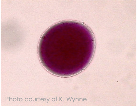
Figure \(\PageIndex{2}\). development in the starfish (Phylum Echinodermata). (CC BY-NC-SA; K. Wynne)
Cleavage
Cleavage– rapid cell divisions following fertilization. Very little growth occurs while the cells are dividing. Each cell called a blastomere.
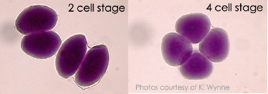
Figure \(\PageIndex{3}\). development in the starfish (Phylum Echinodermata). (CC BY-NC-SA; K. Wynne)
This video shows cleavage in a frog embryo:
Morula
Morula– the name given to the solid ball of cells that results from cleavage. First 5-7 divisions.
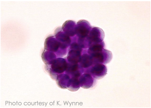
Figure \(\PageIndex{4}\). development in the starfish (Phylum Echinodermata). (CC BY-NC-SA; K. Wynne)
Blastula
As divisions continue, a fluid filled cavity, the blastocoel, forms within the embryo. The resulting hollow ball of cells is now called a blastula.
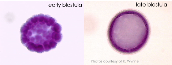
Figure \(\PageIndex{5}\). development in the starfish (Phylum Echinodermata). (CC BY-NC-SA; K. Wynne)
Gastrulation
The morphogenetic process called gastrulation rearranges the cells of a blastula into a three-layered (triploblastic) embryo, called a gastrula, that has a primitive gut (archenteron).
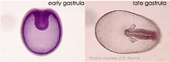
Figure \(\PageIndex{6}\). development in the starfish (Phylum Echinodermata). (CC BY-NC-SA; K. Wynne)
The Blastopore
The blastopore is the first opening in the embryo – the point of invagination during gastrulation. The blastopore will eventually become either the mouth or the anus. One end of the gut-tube or the other. The space that forms during this time is the primitive gut, the archenteron.
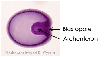
Figure \(\PageIndex{7}\). development in the starfish (Phylum Echinodermata). (CC BY-NC-SA; K. Wynne)
Gastrulation
The three tissue layers produced by gastrulation are called embryonic germlayers. The ectoderm forms the outer layer of the gastrula. Outer surfaces, neural tissue
The endoderm lines the embryonic digestive tract. The mesoderm partly fills the space between the endoderm and ectoderm. Muscles, reproductive system
Gastrulation – Sea Urchin
Gastrulation in a sea urchin produces an embryo with a primitive gut (archenteron) and three germ layers.
Gastrulation - Chick
Gastrulation in the chick is affected by the large amounts of yolk in the egg. Embryo essentially sits on top of large mass of yolk.
Primitive streak– a groove on the surface along the future anterior-posterior axis.
- Functionally equivalent to blastopore lip in frog.
Gastrulation - Mammal
In mammals the blastula is called a blastocyst. Inner cell mass will become the embryo while trophoblast becomes part of the placenta.
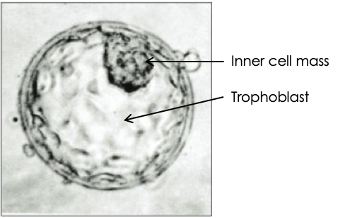
Figure \(\PageIndex{8}\). blastocyst. (CC BY-NC-SA; Wikipedia)
Gastrulation in mammals involves the inner cell mass and is similar to that of the chick due to the fact that mammalian ancestors and early mammals laid eggs. The large mass of yolk may be gone, but the developmental pattern remains.
Suites of Developmental Characters
Two major groups of triploblastic animals:
- Protostomes include flatworms, annelids and molluscs.
- Deuterostomes include echinoderms and chordates.
Protostomes & Deuterostomes
Protostomes & deuterostomes are differentiated by:
- Spiral vs. radial cleavage
- Mosaic vs. regulative cleavage
- Blastopore becomes mouth vs. anus
- Schizocoelousvs. enterocoelous coelom formation.
Spiral vs. Radial Cleavage
Spiral cleavage– occurs in most protostomes. Some ecdysozoans show radial or superficial (insects) cleavage.
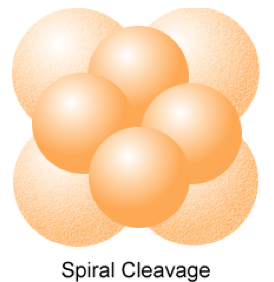
Figure \(\PageIndex{9}\). (CC BY-NC-SA; N. Wheat)
Radial cleavage– is found in most deuterostomes. Tunicates and mammals have specialized cleavage patterns.
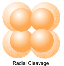
Figure \(\PageIndex{10}\). (CC BY-NC-SA; N. Wheat)
Mosaic vs. Regulative Development
Mosaic development– cell fate is determined by the components of the cytoplasm found in each blastomere. An isolated blastomere can’t develop. Protostomes
Regulative development– the fate of a cell depends on its interactions with neighbors, not what piece of cytoplasm it has. A blastomere isolated early in cleavage is able to from a whole individual (e.g. twins). Deuterostomes
Fate of the Blastopore
Protostome means “first mouth”. Blastopore becomes the mouth. The second opening will become the anus.
Deuterostome means “second mouth”. The blastopore becomes the anus and the mouth develops as the second opening.
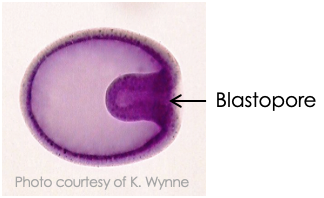
Figure \(\PageIndex{11}\). development in the starfish (Phylum Echinodermata). (CC BY-NC-SA; K. Wynne)
Coelom Formation
The coelom is a body cavity found in many triploblastic organisms that is completely surrounded by mesoderm. Not all protostomes have a true coelom. Pseudocoelomates have a body cavity between mesoderm and endoderm. Acoelomates have no body cavity at all other than the gut.
In protostomes that have a coelom, a mesodermal band of tissue forms before the coelom is formed. In the process of coelom formation called schizocoely, this mesoderm splits to form a coelom.
In enterocoely, the coelom forms as outpocketingof the gut. Typical deuterostomes have coeloms that develop by enterocoely. Vertebrates use a modified version of schizocoely.

This tutorial was funded by the Title V-STEM Grant #P031S090007.


