
11.2: Lytic Life Cycle of Coliphage T4
- Page ID
- 123430
The lytic life cycle of Coliphage T4 consists of the following steps:
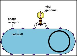
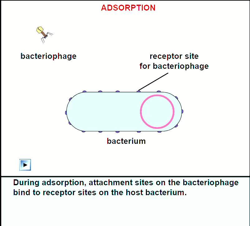
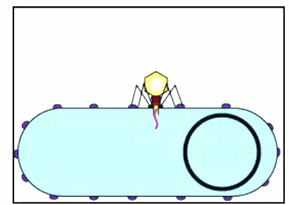
1. Adsorption (Fig. \(\PageIndex{1A}\) and \(\PageIndex{1B}\))
Attachment sites on the bacteriophage tail adsorb to receptor sites on the cell wall of a susceptible host bacterium.
|
Fig \(\PageIndex{1A}\): Adsorption during the Lytic Life Cycle of a Lytic Bacteriophage |
Fig. \(\PageIndex{1B}\): Electron micrograph of Bacteriophages Adsorbing to a Bacterium |
 |
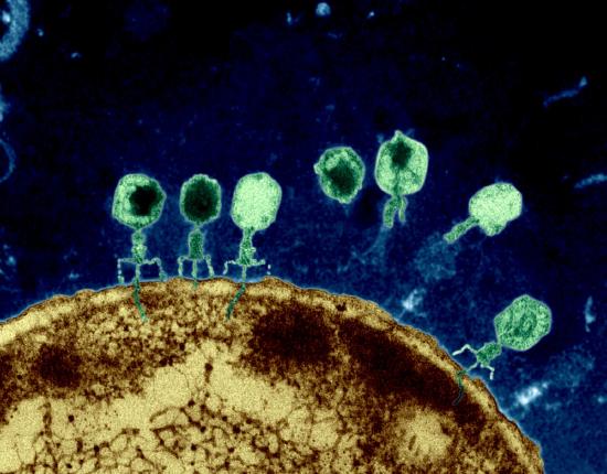 |
| The bacteriophage binds to receptors on the bacterial cell wall. | |
| Copyright; Gary E. Kaiser, Ph.D. The Community College of Baltimore County, Catonsville Campus CC-BY-3.0 | By Dr Graham Beards (en:Image:Phage.jpg) [CC BY-SA 3.0 (https://creativecommons.org/licenses/by-sa/3.0) or GFDL (http://www.gnu.org/copyleft/fdl.html)], via Wikimedia Commons |
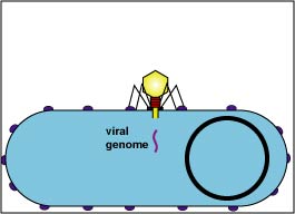
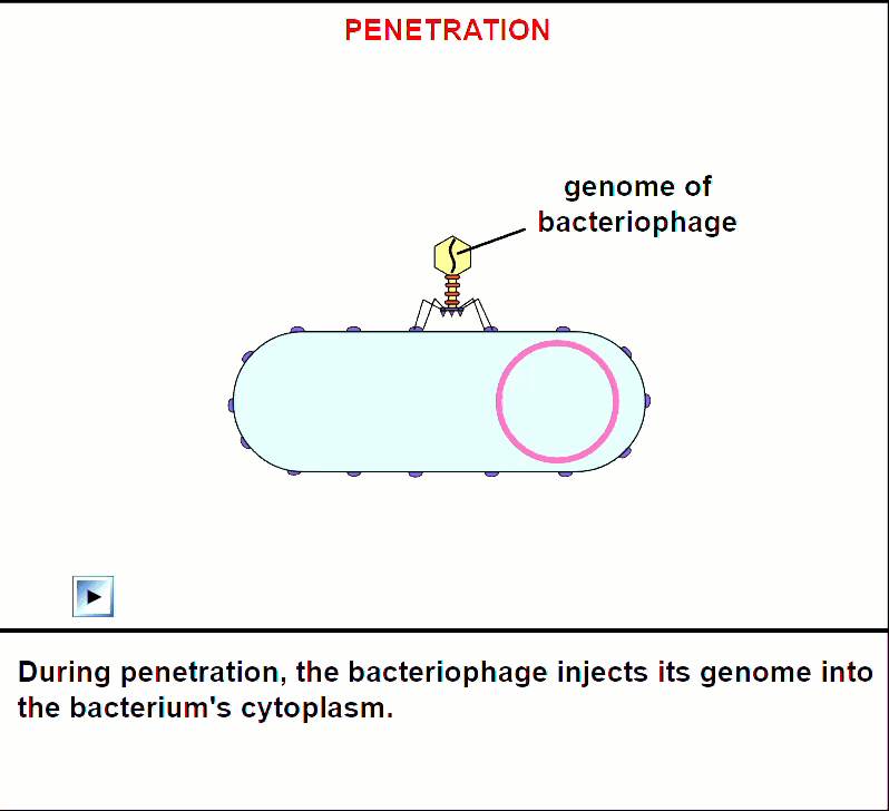
2. Penetration (Fig. \(\PageIndex{1D}\))
A bacteriophage enzyme "drills" a hole in the bacterial cell wall and the bacteriophage injects its genome into the bacterium. This begins the eclipse period, the period in which no intact bacteriophages are seen within the bacterium.
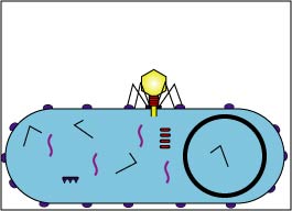
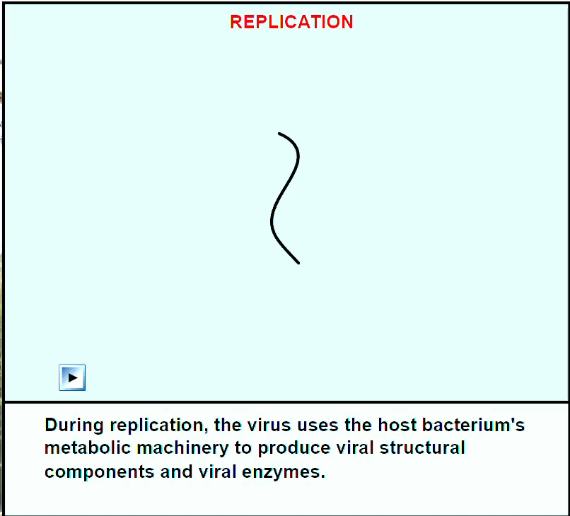
3. Replication (Fig \(\PageIndex{1F}\))
Enzymes coded by the bacteriophage genome shut down the bacterium's macromolecular (protein, RNA, DNA) synthesis. The bacteriophage genome replicates and the bacterium's metabolic machinery is used to synthesize bacteriophage enzymes and bacteriophage structural components.
|
Fig \(\PageIndex{1F}\): Early Replication during the Lytic Life Cycle of a Lytic Bacteriophage |
Fig. \(\PageIndex{1G}\): Late Replication during the Lytic Life Cycle of a Lytic Bacteriophage |
 |
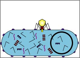 |
| The bacteriophage genome replicates and bacteriophage components begin to be produced by way of the host bacterium's metabolic machinery. | The production of bacteriophage components and enzymes progresses. |
| Copyright; Gary E. Kaiser, Ph.D. The Community College of Baltimore County, Catonsville Campus CC-BY-3.0 | Copyright; Gary E. Kaiser, Ph.D. The Community College of Baltimore County, Catonsville Campus CC-BY-3.0 |
4. Maturation (Fig. \(\PageIndex{1I}\))
The bacteriophage parts assemble around the genome.
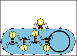
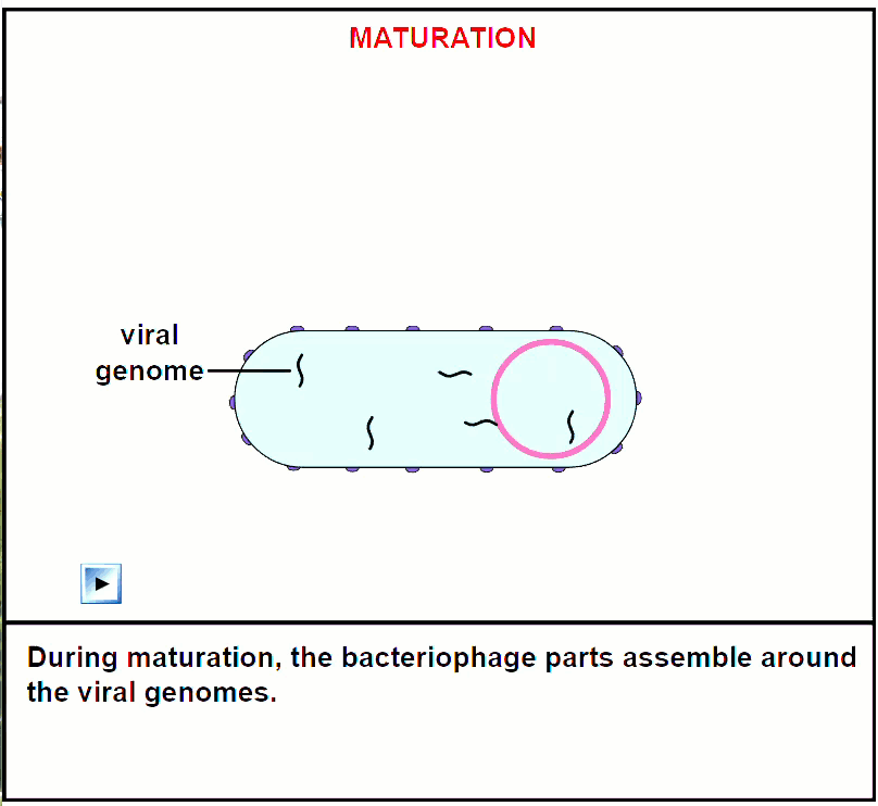
The bacteriophage parts assemble around the genome.
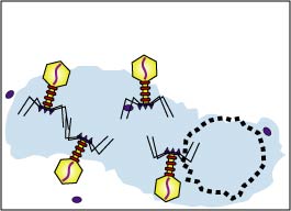
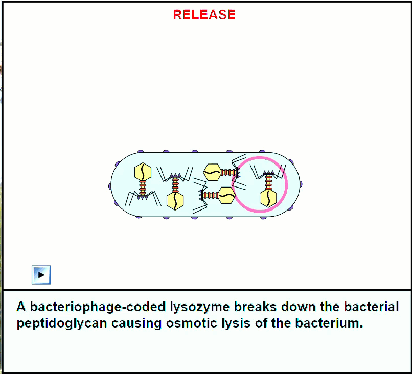
5. Release (Fig. \(\PageIndex{1K}\))
A bacteriophage-coded lysozyme breaks down the bacterial peptidoglycan causing osmotic lysis of the bacterium and release of the intact bacteriophages.
6. Reinfection
From 50-200 bacteriophages may be produced per infected bacterium and they now infect surrounding bacteria.
Some bacteriophages replicate by the lysogenic life cycle and are called temperate bacteriophages. When a temperate bacteriophage infects a bacterium, it can either 1) replicate by the lytic life cycle and cause lysis of the host bacterium, or it can 2) incorporate its DNA into the bacterium's DNA and assume a noninfectious state. In the latter case, the cycle begins by the bacteriophage adsorbing to the host bacterium and injecting its genome, as in the lytic cycle. However, the bacteriophage does not shut down the host bacterium. Instead, the bacteriophage DNA inserts or integrates into the host bacterium's DNA. At this stage, the virus is called a prophage. Expression of the bacteriophage genes controlling bacteriophage replication is repressed by a repressor protein and the bacteriophage DNA replicates as a part of the bacterial nucleoid. However, in approximately one in every million to one in every billion bacteria containing a prophage, spontaneous induction occurs . The bacteriophage genes are activated and bacteriophages are produced as in the lytic life cycle. The lysogenic life cycle is summarized in Figs. \(\PageIndex{1A}\) through \(\PageIndex{1M}\).
Contributors and Attributions
Dr. Gary Kaiser (COMMUNITY COLLEGE OF BALTIMORE COUNTY, CATONSVILLE CAMPUS)