
5.3: Procedure for Direct Staining Using a Basic Dye
- Page ID
- 123350
In direct staining the positively charged color portion of the basic dye combines with the negatively charged bacterium and the organism becomes directly stained.
Videos reviewing techniques used in this lab:
Organisms
Your pure cultures from Lab 3 of Staphylococcus epidermidis (coccus with staphylococcus arrangement) or Micrococcus luteus (coccus with a tetrad or a sarcina arrangement), and Escherichia coli (small bacillus), Pseudomonas aeruginosa (small bacillus), or Klebsiella aerogenes - formerly known as Enterobacter aerogenes (small bacillus).
PROCEDURE (to be done individually)
Organisms
Your pure cultures from Lab 3 of
- Staphylococcus epidermidis (coccus with staphylococcus arrangement) or
- Micrococcus luteus (coccus with a tetrad or a sarcina arrangement), and
- Escherichia coli (small bacillus), Pseudomonas aeruginosa (small bacillus), or
- Klebsiella aerogenes - formerly known as Enterobacter aerogenes (small bacillus).
1. Escherichia coli, Pseudomonas aeruginosa, or Klebsiella aerogenes (formerly known as Enterobacter aerogenes).
a. Fix a smear of either Escherichia coli, Pseudomonas aeruginosa, or Klebsiella aerogenes to the slide as follows:
1. First place a small piece of tape at one end of the slide and label it with the name of the bacterium you will be placing on that slide .
2. Sterilize your inoculating loop and place one loop full of deionized water from your dropper bottle on your slide, as shown 0n Fig, 1 of the previous page Step 1; Step 2; and Step 3 above.
3. Using the edge of your sterile inoculating loop, aseptically remove a small amount of the culture from the agar surface, as shown in Fig. 2 of the previous page , Step 1 above, and gently touch it several times to the drop of water until the water becomes visibly cloudy, as shown in Step 2 and, Step 3 above.
4. Incinerate the remaining bacteria on the inoculating loop. If too much culture is added to the water, you will not see stained individual bacteria.
5. After the inoculating loop cools, spread the suspension over the slide to form a thin film, as shown in Fig. 3 of the previous page above.
6. Allow this thin suspension to completely air dry, as shown in Fig. 4 of the previous page above.. The smear must be completely dry before the slide is fixed!
7. If your professor instructs you to heat-fix the bacteria to the slide, pick up the air-dried slide with provided close pin and hold the bottom of the slide opposite the smear against the opening of the bacto-incinerator for 10 seconds, as shown in Fig. 3 of the previous page above. If the slide is not heated enough, the bacteria will be washed off the slide. If it is overheated, the bacteria structural integrity can be damaged.
8. If your professor instructs you to fix the bacteria to the slide using methanol, add 2-3 drops of 95% methanol to the air-dried smear of bacteria, as shown in Fig. \(\PageIndex{6}\) above and let sit for 2 minutes or until the methanol evaporates. Let the slide again air dry before staining.
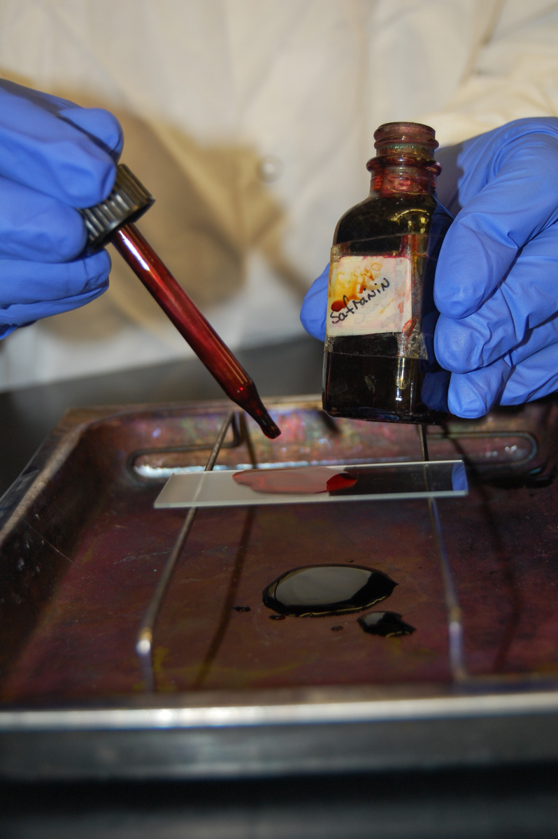
b. Place the slide on a staining tray and cover the entire film with safranin. (See Fig. \(\PageIndex{1}\)A.) Stain for one minute.
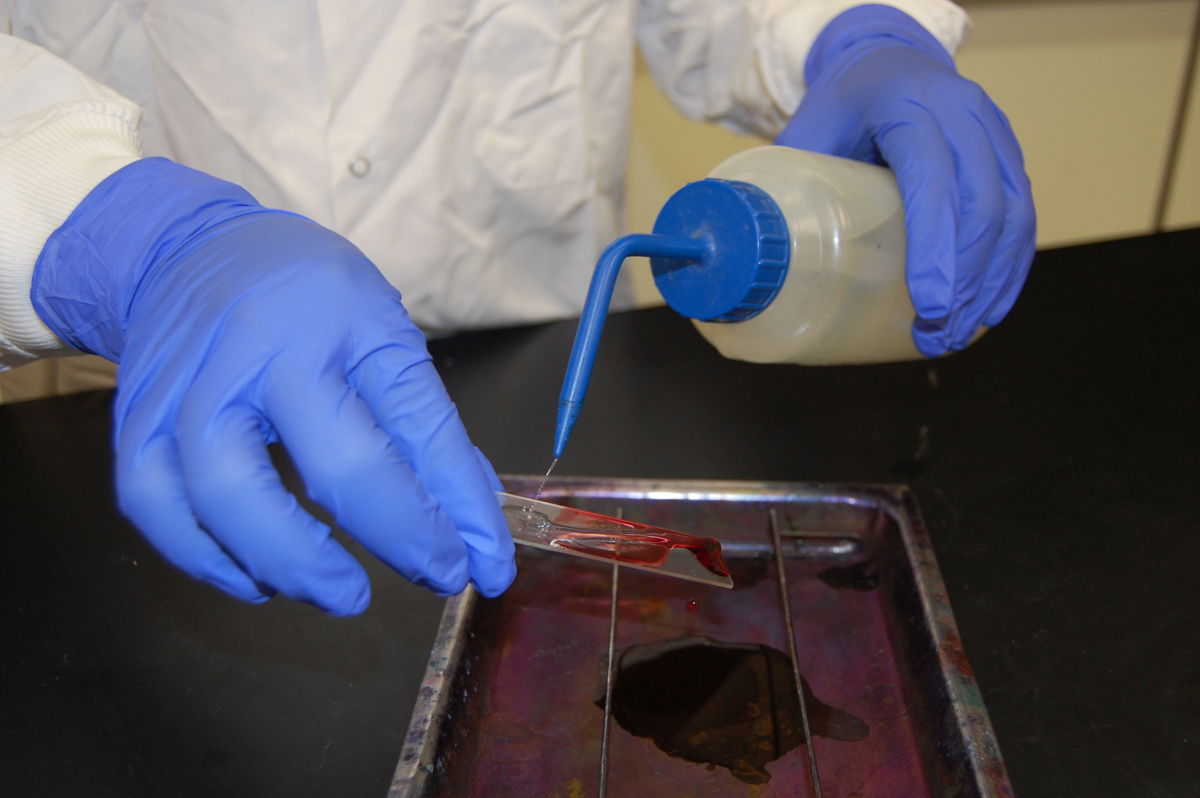
c. Pick up the slide by one end and hold it at an angle over the staining tray. Using the wash bottle on the bench top, gently wash off the excess safranin from the slide. (See Fig. \(\PageIndex{1B}\).) Also wash off any stain that got on the bottom of the slide as well.
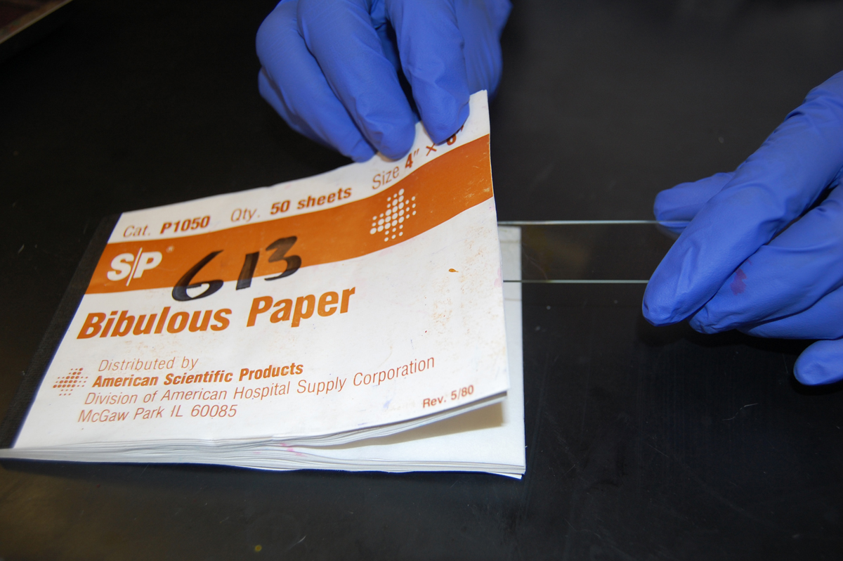
d. Use a book of blotting paper to blot the slide dry. (See Fig. \(\PageIndex{2}\).) Observe using oil immersion microscopy.
Read the Instructions for Focusing a Microscope from Lab 1.
2. Micrococcus luteus or Staphylococcus epidermidis.
a. Fix a smear of either Micrococcus luteus or Staphylococcus epidermidis to the slide as follows:
1. First place a small piece of tape at one end of the slide and label it with the name of the bacterium you will be placing on that slide .
2. Sterilize your inoculating loop and place one loop full of deionized water from your dropper bottle on your slide, as shown in the previous section as Figure 1, Step 1;, Step 2, and Step 3.
3. Using the edge of your sterile inoculating loop, aseptically remove a small amount of the culture from the agar surface, as shown in shown in the previous section as Fig. 2, Step 1, and gently touch it several times to the drop of water until the water becomes visibly cloudy, as shown in shown in the previous section as Fig. 2, Step 2, Step 3.
4. Incinerate the remaining bacteria on the inoculating loop. If too much culture is added to the water, you will not see stained individual bacteria.
5. After the inoculating loop cools, spread the suspension over the slide to form a thin film, as shown in Fig. 3 in the previous section.
6. Allow this thin suspension to completely air dry, as shown in Fig. 4 in the previous section. The smear must be completely dry before the slide is fixed!
7. If your professor instructs you to heat-fix the bacteria to the slide, pick up the air-dried slide with provided close pin and hold the bottom of the slide opposite the smear against the opening of the bacto-incinerator for 10 seconds, as shown in Fig. 5 in the previous section. If the slide is not heated enough, the bacteria will be washed off the slide. If it is overheated, the bacteria structural integrity can be damaged.
8. If your professor instructs you to fix the bacteria to the slide using methanol, add 2-3 drops of 95% methanol to the air-dried smear of bacteria, as shown in Fig. 6 in the previous section and let sit for 2 minutes or until the methanol evaporates. Let the slide again air dry before staining.
b. Stain with crystal violet for one minute.
c. Wash off the excess crystal violet with water.
d. Blot dry and observe using oil immersion microscopy.
3. Prepare a third slide of the normal microbiota and cells of your mouth as follows:
a. Use a sterile cotton swab to vigorously scrape the inside of your mouth and gums.
b. Rub the swab over the slide (do not use water), air dry, and heat-fix.
c. Stain with crystal violet for 30 seconds.
d. Wash off the excess crystal violet with water.
e. Blot dry and observe. Find epithelial cells using your 10X objective, center them in the field, and switch to oil immersion to observe your normal microbiota bacteria on and around your epithelial cells.
4. Make sure you carefully pour the used dye in your staining tray into the designated drains located at the three lab benches and not down the sinks used for hand washing.
RESULTS
Make drawings of your three direct stain preparations and your indirect stain preparation.
|
|
|
| Shape = |
Shape = Arrangement = |

|
|
|
| Shape = Arrangement = |
Contributors and Attributions
Dr. Gary Kaiser (COMMUNITY COLLEGE OF BALTIMORE COUNTY, CATONSVILLE CAMPUS)