
7.8: Laboratory Activities and Assignment


- Last updated
- Jun 7, 2021
- Save as PDF

( \newcommand{\kernel}{\mathrm{null}\,}\)
Laboratory Activities and Assignment
Part 1: Review of the Introduction to Skeleton Anatomy
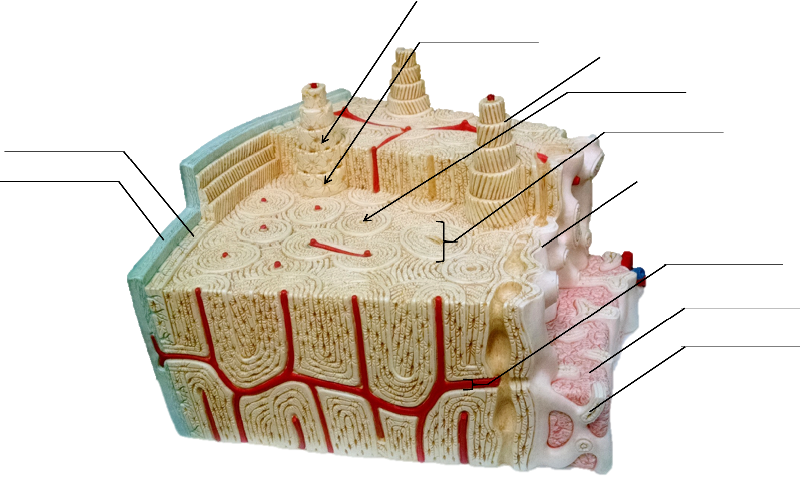
1. Identify and label the bone structures listed using the anatomical model of bone shown below:
|
|
|
|
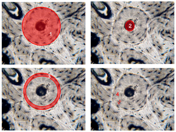
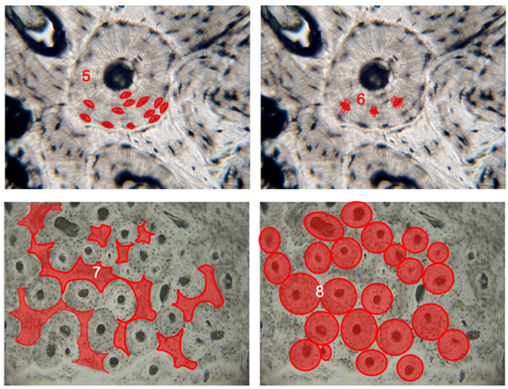
2. Label the structures of compact bone shown in this microscopic images below in the table. Each image is labeled with the number. Place the corresponding anatomical name in the table below next to the number of the image highlighting that structure.
|
|
|
|
Structure # |
Structure Name |
|---|---|
|
1 |
|
|
2 |
|
|
3 |
|
|
4 |
|
|
5 |
|
|
6 |
|
|
7 |
|
|
8 |
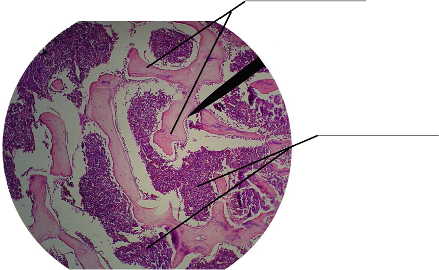
3. Label spongy bone structures shown in this micrograph (arrows):
- trabecula
- bone marrow
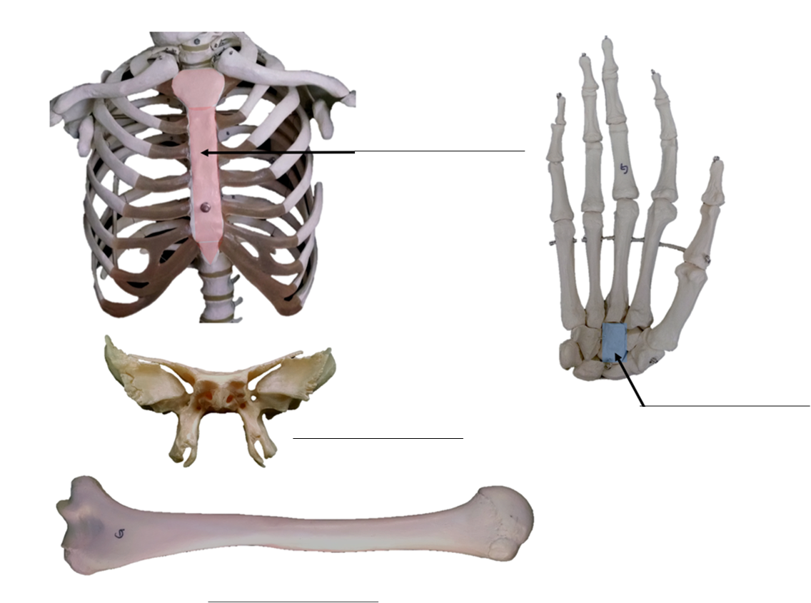
4. Identify the shape of the bones shown below as: long, short, flat, sesamoid or irregular. Write your answers on the spaces provided.
5. Name five bones of the axial skeleton and five bones of the appendicular skeleton.
|
Bones of the Axial Skeleton Include: |
Bones of the Appendicular Skeleton Include: |
|---|---|
|
1. 2. 3. 4. 5. |
1. 2. 3. 4. 5. |
6. Label the anatomy of a long bone with the terms listed below.
|
|
|

Part 2: Skeletal Histology
- Obtain the slides listed below that are available for today’s lab.
- Focus on each sample and identify the structures listed for each type of tissue.
- Indicate the total magnification you make each illustration at in the space provided.
- Illustrate each tissue you observe with the microscope at the magnification you listed.
- Label each illustration with the structures listed for each.
Compact Bone
Label the tissue with: osteon, concentric lamella, osteocyte in lacuna, central canal, canaliculi

Spongy Bone
Label the tissue with: trabeculae, bone marrow
Hyaline Cartilage
Label the tissue with: chondrocyte, lacuna, matrix
Elastic Cartilage
Label the tissue with: chondrocyte, lacuna, elastic fibers, matrix
Fibrocartilage
Label the tissue with: chondrocyte, lacuna, collagen fibers
Part 3: Bone Shape Classifications
Identify bone shapes
- Work in groups of 2-4.
- Obtain a disarticulated bone set.
- Organize the bones according to shape.
- Examine each bone listed below and discuss with your group which shape classification is appropriate for each. You may need to refer your textbook and/or laboratory manual for help identifying each bone.
- Write the shape classification for each bone listed in the table below.
Bone Shapes
|
|
|
|
Bones |
Bone Shape |
|---|---|
|
frontal bone |
|
|
temporal bone |
|
|
parietal bone |
|
|
hyoid bone |
|
|
vertebra |
|
|
mandible |
|
|
sternum |
|
|
clavicle |
|
|
scapula |
|
|
carpal bone |
|
|
metacarpal |
|
|
rib |
|
|
humerus |
|
|
patella |
|
|
radius |
|
|
ulna |
|
|
sacrum |
|
|
coccyx |
|
|
ilium |
|
|
ischium |
|
|
pubis |
|
|
femur |
|
|
tibia |
|
|
fibula |
|
|
calcaneus |
|
|
metatarsal |
|
|
phalanx |
Part 4: Bone Markings
Identify bone markings
- Work in groups of 2-4.
- Obtain a disarticulated bone set.
- Organize the bones according to shape.
- Examine each bone to find bone markings described in this lab exercise. You may need to refer your textbook and/or laboratory manual for help.
- Write at least one bone marking found on each bone in the table below.
Some Common Types of Bone Markings
|
|
|
|
Bones |
Markings on the bone |
|---|---|
|
frontal bone |
|
|
occipital bone |
|
|
temporal bone |
|
|
vertebra |
|
|
mandible |
|
|
sternum |
|
|
clavicle |
|
|
scapula |
|
|
rib |
|
|
humerus |
|
|
radius |
|
|
ulna |
|
|
ilium |
|
|
ischium |
|
|
pubis |
|
|
femur |
|
|
tibia |
|
|
fibula |
Attributions
Part 1: Review of Introduction to Skeleton Anatomy
- "Anatomy and Physiology I Lab" by Victoria Vidal is licensed under CC BY 4.0
- "Human Anatomy Lab Manual" by Malgosia Wilk-Blaszczak, Mavs Open Press, University of Texas at Arlington is licensed under CC BY 4.0
- "Anatomy and Physiology Lab Reference" by Laird C Sheldahl, OpenOregonEducational Resources, Mt. Hood Community College is licensed under CC BY-SA 4.0
Part 2: Skeletal Histology
- "BIOL 250 Human Anatomy Lab Manual SU 19" by Yancy Aquino, Skyline College is licensed under CC BY-NC-SA 4.0
Part 3: Bone Shape Classifications
- "Anatomy and Physiology I Lab" by Victoria Vidal is licensed under CC BY 4.0
Part 4: Bone Markings
- "Anatomy and Physiology I Lab" by Victoria Vidal is licensed under CC BY 4.0