Biology LibreTexts

8.5: Bone Growth, Remodeling, and Repair

  • Page ID
    85251
  • \( \newcommand{\vecs}[1]{\overset { \scriptstyle \rightharpoonup} {\mathbf{#1}} } \)

    \( \newcommand{\vecd}[1]{\overset{-\!-\!\rightharpoonup}{\vphantom{a}\smash {#1}}} \)

    \( \newcommand{\id}{\mathrm{id}}\) \( \newcommand{\Span}{\mathrm{span}}\)

    ( \newcommand{\kernel}{\mathrm{null}\,}\) \( \newcommand{\range}{\mathrm{range}\,}\)

    \( \newcommand{\RealPart}{\mathrm{Re}}\) \( \newcommand{\ImaginaryPart}{\mathrm{Im}}\)

    \( \newcommand{\Argument}{\mathrm{Arg}}\) \( \newcommand{\norm}[1]{\| #1 \|}\)

    \( \newcommand{\inner}[2]{\langle #1, #2 \rangle}\)

    \( \newcommand{\Span}{\mathrm{span}}\)

    \( \newcommand{\id}{\mathrm{id}}\)

    \( \newcommand{\Span}{\mathrm{span}}\)

    \( \newcommand{\kernel}{\mathrm{null}\,}\)

    \( \newcommand{\range}{\mathrm{range}\,}\)

    \( \newcommand{\RealPart}{\mathrm{Re}}\)

    \( \newcommand{\ImaginaryPart}{\mathrm{Im}}\)

    \( \newcommand{\Argument}{\mathrm{Arg}}\)

    \( \newcommand{\norm}[1]{\| #1 \|}\)

    \( \newcommand{\inner}[2]{\langle #1, #2 \rangle}\)

    \( \newcommand{\Span}{\mathrm{span}}\) \( \newcommand{\AA}{\unicode[.8,0]{x212B}}\)

    \( \newcommand{\vectorA}[1]{\vec{#1}}      % arrow\)

    \( \newcommand{\vectorAt}[1]{\vec{\text{#1}}}      % arrow\)

    \( \newcommand{\vectorB}[1]{\overset { \scriptstyle \rightharpoonup} {\mathbf{#1}} } \)

    \( \newcommand{\vectorC}[1]{\textbf{#1}} \)

    \( \newcommand{\vectorD}[1]{\overrightarrow{#1}} \)

    \( \newcommand{\vectorDt}[1]{\overrightarrow{\text{#1}}} \)

    \( \newcommand{\vectE}[1]{\overset{-\!-\!\rightharpoonup}{\vphantom{a}\smash{\mathbf {#1}}}} \)

    \( \newcommand{\vecs}[1]{\overset { \scriptstyle \rightharpoonup} {\mathbf{#1}} } \)

    \( \newcommand{\vecd}[1]{\overset{-\!-\!\rightharpoonup}{\vphantom{a}\smash {#1}}} \)

    \(\newcommand{\avec}{\mathbf a}\) \(\newcommand{\bvec}{\mathbf b}\) \(\newcommand{\cvec}{\mathbf c}\) \(\newcommand{\dvec}{\mathbf d}\) \(\newcommand{\dtil}{\widetilde{\mathbf d}}\) \(\newcommand{\evec}{\mathbf e}\) \(\newcommand{\fvec}{\mathbf f}\) \(\newcommand{\nvec}{\mathbf n}\) \(\newcommand{\pvec}{\mathbf p}\) \(\newcommand{\qvec}{\mathbf q}\) \(\newcommand{\svec}{\mathbf s}\) \(\newcommand{\tvec}{\mathbf t}\) \(\newcommand{\uvec}{\mathbf u}\) \(\newcommand{\vvec}{\mathbf v}\) \(\newcommand{\wvec}{\mathbf w}\) \(\newcommand{\xvec}{\mathbf x}\) \(\newcommand{\yvec}{\mathbf y}\) \(\newcommand{\zvec}{\mathbf z}\) \(\newcommand{\rvec}{\mathbf r}\) \(\newcommand{\mvec}{\mathbf m}\) \(\newcommand{\zerovec}{\mathbf 0}\) \(\newcommand{\onevec}{\mathbf 1}\) \(\newcommand{\real}{\mathbb R}\) \(\newcommand{\twovec}[2]{\left[\begin{array}{r}#1 \\ #2 \end{array}\right]}\) \(\newcommand{\ctwovec}[2]{\left[\begin{array}{c}#1 \\ #2 \end{array}\right]}\) \(\newcommand{\threevec}[3]{\left[\begin{array}{r}#1 \\ #2 \\ #3 \end{array}\right]}\) \(\newcommand{\cthreevec}[3]{\left[\begin{array}{c}#1 \\ #2 \\ #3 \end{array}\right]}\) \(\newcommand{\fourvec}[4]{\left[\begin{array}{r}#1 \\ #2 \\ #3 \\ #4 \end{array}\right]}\) \(\newcommand{\cfourvec}[4]{\left[\begin{array}{c}#1 \\ #2 \\ #3 \\ #4 \end{array}\right]}\) \(\newcommand{\fivevec}[5]{\left[\begin{array}{r}#1 \\ #2 \\ #3 \\ #4 \\ #5 \\ \end{array}\right]}\) \(\newcommand{\cfivevec}[5]{\left[\begin{array}{c}#1 \\ #2 \\ #3 \\ #4 \\ #5 \\ \end{array}\right]}\) \(\newcommand{\mattwo}[4]{\left[\begin{array}{rr}#1 \amp #2 \\ #3 \amp #4 \\ \end{array}\right]}\) \(\newcommand{\laspan}[1]{\text{Span}\{#1\}}\) \(\newcommand{\bcal}{\cal B}\) \(\newcommand{\ccal}{\cal C}\) \(\newcommand{\scal}{\cal S}\) \(\newcommand{\wcal}{\cal W}\) \(\newcommand{\ecal}{\cal E}\) \(\newcommand{\coords}[2]{\left\{#1\right\}_{#2}}\) \(\newcommand{\gray}[1]{\color{gray}{#1}}\) \(\newcommand{\lgray}[1]{\color{lightgray}{#1}}\) \(\newcommand{\rank}{\operatorname{rank}}\) \(\newcommand{\row}{\text{Row}}\) \(\newcommand{\col}{\text{Col}}\) \(\renewcommand{\row}{\text{Row}}\) \(\newcommand{\nul}{\text{Nul}}\) \(\newcommand{\var}{\text{Var}}\) \(\newcommand{\corr}{\text{corr}}\) \(\newcommand{\len}[1]{\left|#1\right|}\) \(\newcommand{\bbar}{\overline{\bvec}}\) \(\newcommand{\bhat}{\widehat{\bvec}}\) \(\newcommand{\bperp}{\bvec^\perp}\) \(\newcommand{\xhat}{\widehat{\xvec}}\) \(\newcommand{\vhat}{\widehat{\vvec}}\) \(\newcommand{\uhat}{\widehat{\uvec}}\) \(\newcommand{\what}{\widehat{\wvec}}\) \(\newcommand{\Sighat}{\widehat{\Sigma}}\) \(\newcommand{\lt}{<}\) \(\newcommand{\gt}{>}\) \(\newcommand{\amp}{&}\) \(\definecolor{fillinmathshade}{gray}{0.9}\)
    Break a Leg

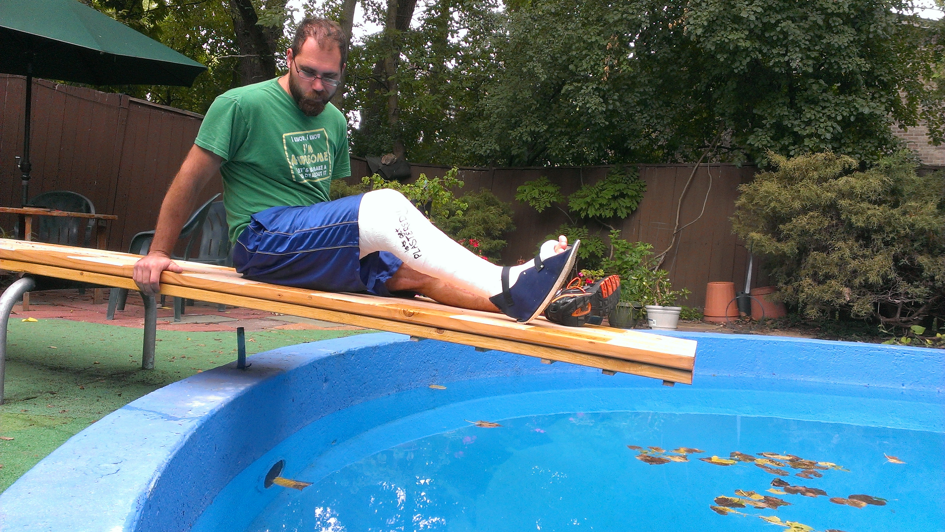
    Did you ever break a leg or other bone, like the man looking longingly at the water in this swimming pool? Having a broken bone can really restrict your activity. Bones are very hard, but they will break, or fracture if enough force is applied to them. Fortunately, bones are highly active organs that can repair themselves if they break. Bones can also remodel themselves and grow. You’ll learn how bones can do all of these things in this concept.

    First plaster long leg cast
    Figure \(\PageIndex{1}\): Broken leg

    Bone Growth

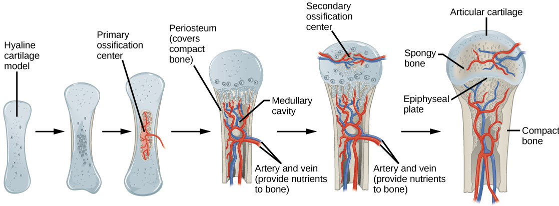
    Early in the development of a human fetus, the skeleton is made almost entirely of cartilage. The relatively soft cartilage gradually turns into hard bone through ossification. Ossification is a process in which bone tissue is created from cartilage. The steps in which bones of the skeleton form from cartilage are illustrated in Figure \(\PageIndex{2}\). The steps include the following:

    1. Cartilage “model” of bone forms; this model continues to grow as ossification takes place.
    2. Ossification begins at a primary ossification center in the middle of the bone.
    3. Ossification then starts to occur at secondary ossification centers at the ends of the bone.
    4. The medullary cavity forms and will contain red bone marrow.
    5. Areas of ossification meet at epiphyseal plates, and articular cartilage forms. Bone growth ends.
    Bone growth
    Figure \(\PageIndex{2}\): The ossification of cartilage in the human skeleton is a process that lasts throughout childhood in some bones.

    Primary and Secondary Ossification Centers

    When bone forms from cartilage, ossification begins with a point in the cartilage called the primary ossification center. This generally appears during fetal development, although a few short bones begin their primary ossification after birth. Ossification occurs toward both ends of the bone from the primary ossification center, and it eventually forms the shaft of the bone in the case of long bones.

    Secondary ossification centers form after birth. Ossification from secondary centers eventually forms the ends of the bones. The shaft and ends of the bone are separated by a growing zone of cartilage until the individual reaches skeletal maturity.

    Skeletal Maturity

    Throughout childhood, the cartilage remaining in the skeleton keeps growing and allows for bones to grow in size. However, once all of the cartilage has been replaced by bone and fusion has taken place at epiphyseal plates, bones can no longer keep growing in length. This is the point at which skeletal maturity has been reached. It generally takes place by age 18 to 25.

    The use of anabolic steroids by teens can speed up the process of skeletal maturity, resulting in a shorter period of cartilage growth before fusion takes place. This means that teens who use steroids are likely to end up shorter as adults than they would otherwise have been.

    Bone Remodeling

    Even after skeletal maturity has been attained, bone is constantly being resorbed and replaced with new bone in a process known as bone remodeling. In this lifelong process, mature bone tissue is continually turned over, with about 10 percent of the skeletal mass of an adult being remodeled each year. Bone remodeling is carried out through the work of osteoclasts, which are bone cells that resorb bone and dissolve its minerals; and osteoblasts, which are bone cells that make the new bone matrix.

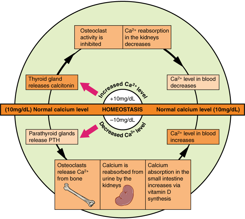
    Bones remodeling serves several functions. It shapes the bones of the skeleton as a child grows, and it repairs tiny flaws in the bone that result from everyday movements. Remodeling also makes bones thicker at points where muscles place the most stress on them. In addition, remodeling helps regulate mineral homeostasis because it either releases minerals from bones into the blood or absorbs minerals from the blood into bones. The figure below shows how osteoclasts in bones are involved in calcium regulation.

    The action of osteoblasts and osteoclasts in bone remodeling and calcium homeostasis is controlled by a number of enzymes, hormones, and other substances that either promote or inhibit the activity of the cells. In this way, these substances control the rate at which bone is made, destroyed, and changed in shape. For example, the rate at which osteoclasts resorb bone and release calcium into the blood is promoted by parathyroid hormone (PTH) and inhibited by calcitonin, which is produced by the thyroid gland (Figure \(\PageIndex{3}\)). The rate at which osteoblasts create new bone is stimulated by growth hormone, which is produced by the anterior lobe of the pituitary gland. Thyroid hormone and sex hormones (estrogens and androgens) also stimulate osteoblasts to create new bone.

    Calcium Homeostasis
    Figure \(\PageIndex{3}\): Keeping the calcium level in homeostasis includes the work of osteoclasts, the bone cells that resorb bone and release calcium into the blood. If calcium levels increase, the thyroid releases calcitonin which inhibits osteoclast activity. This results in less calcium be reabsorbed in the kidneys which results in the lowering of calcium in the blood. If calcium levels decrease below homeostasis, the parathyroid gland releases PTH. This causes osteoclasts to release calcium from bones and increases calcium absorption in the kidney and small intestines. This brings calcium back to homeostasis.

    Bone Repair

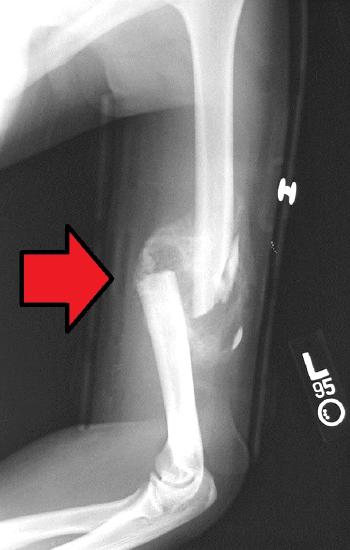
    Bone repair, or healing, is the process in which a bone repairs itself following a bone fracture. You can see an X-ray of bone fracture in Figure \(\PageIndex{4}\). In this fracture, the humerus in the upper arm has been completely broken through its shaft. Before this fracture heals, a physician must push the displaced bone parts back into their correct positions. Then the bone must be stabilized — for example, with a cast and/or pins surgically inserted into the bone — until the bone’s natural healing process is completed. This process may take several weeks.

    The process of bone repair is mainly determined by the periosteum, which is the connective tissue membrane covering the bone. The periosteum is the primary source of precursor cells that develop into osteoblasts, which are essential to the healing process. Bones heal as osteoblasts form new bone tissue.

    Although bone repair is a natural physiological process, it may be promoted or inhibited by several factors. For example, fracture repair is likely to be more successful with adequate nutrient intake. Age, bone type, drug therapy, and pre-existing bone disease are additional factors that may affect healing. Bones that are weakened by diseases, such as osteoporosis or bone cancer, are not only likely to heal more slowly but are also more likely to fracture in the first place.

    Communitive midshaft humeral fracture callus
    Figure \(\PageIndex{4}\): A bone fracture does not always involve a complete break in the bone, as in this X-ray. Sometimes a fracture is just a crack in the bone. In other cases, the bone not only breaks all the way through, as here but also breaks through the soft tissues around it so it protrudes from the skin. This is called an open fracture.
    Feature: Myth vs. Reality

    Bone fractures are fairly common, and there are many myths about them. Knowing the facts is important because fractures generally require emergency medical treatment.

    Myth: A bone fracture is a milder injury than a broken bone.

    Reality: A bone fracture is the same thing as a broken bone.

    Myth: If you still have a full range of motion in a limb, then it must not be fractured.

    Reality: Even if a bone is fractured, the muscles and tendons attached to it may still be able to move the bone normally. This is especially likely if the bone is cracked but not broken into two pieces. Even if a bone is broken all the way through, the range of motion may not be much affected if the bones on either side of the fracture remain properly aligned.

    Myth: A fracture always produces a bruise.

    Reality: Many but not all fractures produce a bruise. If a fracture does produce a bruise, it may take several hours or even a day or more for the bruise to appear.

    Myth: Fractures are so painful that you will immediately know if you break a bone.

    Reality: Ligament sprains and muscle strains are also very painful, sometimes more painful than fractures. Additionally, every person has a different pain tolerance. People with high pain tolerance may continue using a broken bone in spite of the pain.

    Myth: You can tell when a bone is fractured because there will be very localized pain over the break.

    Reality: A broken bone is often accompanied by injuries to surrounding muscles or ligaments. As a result, the pain may extend far beyond the location of the fracture. The pain may be greater directly over the fracture, but the intensity of the pain may make it difficult to pinpoint exactly where the pain originates.

    Review

    1. Outline how bone develops from early in the fetal stage through the age of skeletal maturity.
    2. Describe the process of bone remodeling. When does it occur?
    3. What purposes does bone remodeling serve?
    4. Define bone repair. How long does this process take?
    5. Explain how bone repair occurs.
    6. Identify factors that may affect bone repair.
    7. Parts of bone that have not yet become ossified are made of _________.
    8. If there is a large region between the primary and secondary ossification centers in a bone, is the person young or old? Explain your answer.
    9. The region where the primary and secondary ossification centers meet is called the ________________.
    10. True or False. Most bones are made entirely of cartilage at birth.
    11. True or False. A broken bone is the same as a bone fracture.
    12. If bones can repair themselves, why are casts and pins sometimes needed?
    13. Which bone cell type causes the release of calcium to the bloodstream when calcium levels are low?
    14. Which tissue and bone cell type are mainly involved in bone repair after a fracture?
    15. Describe one way in which hormones are involved in bone remodeling.

    Attributions

    1. Orthopedic cast by 4x4king10, CC BY 2.0 via Wikimedia Commons
    2. Ossification by OpenStax Biology, CC BY 4.0 via Wikimedia Commons
    3. Calcium homeostasis by OpenStax College, CC BY 3.0 via Wikimedia Commons
    4. Communitive midshaft humeral fracture with callus formation by Bill Rhodes, CC BY 2.0 via Wikimedia Commons
    5. Text adapted from Human Biology by CK-12 licensed CC BY-NC 3.0

    This page titled 8.5: Bone Growth, Remodeling, and Repair is shared under a CK-12 license and was authored, remixed, and/or curated by Suzanne Wakim & Mandeep Grewal via source content that was edited to the style and standards of the LibreTexts platform.

    CK-12 Foundation
    LICENSED UNDER
    CK-12 Foundation is licensed under CK-12 Curriculum Materials License