Biology LibreTexts

8.4: Structure of Bone

  • Page ID
    85250
  • \( \newcommand{\vecs}[1]{\overset { \scriptstyle \rightharpoonup} {\mathbf{#1}} } \)

    \( \newcommand{\vecd}[1]{\overset{-\!-\!\rightharpoonup}{\vphantom{a}\smash {#1}}} \)

    \( \newcommand{\id}{\mathrm{id}}\) \( \newcommand{\Span}{\mathrm{span}}\)

    ( \newcommand{\kernel}{\mathrm{null}\,}\) \( \newcommand{\range}{\mathrm{range}\,}\)

    \( \newcommand{\RealPart}{\mathrm{Re}}\) \( \newcommand{\ImaginaryPart}{\mathrm{Im}}\)

    \( \newcommand{\Argument}{\mathrm{Arg}}\) \( \newcommand{\norm}[1]{\| #1 \|}\)

    \( \newcommand{\inner}[2]{\langle #1, #2 \rangle}\)

    \( \newcommand{\Span}{\mathrm{span}}\)

    \( \newcommand{\id}{\mathrm{id}}\)

    \( \newcommand{\Span}{\mathrm{span}}\)

    \( \newcommand{\kernel}{\mathrm{null}\,}\)

    \( \newcommand{\range}{\mathrm{range}\,}\)

    \( \newcommand{\RealPart}{\mathrm{Re}}\)

    \( \newcommand{\ImaginaryPart}{\mathrm{Im}}\)

    \( \newcommand{\Argument}{\mathrm{Arg}}\)

    \( \newcommand{\norm}[1]{\| #1 \|}\)

    \( \newcommand{\inner}[2]{\langle #1, #2 \rangle}\)

    \( \newcommand{\Span}{\mathrm{span}}\) \( \newcommand{\AA}{\unicode[.8,0]{x212B}}\)

    \( \newcommand{\vectorA}[1]{\vec{#1}}      % arrow\)

    \( \newcommand{\vectorAt}[1]{\vec{\text{#1}}}      % arrow\)

    \( \newcommand{\vectorB}[1]{\overset { \scriptstyle \rightharpoonup} {\mathbf{#1}} } \)

    \( \newcommand{\vectorC}[1]{\textbf{#1}} \)

    \( \newcommand{\vectorD}[1]{\overrightarrow{#1}} \)

    \( \newcommand{\vectorDt}[1]{\overrightarrow{\text{#1}}} \)

    \( \newcommand{\vectE}[1]{\overset{-\!-\!\rightharpoonup}{\vphantom{a}\smash{\mathbf {#1}}}} \)

    \( \newcommand{\vecs}[1]{\overset { \scriptstyle \rightharpoonup} {\mathbf{#1}} } \)

    \( \newcommand{\vecd}[1]{\overset{-\!-\!\rightharpoonup}{\vphantom{a}\smash {#1}}} \)

    \(\newcommand{\avec}{\mathbf a}\) \(\newcommand{\bvec}{\mathbf b}\) \(\newcommand{\cvec}{\mathbf c}\) \(\newcommand{\dvec}{\mathbf d}\) \(\newcommand{\dtil}{\widetilde{\mathbf d}}\) \(\newcommand{\evec}{\mathbf e}\) \(\newcommand{\fvec}{\mathbf f}\) \(\newcommand{\nvec}{\mathbf n}\) \(\newcommand{\pvec}{\mathbf p}\) \(\newcommand{\qvec}{\mathbf q}\) \(\newcommand{\svec}{\mathbf s}\) \(\newcommand{\tvec}{\mathbf t}\) \(\newcommand{\uvec}{\mathbf u}\) \(\newcommand{\vvec}{\mathbf v}\) \(\newcommand{\wvec}{\mathbf w}\) \(\newcommand{\xvec}{\mathbf x}\) \(\newcommand{\yvec}{\mathbf y}\) \(\newcommand{\zvec}{\mathbf z}\) \(\newcommand{\rvec}{\mathbf r}\) \(\newcommand{\mvec}{\mathbf m}\) \(\newcommand{\zerovec}{\mathbf 0}\) \(\newcommand{\onevec}{\mathbf 1}\) \(\newcommand{\real}{\mathbb R}\) \(\newcommand{\twovec}[2]{\left[\begin{array}{r}#1 \\ #2 \end{array}\right]}\) \(\newcommand{\ctwovec}[2]{\left[\begin{array}{c}#1 \\ #2 \end{array}\right]}\) \(\newcommand{\threevec}[3]{\left[\begin{array}{r}#1 \\ #2 \\ #3 \end{array}\right]}\) \(\newcommand{\cthreevec}[3]{\left[\begin{array}{c}#1 \\ #2 \\ #3 \end{array}\right]}\) \(\newcommand{\fourvec}[4]{\left[\begin{array}{r}#1 \\ #2 \\ #3 \\ #4 \end{array}\right]}\) \(\newcommand{\cfourvec}[4]{\left[\begin{array}{c}#1 \\ #2 \\ #3 \\ #4 \end{array}\right]}\) \(\newcommand{\fivevec}[5]{\left[\begin{array}{r}#1 \\ #2 \\ #3 \\ #4 \\ #5 \\ \end{array}\right]}\) \(\newcommand{\cfivevec}[5]{\left[\begin{array}{c}#1 \\ #2 \\ #3 \\ #4 \\ #5 \\ \end{array}\right]}\) \(\newcommand{\mattwo}[4]{\left[\begin{array}{rr}#1 \amp #2 \\ #3 \amp #4 \\ \end{array}\right]}\) \(\newcommand{\laspan}[1]{\text{Span}\{#1\}}\) \(\newcommand{\bcal}{\cal B}\) \(\newcommand{\ccal}{\cal C}\) \(\newcommand{\scal}{\cal S}\) \(\newcommand{\wcal}{\cal W}\) \(\newcommand{\ecal}{\cal E}\) \(\newcommand{\coords}[2]{\left\{#1\right\}_{#2}}\) \(\newcommand{\gray}[1]{\color{gray}{#1}}\) \(\newcommand{\lgray}[1]{\color{lightgray}{#1}}\) \(\newcommand{\rank}{\operatorname{rank}}\) \(\newcommand{\row}{\text{Row}}\) \(\newcommand{\col}{\text{Col}}\) \(\renewcommand{\row}{\text{Row}}\) \(\newcommand{\nul}{\text{Nul}}\) \(\newcommand{\var}{\text{Var}}\) \(\newcommand{\corr}{\text{corr}}\) \(\newcommand{\len}[1]{\left|#1\right|}\) \(\newcommand{\bbar}{\overline{\bvec}}\) \(\newcommand{\bhat}{\widehat{\bvec}}\) \(\newcommand{\bperp}{\bvec^\perp}\) \(\newcommand{\xhat}{\widehat{\xvec}}\) \(\newcommand{\vhat}{\widehat{\vvec}}\) \(\newcommand{\uhat}{\widehat{\uvec}}\) \(\newcommand{\what}{\widehat{\wvec}}\) \(\newcommand{\Sighat}{\widehat{\Sigma}}\) \(\newcommand{\lt}{<}\) \(\newcommand{\gt}{>}\) \(\newcommand{\amp}{&}\) \(\definecolor{fillinmathshade}{gray}{0.9}\)
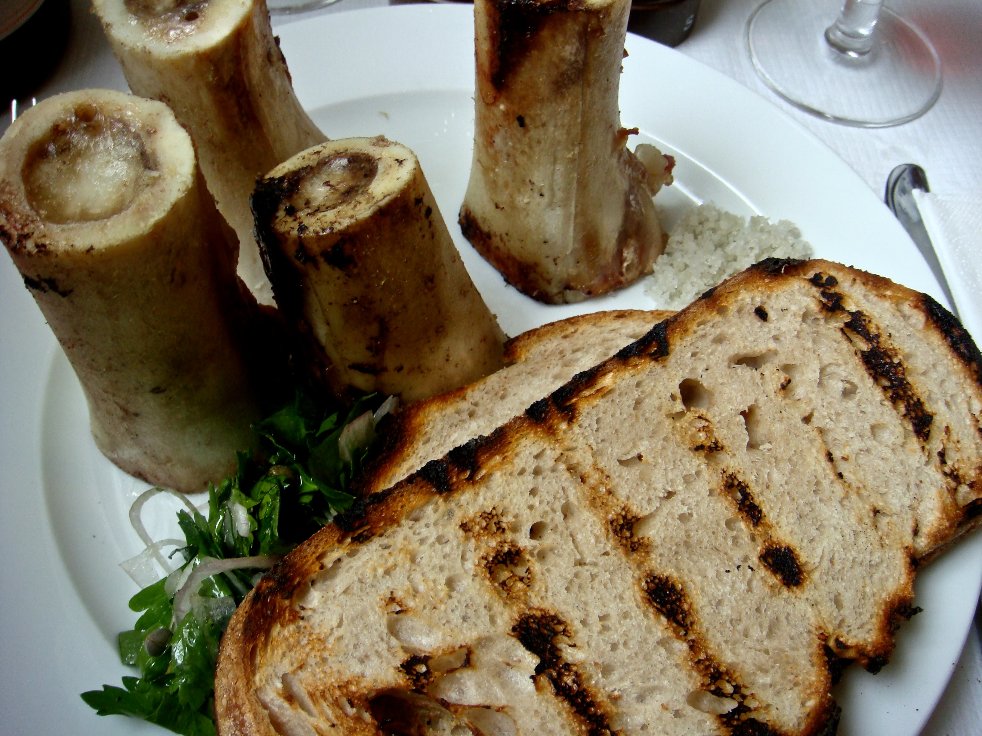
    Roasted Bone Marrow

    Do you recognize the food item in the top left of this photo in Figure \(\PageIndex{1}\)? It’s roasted bone marrow, still inside the bones. It’s considered a delicacy in some cuisines. Marrow is a type of tissue found inside many animal bones, including our own. It’s a soft tissue that in adults may be mostly fat. You’ll learn more about bone marrow and other tissues that make up bones when you read this concept.

    Roast Bone Marrow & Parsley Salad
    Figure \(\PageIndex{1}\):Roasted Bone Marrow

    Bones are organs that consist primarily of bone tissue, also called osseous tissue. Bone tissue is a type of connective tissue consisting mainly of a collagen matrix that is mineralized with calcium and phosphorus crystals. The combination of flexible collagen and hard mineral crystals makes bone tissue hard without making it brittle.

    Bone Anatomy

    There are several different types of tissues in bones, including two types of osseous tissues.

    Types of Osseous Tissue

    The two different types of osseous tissue are compact bone tissue (also called hard or cortical bone) tissue and spongy bone tissue (also called cancellous or trabecular bone).

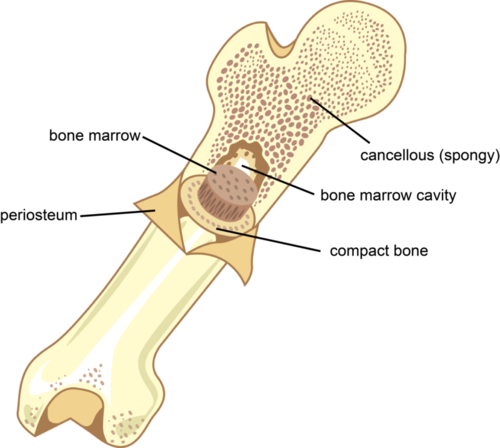
    long bone longitudinal section
    Figure \(\PageIndex{2}\): Bones are more complex on the inside than you would expect from their outer appearance. There is bone marrow in the hollow part of the long bone. The outer boundary of bone is composed of compact bone tissue, and from the ends, and the rest of the bone interior is composed of cancellous or spongy bone tissue. The bone is covered in connecting tissue called the periosteum.

    Compact bone tissue forms the extremely hard outside layer of bones. Cortical bone tissue gives bone its smooth, dense, solid appearance. It accounts for about 80 percent of the total bone mass of the adult skeleton. Spongy bone tissue fills part or all of the interior of many bones. As its name suggests, spongy bone is porous like a sponge, containing an irregular network of spaces. This makes spongy bone much less dense than compact bone. Spongy bone has a greater surface area than cortical bone but makes up only 20 percent of bone mass.

    Both compact and spongy bone tissues have the same types of cells, but they differ in how the cells are arranged. The cells in the compact bone are arranged in multiple microscopic columns, whereas the cells in the spongy bone are arranged in a looser, more open network. These cellular differences explain why cortical and spongy bone tissues have such different structures.

    Other Tissues in Bones

    Besides cortical and spongy bone tissues, bones contain several other tissues, including blood vessels and nerves. In addition, bones contain bone marrow and periosteum. You can see these tissues in Figure \(\PageIndex{2}\).

    • Bone marrow is a soft connective tissue that is found inside a cavity, called the marrow cavity. There are two types of marrow in adults, yellow bone marrow, which consists mostly of fat, and red bone marrow. All marrow is red in newborns, but by adulthood, much of the red marrow has changed to yellow marrow. In adults, red marrow is found mainly in the femur, ribs, vertebrae, and pelvic bones. Red bone marrow contains hematopoietic stem cells that give rise to red blood cells, white blood cells, and platelets in the process of hematopoiesis.
    • Periosteum is a tough, fibrous membrane that covers the outer surface of bones. It provides a protective covering for cortical bone tissue. It is also the source of new bone cells.

    Bone Cells

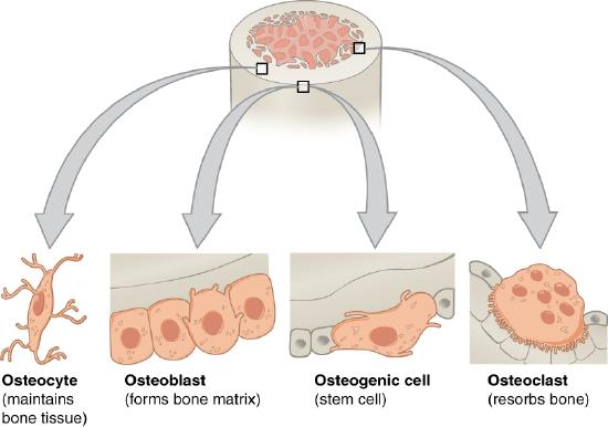
    As shown in Figure \(\PageIndex{3}\), bone tissues are composed of four different types of bone cells: osteoblasts, osteocytes, osteoclasts, and osteogenic cells.

    • Osteoblasts are bone cells with a single nucleus that make and mineralize bone matrix. They make a protein mixture that is composed primarily of collagen and creates the organic part of the matrix. They also release calcium and phosphate ions that form mineral crystals within the matrix. In addition, they produce hormones that also play a role in the mineralization of the matrix.
    • Osteocytes are mainly inactive bone cells that form from osteoblasts that have become entrapped within their own bone matrix. Osteocytes help regulate the formation and breakdown of bone tissue. They have multiple cell projections that are thought to be involved in communication with other bone cells.
    • Osteoclasts are bone cells with multiple nuclei that resorb bone tissue and break down bone. They dissolve the minerals in bone and release them into the blood.
    • Osteogenic cells are undifferentiated stem cells. They are the only bone cells that can divide. When they do, they differentiate and develop into osteoblasts.

    Bone is a very active tissue. It is constantly remodeled by the work of osteoblasts and osteoclasts. Osteoblasts continuously make new bone, and osteoclasts keep breaking down bone. This allows for minor repair of bones as well as homeostasis of mineral ions in the blood.

    Bone cells
    Figure \(\PageIndex{3}\): Different types of bones cells have different functions.

    Microscopic Anatomy of The Compact Bone

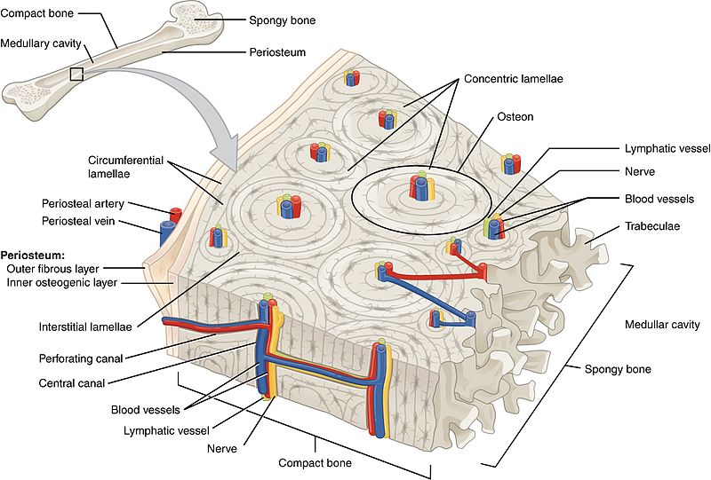
    Compact Bone
    Figure \(\PageIndex{4}\): Macroscopic and microscopic structures of the compact bone tissue.

    The basic microscopic unit of bone is an osteon (or Haversian system). Osteons are roughly cylindrical structures that can measure several millimeters long and around 0.2 mm in diameter. Each osteon consists of lamellae of compact bone tissue that surround a central canal (Haversian canal). The Haversian canal contains the bone's blood supplies. The boundary of an osteon is called the cement line. Osteons can be arranged into woven bone or lamellar bone. Osteoblasts make the matrix of bone which calcifies hardens. This entraps the mature bone cells, osteocytes, in a little chamber called lacunae. The osteocytes receive their nutrition from the central (Haversian) canal via little canals called canaliculi. All of these structures plus more are visible in Figure \(\PageIndex{4}\).

    Types of Bones

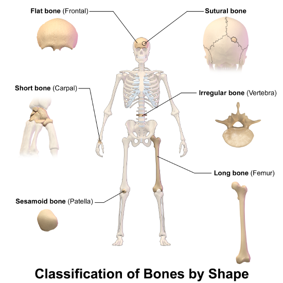
    There are six types of bones in the human body based on their shape or location: long, short, flat, sesamoid, sutural, and irregular bones. You can see an example of each type of bone in Figure \(\PageIndex{5}\).

    • Long bones are characterized by a shaft that is much longer than it is wide and by a rounded head at each end of the shaft. Long bones are made mostly of compact bone, with lesser amounts of spongy bone and marrow. Most bones of the limbs, including those of the fingers and toes, are long bones.
    • Short bones are roughly cube-shaped and have only a thin layer of cortical bone surrounding a spongy bone interior. The bones of the wrists and ankles are short bones.
    • Flat bones are thin and generally curved, with two parallel layers of compact bone sandwiching a layer of spongy bone. Most of the bones of the skull are flat bones, as is the sternum (breast bone).
    • Sesamoid bones are embedded in tendons, the connective tissues that bind muscles to bones. Sesamoid bones hold tendons farther away from joints so the angle of the tendons is increased, thus increasing the leverage of muscles. The patella (knee cap) is an example of a sesamoid bone.
    • Sutural bones are very small bones that are located between the major bones of the skull, within the joints (sutures) between the larger bones. They are not always present.
    • Irregular bones are those that do not fit into any of the above categories. They generally consist of thin layers of cortical bone surrounding a spongy bone interior. Their shapes are irregular and complicated. Examples of irregular bones include the vertebrae and the bones of the pelvis.
    Classification of Bones
    Figure \(\PageIndex{5}\): six types of bones classified by shape or location.

    Feature: Reliable Sources

    Diseased or damaged bone marrow can be replaced by donated bone marrow cells, which help treat and often cure many life-threatening conditions, including leukemia, lymphoma, sickle cell anemia, and thalassemia. If a bone marrow transplant is successful, the new bone marrow will start making healthy blood cells and improve the patient’s condition.

    Learn more about bone marrow donation, and consider whether you might want to do it yourself. Find reliable sources to answer the following questions:

    1. How does one become a potential bone marrow donor?
    2. Who can and who cannot donate bone marrow?
    3. How is a bone marrow donation made?
    4. What risks are there in donating bone marrow?

    Review

    1. Describe osseous tissue.
    2. Why are bones hard but not brittle?
    3. Compare and contrast the two main types of osseous tissue.
    4. What non-osseous tissues are found in bones?
    5. List four types of bone cells and their functions.
    6. Identify six types of bones, and give an example of each type.
    7. True or False. Spongy bone tissue is another name for bone marrow.
    8. True or False. Periosteum covers osseous tissue.
    9. Compare and contrast yellow bone marrow and red bone marrow.
    10. Which bone is mostly made of cortical bone tissue?

      A. Pelvis

      B. Vertebrae

      C. Femur

      D. Carpal

    11. a. Which type of bone cell divides to produce new bone cells?

      b. Where is this cell type located?

    12. Where do osteoblasts and osteocytes come from, and how are they related to each other?
    13. Which type of bone is embedded in tendons?
    14. True or False. Calcium is the only mineral in bones.

    Explore More

    Watch this entertaining and fast-paced Crash Course video to further explore bone structure:

    Check out this video to learn more about bone remodeling:

    Attributions

    1. Roast Bone Marrow by Simon Doggett, CC BY 2.0 via Wikimedia Commons
    2. Bone structure by Christopher Auyeung via CK-12 licensed CC BY-NC 3.0
    3. Bone cells by OpenStax College, CC BY 3.0 via Wikimedia Commons
    4. Compact bone by OpenStax Anatomy and Physiology, CC BY 3.0 via Wikimedia Commons
    5. Types of bone by BruceBlaus, CC BY 3.0 via Wikimedia Commons
    6. Text adapted from Human Biology by CK-12 licensed CC BY-NC 3.0

    This page titled 8.4: Structure of Bone is shared under a CK-12 license and was authored, remixed, and/or curated by Suzanne Wakim & Mandeep Grewal via source content that was edited to the style and standards of the LibreTexts platform.

    CK-12 Foundation
    LICENSED UNDER
    CK-12 Foundation is licensed under CK-12 Curriculum Materials License