
6.4: Bacteriophages
- Page ID
- 52252
\( \newcommand{\vecs}[1]{\overset { \scriptstyle \rightharpoonup} {\mathbf{#1}} } \)
\( \newcommand{\vecd}[1]{\overset{-\!-\!\rightharpoonup}{\vphantom{a}\smash {#1}}} \)
\( \newcommand{\dsum}{\displaystyle\sum\limits} \)
\( \newcommand{\dint}{\displaystyle\int\limits} \)
\( \newcommand{\dlim}{\displaystyle\lim\limits} \)
\( \newcommand{\id}{\mathrm{id}}\) \( \newcommand{\Span}{\mathrm{span}}\)
( \newcommand{\kernel}{\mathrm{null}\,}\) \( \newcommand{\range}{\mathrm{range}\,}\)
\( \newcommand{\RealPart}{\mathrm{Re}}\) \( \newcommand{\ImaginaryPart}{\mathrm{Im}}\)
\( \newcommand{\Argument}{\mathrm{Arg}}\) \( \newcommand{\norm}[1]{\| #1 \|}\)
\( \newcommand{\inner}[2]{\langle #1, #2 \rangle}\)
\( \newcommand{\Span}{\mathrm{span}}\)
\( \newcommand{\id}{\mathrm{id}}\)
\( \newcommand{\Span}{\mathrm{span}}\)
\( \newcommand{\kernel}{\mathrm{null}\,}\)
\( \newcommand{\range}{\mathrm{range}\,}\)
\( \newcommand{\RealPart}{\mathrm{Re}}\)
\( \newcommand{\ImaginaryPart}{\mathrm{Im}}\)
\( \newcommand{\Argument}{\mathrm{Arg}}\)
\( \newcommand{\norm}[1]{\| #1 \|}\)
\( \newcommand{\inner}[2]{\langle #1, #2 \rangle}\)
\( \newcommand{\Span}{\mathrm{span}}\) \( \newcommand{\AA}{\unicode[.8,0]{x212B}}\)
\( \newcommand{\vectorA}[1]{\vec{#1}} % arrow\)
\( \newcommand{\vectorAt}[1]{\vec{\text{#1}}} % arrow\)
\( \newcommand{\vectorB}[1]{\overset { \scriptstyle \rightharpoonup} {\mathbf{#1}} } \)
\( \newcommand{\vectorC}[1]{\textbf{#1}} \)
\( \newcommand{\vectorD}[1]{\overrightarrow{#1}} \)
\( \newcommand{\vectorDt}[1]{\overrightarrow{\text{#1}}} \)
\( \newcommand{\vectE}[1]{\overset{-\!-\!\rightharpoonup}{\vphantom{a}\smash{\mathbf {#1}}}} \)
\( \newcommand{\vecs}[1]{\overset { \scriptstyle \rightharpoonup} {\mathbf{#1}} } \)
\(\newcommand{\longvect}{\overrightarrow}\)
\( \newcommand{\vecd}[1]{\overset{-\!-\!\rightharpoonup}{\vphantom{a}\smash {#1}}} \)
\(\newcommand{\avec}{\mathbf a}\) \(\newcommand{\bvec}{\mathbf b}\) \(\newcommand{\cvec}{\mathbf c}\) \(\newcommand{\dvec}{\mathbf d}\) \(\newcommand{\dtil}{\widetilde{\mathbf d}}\) \(\newcommand{\evec}{\mathbf e}\) \(\newcommand{\fvec}{\mathbf f}\) \(\newcommand{\nvec}{\mathbf n}\) \(\newcommand{\pvec}{\mathbf p}\) \(\newcommand{\qvec}{\mathbf q}\) \(\newcommand{\svec}{\mathbf s}\) \(\newcommand{\tvec}{\mathbf t}\) \(\newcommand{\uvec}{\mathbf u}\) \(\newcommand{\vvec}{\mathbf v}\) \(\newcommand{\wvec}{\mathbf w}\) \(\newcommand{\xvec}{\mathbf x}\) \(\newcommand{\yvec}{\mathbf y}\) \(\newcommand{\zvec}{\mathbf z}\) \(\newcommand{\rvec}{\mathbf r}\) \(\newcommand{\mvec}{\mathbf m}\) \(\newcommand{\zerovec}{\mathbf 0}\) \(\newcommand{\onevec}{\mathbf 1}\) \(\newcommand{\real}{\mathbb R}\) \(\newcommand{\twovec}[2]{\left[\begin{array}{r}#1 \\ #2 \end{array}\right]}\) \(\newcommand{\ctwovec}[2]{\left[\begin{array}{c}#1 \\ #2 \end{array}\right]}\) \(\newcommand{\threevec}[3]{\left[\begin{array}{r}#1 \\ #2 \\ #3 \end{array}\right]}\) \(\newcommand{\cthreevec}[3]{\left[\begin{array}{c}#1 \\ #2 \\ #3 \end{array}\right]}\) \(\newcommand{\fourvec}[4]{\left[\begin{array}{r}#1 \\ #2 \\ #3 \\ #4 \end{array}\right]}\) \(\newcommand{\cfourvec}[4]{\left[\begin{array}{c}#1 \\ #2 \\ #3 \\ #4 \end{array}\right]}\) \(\newcommand{\fivevec}[5]{\left[\begin{array}{r}#1 \\ #2 \\ #3 \\ #4 \\ #5 \\ \end{array}\right]}\) \(\newcommand{\cfivevec}[5]{\left[\begin{array}{c}#1 \\ #2 \\ #3 \\ #4 \\ #5 \\ \end{array}\right]}\) \(\newcommand{\mattwo}[4]{\left[\begin{array}{rr}#1 \amp #2 \\ #3 \amp #4 \\ \end{array}\right]}\) \(\newcommand{\laspan}[1]{\text{Span}\{#1\}}\) \(\newcommand{\bcal}{\cal B}\) \(\newcommand{\ccal}{\cal C}\) \(\newcommand{\scal}{\cal S}\) \(\newcommand{\wcal}{\cal W}\) \(\newcommand{\ecal}{\cal E}\) \(\newcommand{\coords}[2]{\left\{#1\right\}_{#2}}\) \(\newcommand{\gray}[1]{\color{gray}{#1}}\) \(\newcommand{\lgray}[1]{\color{lightgray}{#1}}\) \(\newcommand{\rank}{\operatorname{rank}}\) \(\newcommand{\row}{\text{Row}}\) \(\newcommand{\col}{\text{Col}}\) \(\renewcommand{\row}{\text{Row}}\) \(\newcommand{\nul}{\text{Nul}}\) \(\newcommand{\var}{\text{Var}}\) \(\newcommand{\corr}{\text{corr}}\) \(\newcommand{\len}[1]{\left|#1\right|}\) \(\newcommand{\bbar}{\overline{\bvec}}\) \(\newcommand{\bhat}{\widehat{\bvec}}\) \(\newcommand{\bperp}{\bvec^\perp}\) \(\newcommand{\xhat}{\widehat{\xvec}}\) \(\newcommand{\vhat}{\widehat{\vvec}}\) \(\newcommand{\uhat}{\widehat{\uvec}}\) \(\newcommand{\what}{\widehat{\wvec}}\) \(\newcommand{\Sighat}{\widehat{\Sigma}}\) \(\newcommand{\lt}{<}\) \(\newcommand{\gt}{>}\) \(\newcommand{\amp}{&}\) \(\definecolor{fillinmathshade}{gray}{0.9}\)Learning Objectives
- Learn how to culture viruses in a host cell
- Quantitate viruses in a specimen
- Identify viral plaques in a bacterial lawn
Viruses are obligate intracellular parasites that multiple within the host cytoplasm. Bacteriophages are viruses which infect bacteria. PHAGE (as in phagocytosis) means "to eat", and generally refers to a virus. Viruses multiply within host cells and rely upon the host's metabolic machinery for replication.
Most bacteria have phages that are able to parasitize them. In fact, the ability to be infected with a known phage type is used to identify some strains of bacteria (like Staph), known as phage typing. As the virus infects bacterial cells that it has been mixed with, the lytic infection destroys the bacteria. The bacteria have been poured into what is called a bacterial lawn on the agar plate. As the surrounding cells are infected and killed by the released viruses, a clear spot on the agar---in the bacterial lawn--develops, called a plaque.The plaques can be counted and the number of virus particles or virions in the original specimen, can be quantified as viruses/ ml or plaque-forming units/ml (PFUs).
One common phage is called "T4" and it is capable of infecting its host, Escherichia coli. This type of bacteriophage is called a "coliphage. " Viruses have high specificity for its host, and will only infect a specific bacterium host.
Watch this video animation on T4 phage infecting E. coli
Watch Video 1: T4 phage infecting E. coli. URL: https://youtu.be/V73nEGXUeBY
Method for estimating the number of bacteriophages
Knowing how to determine the number of microorganisms in a sample is extremely important in microbiology, and requires accurate pipetting, aseptic technique, and calculation skills. Similar to enumeration of bacteria, one has to prepare serial dilutions of a sample and plate these samples out. Although the principles are the same, you will be enumerating viruses instead of bacteria and conserving on materials by using micropipettes and smaller volumes in your dilutions.
The number of viruses in a sample can be determined by direct count using electron microscopy or by determining the number of infectious virus particles using a plaque assay. Viruses are obligate intracellular parasites. Therefore, they require a host cell in which to grow. Some viruses lyse the cells in which they have replicated while others appear to cause little cell damage.
A plaque assay can be used to enumerate viruses that lyse their host cells. In a plaque assay the host cells and virus are incubated together for a short time to allow the virus to attach to and enter the host cell. Then the mixture in plated within a semi-solid agar. This semi-solid agar is poured onto a "bottom agar" that serves to supply adequate nutrients for the host cell. At the end of one cycle of virus replication, a cell infected with a single virus particle will lyse, releasing hundreds of new viruses.
In the semi-solid medium these newly released viruses can only infect neighboring cells; after a second cycle of replication these neighboring cells will be lysed. Those cells that escape infection will continue to grow. After 24 to 48 hours plates that were not infected with a virus will contain a confluent layer of cells (they will resemble the too numerous to count/TNTC plates from bacterial standard/viable plate counts). Those plates that were infected with several hundred viruses may actually appear clear- the viruses will have infected and lysed all of the host cells. Those plates that contain an intermediate number of viruses will have plaques, clear or partially clear circular areas in an otherwise turbid background of cellular growth. Each plaque represents the result of one infectious virus, called a plaque forming unit, or PFU. Many animal and bacterial viruses can be enumerated using a plaque assay. Bacterial viruses are also referred to as phage; in our lab, we typically use a well-characterized bacterial phage, such as T4, and its host cell, Escherichia coli.
General procedure
- The phage specimen you will use is already diluted to 1/ 1000, and you will dilute further.
- Bacteria and phage are mixed together in tubes of soft agar. The mix is incubated in the water bath.
- After incubation the mix is added to the soft agar and poured over the tryptone agar plates.
- Be SURE to mix the dilutions.
- Change pipettes between dilutions.
- Each table will use a different combination of a phage and an E. coli host.
Detailed procedure with diagram

Image 1: Plaque Assay diagram
Set up 5 saline (0.85% NaCl) dilution tubes labeled 10-4, 10-5,10-6, 10-7, and 10-8. Into each tube, place 9ml pf into which you will dilute the viral solution. You will be making 1/10 dilutions.
- Starting with the 10-3 dilution of the virus that you picked up (or were given by your instructor), transfer 1 ml to the dilution tube marked 10-4 and mix.
- Make 4 more dilutions out to 10-8.
- Into 6 microtubes, add 100 microliters of each viral dilution, plus 300 microliters of E. coli. Let sit at room temperature for 10 minutes while the virus infects the bacteria. Mix these well.
- Take the tubes over to the water bath and transfer the entire contents of E. coli - phage tubes into 6 soft agar tubes, using a sterile plastic transfer pipet. Mix well. KEEP SOFT AGARS INSIDE OF WATER BATH SO THEY DO NOT SOLIDIFY.
- Remove 1 soft agar tube at a time and pour it onto the TSA agar plates, gently rotating the plate WELL so as to distribute the phage - bacteria all over the agar.
- Allow the plates to harden and incubate at 37º C right side up.
INTERPRETATION
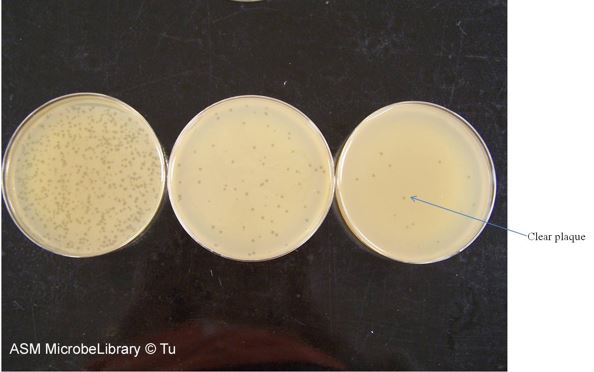
Image 2: Determination of bacteriophage T4 titer by serial dilution and plating on Escherichia coli B host. These series of plates show viral plaques--note the round, clear zones on the agar bacterial lawn on each plate (see arrow pointing to one). Image by Anh-Hue T. Tu, Georgia Southwestern State University, Americus, GA.
- Lay the 6 plates right side up, from lowest dilution towards highest dilution.
- Pick each plate up, hold it up to the light, and determine which one has between 30 - 300 plaques (you can also use the Quebec colony counters---good backlighting!)
- Get an accurate count of that plate. Fill in the formula for viral counts.

- Calculate the number of viruses per ml. of original specimen.
Watch Video 2: Plaque Assay
In this video, the scientist has already completed the serial dilutions and is demonstrating the mixing of the phage/bacterium solution with the soft top agar and pouring this mixture onto the bottom agar plate.
Watch Video 2: Plaque assay. URL: https://youtu.be/uAlRSB6iCt0
Contributors and Attributions
Jackie Reynolds, Professor of Biology (Richland College)