
5.3: Reading- Seed Plants
- Page ID
- 41798

\( \newcommand{\vecs}[1]{\overset { \scriptstyle \rightharpoonup} {\mathbf{#1}} } \)
\( \newcommand{\vecd}[1]{\overset{-\!-\!\rightharpoonup}{\vphantom{a}\smash {#1}}} \)
\( \newcommand{\id}{\mathrm{id}}\) \( \newcommand{\Span}{\mathrm{span}}\)
( \newcommand{\kernel}{\mathrm{null}\,}\) \( \newcommand{\range}{\mathrm{range}\,}\)
\( \newcommand{\RealPart}{\mathrm{Re}}\) \( \newcommand{\ImaginaryPart}{\mathrm{Im}}\)
\( \newcommand{\Argument}{\mathrm{Arg}}\) \( \newcommand{\norm}[1]{\| #1 \|}\)
\( \newcommand{\inner}[2]{\langle #1, #2 \rangle}\)
\( \newcommand{\Span}{\mathrm{span}}\)
\( \newcommand{\id}{\mathrm{id}}\)
\( \newcommand{\Span}{\mathrm{span}}\)
\( \newcommand{\kernel}{\mathrm{null}\,}\)
\( \newcommand{\range}{\mathrm{range}\,}\)
\( \newcommand{\RealPart}{\mathrm{Re}}\)
\( \newcommand{\ImaginaryPart}{\mathrm{Im}}\)
\( \newcommand{\Argument}{\mathrm{Arg}}\)
\( \newcommand{\norm}[1]{\| #1 \|}\)
\( \newcommand{\inner}[2]{\langle #1, #2 \rangle}\)
\( \newcommand{\Span}{\mathrm{span}}\) \( \newcommand{\AA}{\unicode[.8,0]{x212B}}\)
\( \newcommand{\vectorA}[1]{\vec{#1}} % arrow\)
\( \newcommand{\vectorAt}[1]{\vec{\text{#1}}} % arrow\)
\( \newcommand{\vectorB}[1]{\overset { \scriptstyle \rightharpoonup} {\mathbf{#1}} } \)
\( \newcommand{\vectorC}[1]{\textbf{#1}} \)
\( \newcommand{\vectorD}[1]{\overrightarrow{#1}} \)
\( \newcommand{\vectorDt}[1]{\overrightarrow{\text{#1}}} \)
\( \newcommand{\vectE}[1]{\overset{-\!-\!\rightharpoonup}{\vphantom{a}\smash{\mathbf {#1}}}} \)
\( \newcommand{\vecs}[1]{\overset { \scriptstyle \rightharpoonup} {\mathbf{#1}} } \)
\( \newcommand{\vecd}[1]{\overset{-\!-\!\rightharpoonup}{\vphantom{a}\smash {#1}}} \)
\(\newcommand{\avec}{\mathbf a}\) \(\newcommand{\bvec}{\mathbf b}\) \(\newcommand{\cvec}{\mathbf c}\) \(\newcommand{\dvec}{\mathbf d}\) \(\newcommand{\dtil}{\widetilde{\mathbf d}}\) \(\newcommand{\evec}{\mathbf e}\) \(\newcommand{\fvec}{\mathbf f}\) \(\newcommand{\nvec}{\mathbf n}\) \(\newcommand{\pvec}{\mathbf p}\) \(\newcommand{\qvec}{\mathbf q}\) \(\newcommand{\svec}{\mathbf s}\) \(\newcommand{\tvec}{\mathbf t}\) \(\newcommand{\uvec}{\mathbf u}\) \(\newcommand{\vvec}{\mathbf v}\) \(\newcommand{\wvec}{\mathbf w}\) \(\newcommand{\xvec}{\mathbf x}\) \(\newcommand{\yvec}{\mathbf y}\) \(\newcommand{\zvec}{\mathbf z}\) \(\newcommand{\rvec}{\mathbf r}\) \(\newcommand{\mvec}{\mathbf m}\) \(\newcommand{\zerovec}{\mathbf 0}\) \(\newcommand{\onevec}{\mathbf 1}\) \(\newcommand{\real}{\mathbb R}\) \(\newcommand{\twovec}[2]{\left[\begin{array}{r}#1 \\ #2 \end{array}\right]}\) \(\newcommand{\ctwovec}[2]{\left[\begin{array}{c}#1 \\ #2 \end{array}\right]}\) \(\newcommand{\threevec}[3]{\left[\begin{array}{r}#1 \\ #2 \\ #3 \end{array}\right]}\) \(\newcommand{\cthreevec}[3]{\left[\begin{array}{c}#1 \\ #2 \\ #3 \end{array}\right]}\) \(\newcommand{\fourvec}[4]{\left[\begin{array}{r}#1 \\ #2 \\ #3 \\ #4 \end{array}\right]}\) \(\newcommand{\cfourvec}[4]{\left[\begin{array}{c}#1 \\ #2 \\ #3 \\ #4 \end{array}\right]}\) \(\newcommand{\fivevec}[5]{\left[\begin{array}{r}#1 \\ #2 \\ #3 \\ #4 \\ #5 \\ \end{array}\right]}\) \(\newcommand{\cfivevec}[5]{\left[\begin{array}{c}#1 \\ #2 \\ #3 \\ #4 \\ #5 \\ \end{array}\right]}\) \(\newcommand{\mattwo}[4]{\left[\begin{array}{rr}#1 \amp #2 \\ #3 \amp #4 \\ \end{array}\right]}\) \(\newcommand{\laspan}[1]{\text{Span}\{#1\}}\) \(\newcommand{\bcal}{\cal B}\) \(\newcommand{\ccal}{\cal C}\) \(\newcommand{\scal}{\cal S}\) \(\newcommand{\wcal}{\cal W}\) \(\newcommand{\ecal}{\cal E}\) \(\newcommand{\coords}[2]{\left\{#1\right\}_{#2}}\) \(\newcommand{\gray}[1]{\color{gray}{#1}}\) \(\newcommand{\lgray}[1]{\color{lightgray}{#1}}\) \(\newcommand{\rank}{\operatorname{rank}}\) \(\newcommand{\row}{\text{Row}}\) \(\newcommand{\col}{\text{Col}}\) \(\renewcommand{\row}{\text{Row}}\) \(\newcommand{\nul}{\text{Nul}}\) \(\newcommand{\var}{\text{Var}}\) \(\newcommand{\corr}{\text{corr}}\) \(\newcommand{\len}[1]{\left|#1\right|}\) \(\newcommand{\bbar}{\overline{\bvec}}\) \(\newcommand{\bhat}{\widehat{\bvec}}\) \(\newcommand{\bperp}{\bvec^\perp}\) \(\newcommand{\xhat}{\widehat{\xvec}}\) \(\newcommand{\vhat}{\widehat{\vvec}}\) \(\newcommand{\uhat}{\widehat{\uvec}}\) \(\newcommand{\what}{\widehat{\wvec}}\) \(\newcommand{\Sighat}{\widehat{\Sigma}}\) \(\newcommand{\lt}{<}\) \(\newcommand{\gt}{>}\) \(\newcommand{\amp}{&}\) \(\definecolor{fillinmathshade}{gray}{0.9}\)Gymnosperms
The four phyla of gymnosperms are cycads, ginkgo, gnetophytes, and conifers.
Gymnosperms have naked seeds. The seeds of angiosperms are contained within a fruit.
Gymnosperm Diversity
We will examine conifers in some detail during this lab class but will use photographs on the Internet to study the other three divisions. Click on the links below to view photographs of them.
Cycads
Cycads re cone-bearing palmlike plants found mainly in tropical and subtropical regions today. They were very numerous in the Mesozoic Era.
Ginkgo
There is only one species of Ginkgo left. It survived due to Chinese planting them along roadsides. Click here for information and photographs.
Gnetophytes
Welwitschia and Ephedra (information and photographs)
Conifers
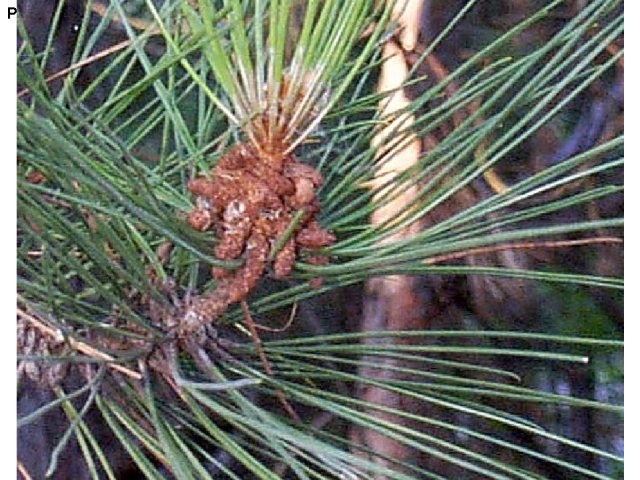
Conifers are the largest group of gymnosperms. They include evergreen trees such as pine, cedar, spruce, fir, and redwood trees. Examine the leaves of pine on display.
The leaves of conifers are needle-like and are adapted for dry conditions such as hot summers or freezing winters. Needles lose water slower than broad, flat leaves and therefore do not need to be shed during seasons when water is scarce, so most conifers are evergreen.
Reproduction in Pine
Life Cycle of Seed Plants
Seed plants are heterosporous—they have two different spore sizes: megaspores and microspores.
The generalized life cycle of plants has been modified (below) to illustrate plants which have separate male and female gametophytes (megagametophyte and microgametophyte) produced by different sized spores (megaspores and microspores).
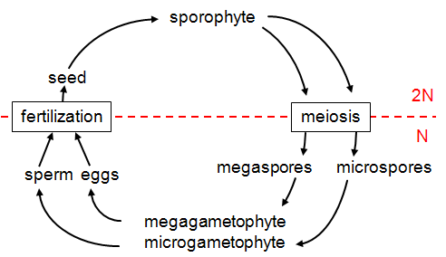
The evolutionary trend from nonvascular plants to seedless vascular plants to seed plants has been a reduction in the size of the gametophyte. In seed plants, the gametophyte is usually microscopic and is retained within the tissues of the sporophyte.
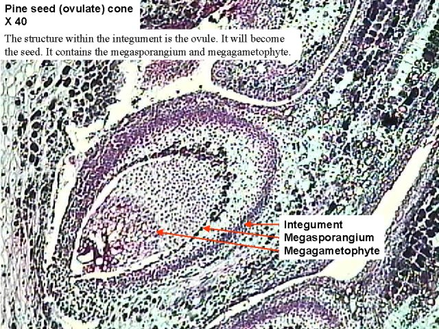
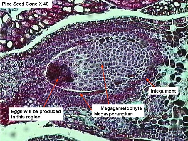
The megasporangium is surrounded by layers of sporophyte tissue called the integument. The integument and structures within (megasporangium, megaspore) are the ovule.
Microspores germinate within the sporophyte tissue and become pollen grains. The microgametophyte is contained within the tough, protective coat of the pollen grain.
The entire microgametophyte (pollen grain) is transferred to the vicinity of the megagametophyte by a process of pollination. Wind or animals usually accomplish this transfer.
When pollen reaches the female gametophyte, it produces an elongate structure (pollen tube) that grows to the egg cell. Sperm are transferred directly through this tube to the egg. The advantage of this process is that sperm do not have to swim long distances as they do in seedless plants.
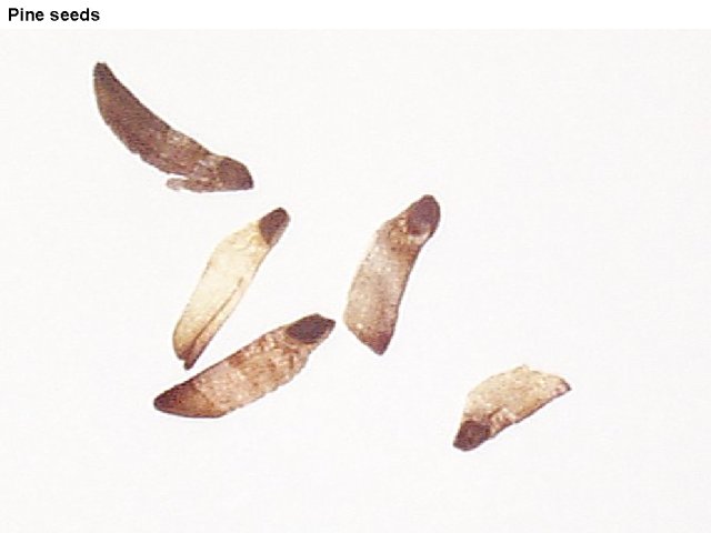
Seeds
Seeds contain the sporophyte embryo, food for the embryo, and a protective coat.
The embryo within the seed is dormant; it can survive for long periods without additional food or water. When conditions become favorable, the embryo resumes growth as the seed germinates.
- Draw the life cycle of pine and include the following terms: eggs, embryo, fertilization, megagametophyte, megasporangium, megaspore, meiosis, microgametophyte, microsporangium, microspores, and zygote.
- Observe the pine pollen cones on display. Is this structure haploid or diploid?
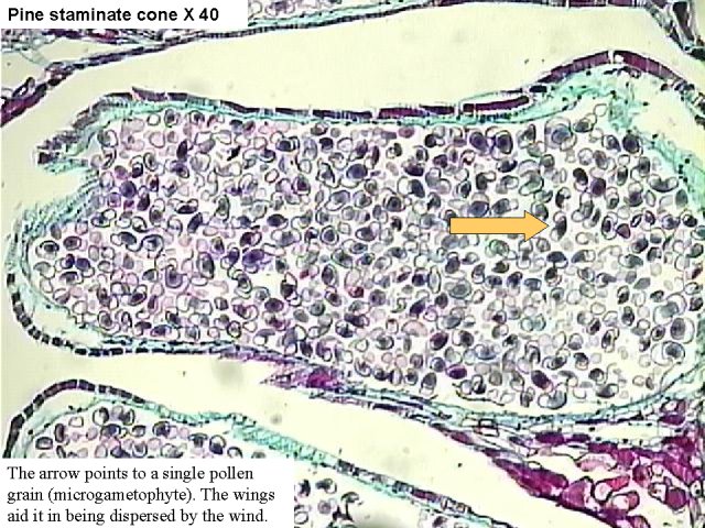
- View a slide showing a section (l.s.) of a pine pollen cone. Identify the microsporangium. Identify the microgametophytes. What is another name for microgametophyte?
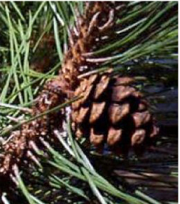
- View a pine seed cone on display. Are there any seeds within the cone?
- View a slide showing a longitudinal section of a pine seed cone. Identify the integument, ovule, megasporangium, and megagametophyte. Which of these structures is part of the sporophyte? Which are haploid? Which are diploid?
- View the pine seeds on display. From your drawing of the life cycle of pine, identify the structures that are part of the seed.
Angiosperms
Create another diagram of the life cycle of seed plants that includes the following terms: eggs, embryo, fertilization, megagametophyte, megasporangium, megaspore, meiosis, microgametophyte, microsporangium, microspores, and zygote. This diagram will be used as a reference when viewing the reproductive structures of angiosperms.
Flower Parts
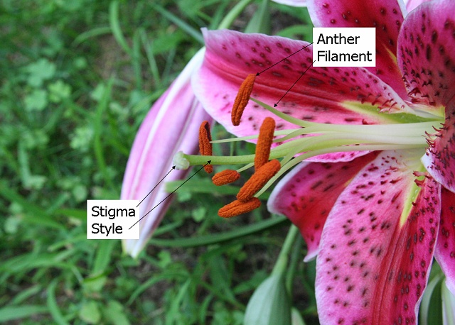
- Obtain a monocot flower such as lily and identify the following structures: anther, filament, stamen, stigma, style, ovary, pistil, petals, sepals. State the function of each of these structures.
- Remove the petals, stamens and pistil.
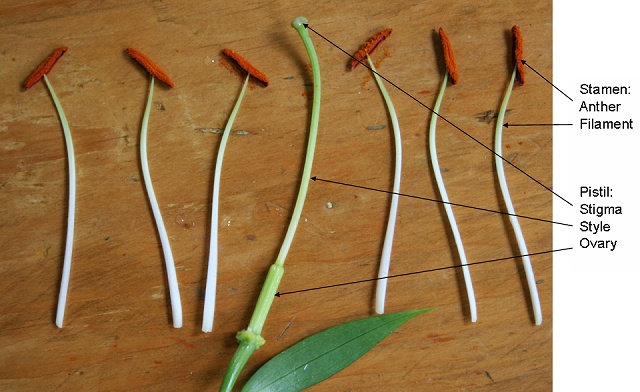
- How many petals are present? How many sepals? Is lily a monocot or a eudicot? List three characteristics that can be used to distinguish between monocots and eudicots.
Within the Ovary
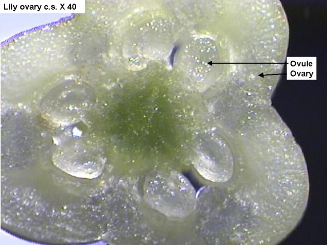
- Use a scalpel to cut a thin cross section slice from the ovary. This can be done by cutting across the ovary and then slicing a thin section next to the first cut. Use a dissecting microscope to determine the number of carpels within the ovary. Identify the ovules. Which structures on the life cycle diagram are found within the ovules?
- View a prepared slide of a lily mature female gametophyte. Identify the megagametophyte, Find the megagametophyte on the life cycle diagram. Try to find an egg and polar nuclei.
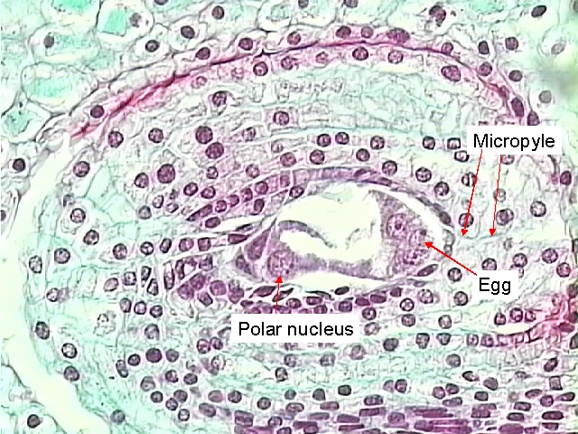
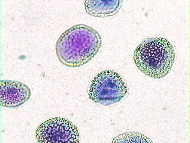
The photograph below shows a megaspore mother cell. It will divide by meiosis to produce megaspores.
Within the Anther
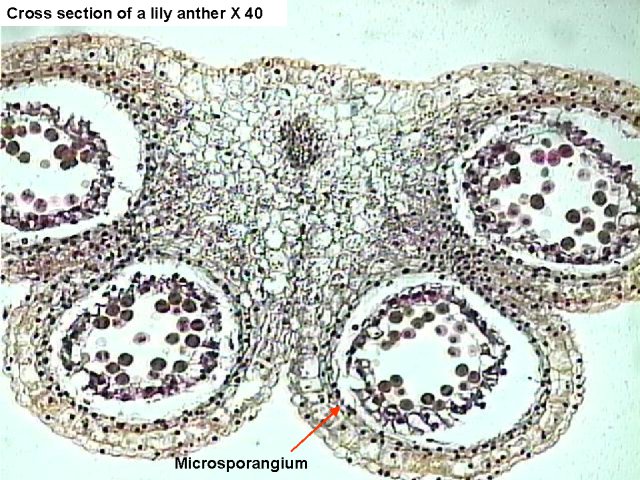
- Use a scalpel to cut a thin cross-section of a lily anther and view it under a dissecting microscope. Identify the microsporangium. Are pollen grains visible? What structures on the life cycle diagram are contained within the anther?
Meiosis occurs within the anther to produce microspores. Microspores undergo mitosis to produce microgametophytes (pollen grains). - If you were unable to get a good view of a lily anther in the dissection above, view a prepared slide of a lily anther c.s. and identify the microsporangium and pollen grains. Find where these two structures are located on your life cycle diagram.
- View a slide of lily pollen. identify the two nuclei.
- View slides of germinated pollen. Note the three nuclei within the pollen tubes. One is a tube nucleus. It directs the growth of the pollen tube. The other two are sperm.
After Fertilization: Embryonic Development
- View a slide of a Capsella early embryo. Identify the suspensor and cotyledons. Is Capsella a monocot or a eudicot?
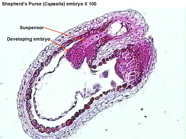
- View a slide of a Capsella mature embryo. Identify the cotyledons, the tip of the growing root (root apical meristem) and the tip of the growing shoot (shoot apical meristem).
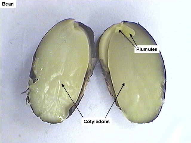
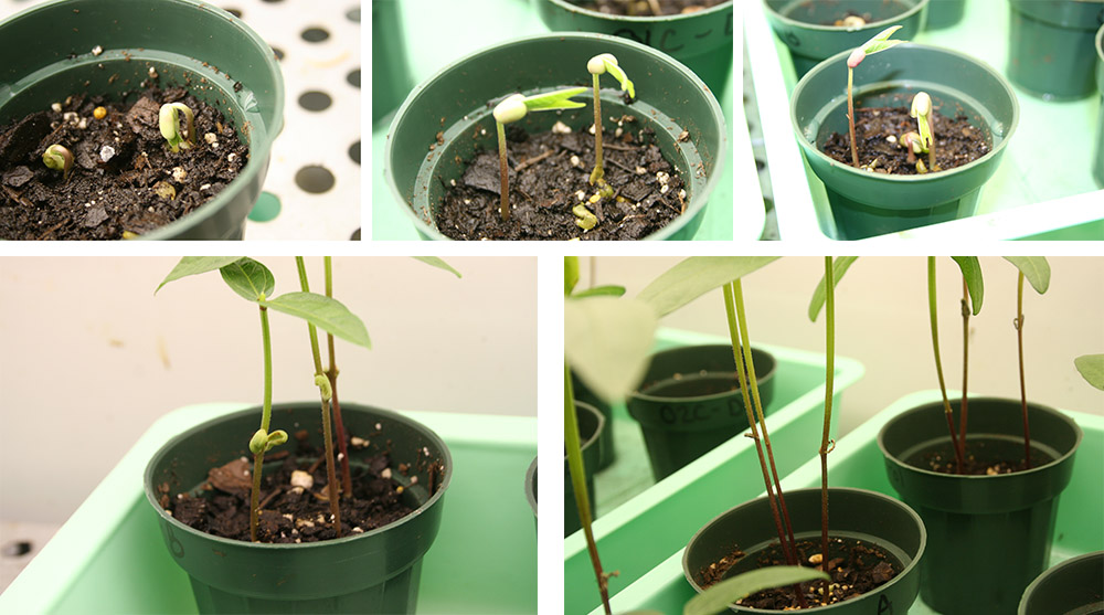
- Obtain a bean seed that has been soaking in water. Cut the seed in half so that each cotyledon is visible and examine it using a dissecting microscope. Identify the embryo.
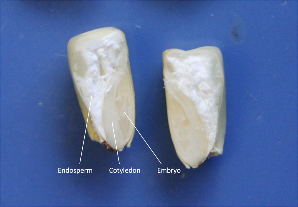
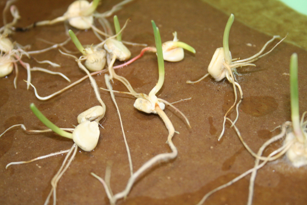
- Obtain a corn that has been soaking in water and cut it lengthwise. Use a dissecting microscope to identify the embryo, the cotyledon, and the endosperm.
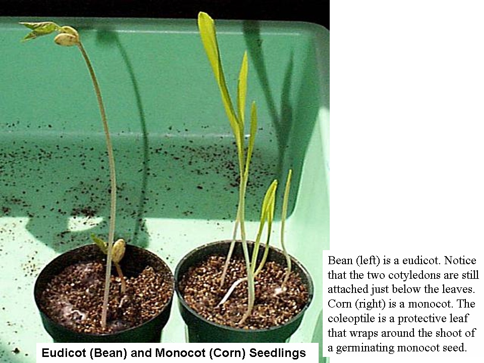
- Observe beans and corn on display that have been recently germinated. Identify the cotyledons on the beans. Can you see cotyledons on the corn? Can you identify the coleoptile?
Fruits
Angiosperms are distinguished from Gymnosperms in that the seeds are enclosed in a covering called the fruit.
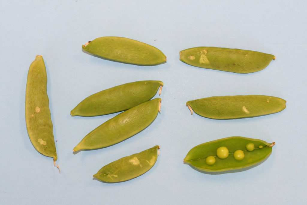
Observe peas. Peas are seeds contained within a pod (fruit).
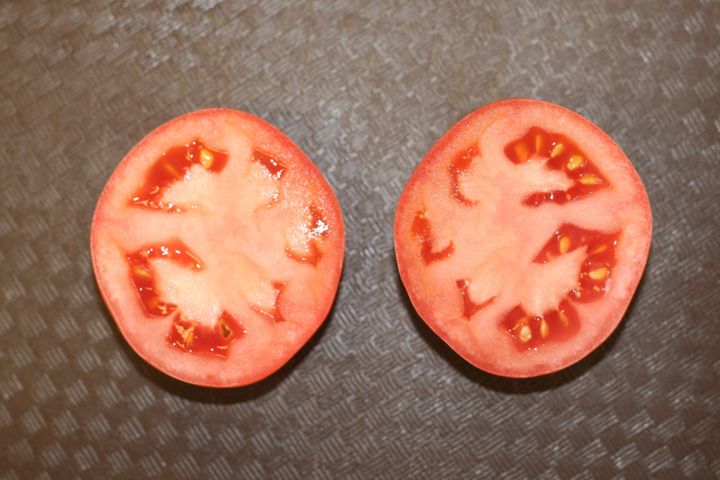
Observe the sliced tomato. It is produced from several fused carpels. Can you see the carpels? How many are there?
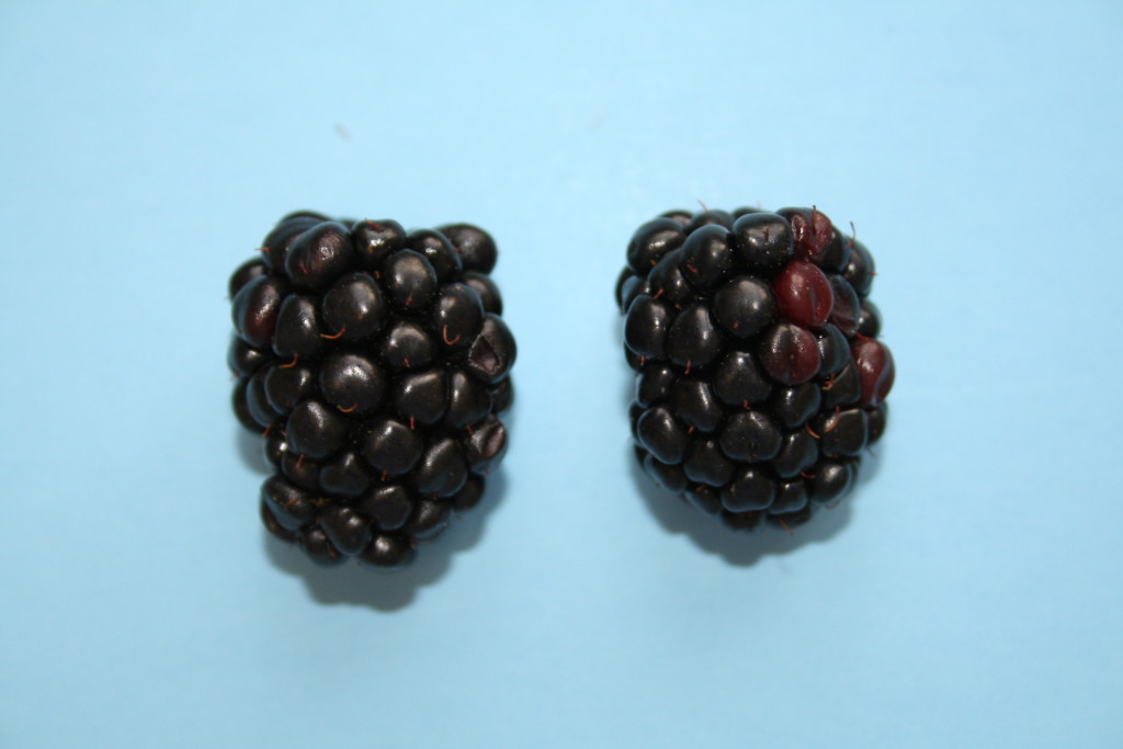
Observe a strawberry or a blackberry. These fruits are formed from a single flower that contained many pistals.
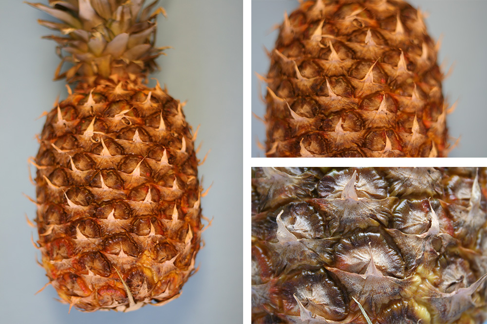
Observe a pineapple. This fruit is produced by the fusion of many flowers. Can you see each individual fruit?
Contributors and Attributions
- Seed Plants (Kingdom: Plantae), Biology 102. Authored by: Michael J. Gregory, Ph.D.. Provided by: LibreTexts. Located at: bio.libretexts.org/Under_Construction/BioStuff/BIO_102/Laboratory_Exercises/Seed_Plants. Project: The Biology Web. License: CC BY-NC-SA: Attribution-NonCommercial-ShareAlike