Biology LibreTexts

Chapter 1: Introduction to Microbiology

  • Page ID
    142369
  • \( \newcommand{\vecs}[1]{\overset { \scriptstyle \rightharpoonup} {\mathbf{#1}} } \)

    \( \newcommand{\vecd}[1]{\overset{-\!-\!\rightharpoonup}{\vphantom{a}\smash {#1}}} \)

    \( \newcommand{\id}{\mathrm{id}}\) \( \newcommand{\Span}{\mathrm{span}}\)

    ( \newcommand{\kernel}{\mathrm{null}\,}\) \( \newcommand{\range}{\mathrm{range}\,}\)

    \( \newcommand{\RealPart}{\mathrm{Re}}\) \( \newcommand{\ImaginaryPart}{\mathrm{Im}}\)

    \( \newcommand{\Argument}{\mathrm{Arg}}\) \( \newcommand{\norm}[1]{\| #1 \|}\)

    \( \newcommand{\inner}[2]{\langle #1, #2 \rangle}\)

    \( \newcommand{\Span}{\mathrm{span}}\)

    \( \newcommand{\id}{\mathrm{id}}\)

    \( \newcommand{\Span}{\mathrm{span}}\)

    \( \newcommand{\kernel}{\mathrm{null}\,}\)

    \( \newcommand{\range}{\mathrm{range}\,}\)

    \( \newcommand{\RealPart}{\mathrm{Re}}\)

    \( \newcommand{\ImaginaryPart}{\mathrm{Im}}\)

    \( \newcommand{\Argument}{\mathrm{Arg}}\)

    \( \newcommand{\norm}[1]{\| #1 \|}\)

    \( \newcommand{\inner}[2]{\langle #1, #2 \rangle}\)

    \( \newcommand{\Span}{\mathrm{span}}\) \( \newcommand{\AA}{\unicode[.8,0]{x212B}}\)

    \( \newcommand{\vectorA}[1]{\vec{#1}}      % arrow\)

    \( \newcommand{\vectorAt}[1]{\vec{\text{#1}}}      % arrow\)

    \( \newcommand{\vectorB}[1]{\overset { \scriptstyle \rightharpoonup} {\mathbf{#1}} } \)

    \( \newcommand{\vectorC}[1]{\textbf{#1}} \)

    \( \newcommand{\vectorD}[1]{\overrightarrow{#1}} \)

    \( \newcommand{\vectorDt}[1]{\overrightarrow{\text{#1}}} \)

    \( \newcommand{\vectE}[1]{\overset{-\!-\!\rightharpoonup}{\vphantom{a}\smash{\mathbf {#1}}}} \)

    \( \newcommand{\vecs}[1]{\overset { \scriptstyle \rightharpoonup} {\mathbf{#1}} } \)

    \( \newcommand{\vecd}[1]{\overset{-\!-\!\rightharpoonup}{\vphantom{a}\smash {#1}}} \)

    \(\newcommand{\avec}{\mathbf a}\) \(\newcommand{\bvec}{\mathbf b}\) \(\newcommand{\cvec}{\mathbf c}\) \(\newcommand{\dvec}{\mathbf d}\) \(\newcommand{\dtil}{\widetilde{\mathbf d}}\) \(\newcommand{\evec}{\mathbf e}\) \(\newcommand{\fvec}{\mathbf f}\) \(\newcommand{\nvec}{\mathbf n}\) \(\newcommand{\pvec}{\mathbf p}\) \(\newcommand{\qvec}{\mathbf q}\) \(\newcommand{\svec}{\mathbf s}\) \(\newcommand{\tvec}{\mathbf t}\) \(\newcommand{\uvec}{\mathbf u}\) \(\newcommand{\vvec}{\mathbf v}\) \(\newcommand{\wvec}{\mathbf w}\) \(\newcommand{\xvec}{\mathbf x}\) \(\newcommand{\yvec}{\mathbf y}\) \(\newcommand{\zvec}{\mathbf z}\) \(\newcommand{\rvec}{\mathbf r}\) \(\newcommand{\mvec}{\mathbf m}\) \(\newcommand{\zerovec}{\mathbf 0}\) \(\newcommand{\onevec}{\mathbf 1}\) \(\newcommand{\real}{\mathbb R}\) \(\newcommand{\twovec}[2]{\left[\begin{array}{r}#1 \\ #2 \end{array}\right]}\) \(\newcommand{\ctwovec}[2]{\left[\begin{array}{c}#1 \\ #2 \end{array}\right]}\) \(\newcommand{\threevec}[3]{\left[\begin{array}{r}#1 \\ #2 \\ #3 \end{array}\right]}\) \(\newcommand{\cthreevec}[3]{\left[\begin{array}{c}#1 \\ #2 \\ #3 \end{array}\right]}\) \(\newcommand{\fourvec}[4]{\left[\begin{array}{r}#1 \\ #2 \\ #3 \\ #4 \end{array}\right]}\) \(\newcommand{\cfourvec}[4]{\left[\begin{array}{c}#1 \\ #2 \\ #3 \\ #4 \end{array}\right]}\) \(\newcommand{\fivevec}[5]{\left[\begin{array}{r}#1 \\ #2 \\ #3 \\ #4 \\ #5 \\ \end{array}\right]}\) \(\newcommand{\cfivevec}[5]{\left[\begin{array}{c}#1 \\ #2 \\ #3 \\ #4 \\ #5 \\ \end{array}\right]}\) \(\newcommand{\mattwo}[4]{\left[\begin{array}{rr}#1 \amp #2 \\ #3 \amp #4 \\ \end{array}\right]}\) \(\newcommand{\laspan}[1]{\text{Span}\{#1\}}\) \(\newcommand{\bcal}{\cal B}\) \(\newcommand{\ccal}{\cal C}\) \(\newcommand{\scal}{\cal S}\) \(\newcommand{\wcal}{\cal W}\) \(\newcommand{\ecal}{\cal E}\) \(\newcommand{\coords}[2]{\left\{#1\right\}_{#2}}\) \(\newcommand{\gray}[1]{\color{gray}{#1}}\) \(\newcommand{\lgray}[1]{\color{lightgray}{#1}}\) \(\newcommand{\rank}{\operatorname{rank}}\) \(\newcommand{\row}{\text{Row}}\) \(\newcommand{\col}{\text{Col}}\) \(\renewcommand{\row}{\text{Row}}\) \(\newcommand{\nul}{\text{Nul}}\) \(\newcommand{\var}{\text{Var}}\) \(\newcommand{\corr}{\text{corr}}\) \(\newcommand{\len}[1]{\left|#1\right|}\) \(\newcommand{\bbar}{\overline{\bvec}}\) \(\newcommand{\bhat}{\widehat{\bvec}}\) \(\newcommand{\bperp}{\bvec^\perp}\) \(\newcommand{\xhat}{\widehat{\xvec}}\) \(\newcommand{\vhat}{\widehat{\vvec}}\) \(\newcommand{\uhat}{\widehat{\uvec}}\) \(\newcommand{\what}{\widehat{\wvec}}\) \(\newcommand{\Sighat}{\widehat{\Sigma}}\) \(\newcommand{\lt}{<}\) \(\newcommand{\gt}{>}\) \(\newcommand{\amp}{&}\) \(\definecolor{fillinmathshade}{gray}{0.9}\)

    From boiling thermal hot springs to deep beneath the Antarctic ice, microorganisms can be found almost everywhere on earth in great quantities. Microorganisms (or microbes, as they are also called) are small organisms. Most are so small that they cannot be seen without a microscope.

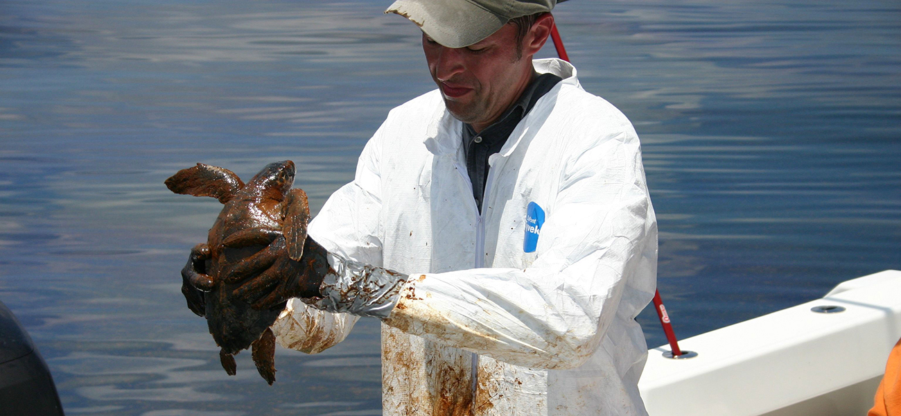
    Most microorganisms are harmless to humans and, in fact, many are helpful. They play fundamental roles in ecosystems everywhere on earth, forming the backbone of many food webs. People use them to make biofuels, medicines, and even foods. Without microbes, there would be no bread, cheese, or beer. Our bodies are filled with microbes, and our skin alone is home to trillions of them1. Some of them we can’t live without; others cause diseases that can make us sick or even kill us.

    OSC_Microbio_01_00_splash.jpg
    Figure \(\PageIndex{1}\): A veterinarian gets ready to clean a sea turtle covered in oil following the Deepwater Horizon oil spill in the Gulf of Mexico in 2010. After the spill, the population of a naturally occurring oil-eating marine bacterium called Alcanivorax borkumensis skyrocketed, helping to get rid of the oil. Scientists are working on ways to genetically engineer this bacterium to be more efficient in cleaning up future spills. (credit: modification of work by NOAA’s National Ocean Service)

    Although much more is known today about microbial life than ever before, the vast majority of this invisible world remains unexplored. Microbiologists continue to identify new ways that microbes benefit and threaten humans.

    • Section 1.1: Introduction to Microbiology
      Microorganisms are typically too small to be seen with the naked eye. Bacteria, fungi, viruses, protozoa, and algae are the major groups of microorganisms. The vast majority of microorganisms are not harmful but rather beneficial. Microbiota refers to all of the microorganisms that live in a particular environment. A microbiome is the entire collection of genes found in all of the microbes associated with a particular host.
    • Section 1.2: Introduction to Microbiology
      Microorganisms are typically too small to be seen with the naked eye. Bacteria, fungi, viruses, protozoa, and algae are the major groups of microorganisms. The vast majority of microorganisms are not harmful but rather beneficial. Microbiota refers to all of the microorganisms that live in a particular environment. A microbiome is the entire collection of genes found in all of the microbes associated with a particular host.
    • Section 1.3: Cellular Organization - Prokaryotic and Eukaryotic Cells
      here are two basic types of cells in nature: prokaryotic and eukaryotic. Prokaryotic cells are structurally simpler than eukaryotic cells. The smaller a cell, the greater its surface to volume ratio. The smaller the surface to volume ratio, the more structurally complex (compartmentalized) a cell needs to be in order to carry out life functions. There are fundamental differences between prokaryotic and eukaryotic cells.
    • Section 1.4: Classification - The Three Domain System
      Phylogeny refers to the evolutionary relationships between organisms. Organisms can be classified into one of three domains based on differences in the sequences of nucleotides in the cell's ribosomal RNAs (rRNA), the cell's membrane lipid structure, and its sensitivity to antibiotics. The three domains are the Archaea, the Bacteria, and the Eukarya. Prokaryotic organisms belong either to the domain Archaea or the domain Bacteria; organisms with eukaryotic cells belong to the domain Eukarya.
    • Section 1.5: What Our Ancestors Knew
      Microorganisms (or microbes) are living organisms that are generally too small to be seen without a microscope. Throughout history, humans have used microbes to make fermented foods such as beer, bread, cheese, and wine. Long before the invention of the microscope, some people theorized that infection and disease were spread by living things that were too small to be seen. They also correctly intuited certain principles regarding the spread of disease and immunity.
    • Section 1.6: A Systematic Approach
      Carolus Linnaeus developed a taxonomic system for categorizing organisms into related groups. Binomial nomenclature assigns organisms Latinized scientific names with a genus and species designation. A phylogenetic tree is a way of showing how different organisms are thought to be related to one another from an evolutionary standpoint. The first phylogenetic tree contained kingdoms for plants and animals; Ernst Haeckel proposed adding a kingdom for protists.
    • Section 1.7: Types of Microorganisms
      Microorganisms are very diverse and are found in all three domains of life: Archaea, Bacteria, and Eukarya. Archaea and bacteria are classified as prokaryotes because they lack a cellular nucleus. Archaea differ from bacteria in evolutionary history, genetics, metabolic pathways, and cell wall and membrane composition. Archaea inhabit nearly every environment on earth, but no archaea have been identified as human pathogens.
    • Section 1.8: Classification - The Three Domain System
      Phylogeny refers to the evolutionary relationships between organisms. Organisms can be classified into one of three domains based on differences in the sequences of nucleotides in the cell's ribosomal RNAs (rRNA), the cell's membrane lipid structure, and its sensitivity to antibiotics. The three domains are the Archaea, the Bacteria, and the Eukarya. Prokaryotic organisms belong either to the domain Archaea or the domain Bacteria; organisms with eukaryotic cells belong to the domain Eukarya.
    • Section 1.9: How We See the Invisible World
      Through a microscope, we can examine microbial cells and colonies, using various techniques to manipulate color, size, and contrast in ways that help us identify species and diagnose disease. This chapter explores how various types of microscopes manipulate light in order to provide a window into the world of microorganisms. By understanding how various kinds of microscopes work, we can produce highly detailed images of microbes that can be useful for both research and clinical applications.
    • Section 1.E: An Invisible World (Exercises)
    • Section 1.E: Fundamentals of Microbiology (Exercises)
      These are homework exercises to accompany Kaiser's "Microbiology" TextMap. Microbiology is the study of microorganisms, which are defined as any microscopic organism that comprises either a single cell (unicellular), cell clusters or no cell at all (acellular). This includes eukaryotes, such as fungi and protists, and prokaryotes. Viruses and prions, though not strictly classed as living organisms, are also studied.

    Footnotes

    1. 1 J. Hulcr et al. “A Jungle in There: Bacteria in Belly Buttons are Highly Diverse, but Predictable.” PLoS ONE 7 no. 11 (2012): e47712. doi:10.1371/journal.pone.0047712.

    Thumbnail: A cluster of Escherichia coli bacteria magnified 10,000 times. (Public Domain; Eric Erbe, digital colorization by Christopher Pooley, both of USDA, ARS, EMU).


    This page titled Chapter 1: Introduction to Microbiology is shared under a CC BY 4.0 license and was authored, remixed, and/or curated by Ying Liu via source content that was edited to the style and standards of the LibreTexts platform.

    • Was this article helpful?