
12.4: The Lymphoid System
- Page ID
- 3301

\( \newcommand{\vecs}[1]{\overset { \scriptstyle \rightharpoonup} {\mathbf{#1}} } \)
\( \newcommand{\vecd}[1]{\overset{-\!-\!\rightharpoonup}{\vphantom{a}\smash {#1}}} \)
\( \newcommand{\id}{\mathrm{id}}\) \( \newcommand{\Span}{\mathrm{span}}\)
( \newcommand{\kernel}{\mathrm{null}\,}\) \( \newcommand{\range}{\mathrm{range}\,}\)
\( \newcommand{\RealPart}{\mathrm{Re}}\) \( \newcommand{\ImaginaryPart}{\mathrm{Im}}\)
\( \newcommand{\Argument}{\mathrm{Arg}}\) \( \newcommand{\norm}[1]{\| #1 \|}\)
\( \newcommand{\inner}[2]{\langle #1, #2 \rangle}\)
\( \newcommand{\Span}{\mathrm{span}}\)
\( \newcommand{\id}{\mathrm{id}}\)
\( \newcommand{\Span}{\mathrm{span}}\)
\( \newcommand{\kernel}{\mathrm{null}\,}\)
\( \newcommand{\range}{\mathrm{range}\,}\)
\( \newcommand{\RealPart}{\mathrm{Re}}\)
\( \newcommand{\ImaginaryPart}{\mathrm{Im}}\)
\( \newcommand{\Argument}{\mathrm{Arg}}\)
\( \newcommand{\norm}[1]{\| #1 \|}\)
\( \newcommand{\inner}[2]{\langle #1, #2 \rangle}\)
\( \newcommand{\Span}{\mathrm{span}}\) \( \newcommand{\AA}{\unicode[.8,0]{x212B}}\)
\( \newcommand{\vectorA}[1]{\vec{#1}} % arrow\)
\( \newcommand{\vectorAt}[1]{\vec{\text{#1}}} % arrow\)
\( \newcommand{\vectorB}[1]{\overset { \scriptstyle \rightharpoonup} {\mathbf{#1}} } \)
\( \newcommand{\vectorC}[1]{\textbf{#1}} \)
\( \newcommand{\vectorD}[1]{\overrightarrow{#1}} \)
\( \newcommand{\vectorDt}[1]{\overrightarrow{\text{#1}}} \)
\( \newcommand{\vectE}[1]{\overset{-\!-\!\rightharpoonup}{\vphantom{a}\smash{\mathbf {#1}}}} \)
\( \newcommand{\vecs}[1]{\overset { \scriptstyle \rightharpoonup} {\mathbf{#1}} } \)
\( \newcommand{\vecd}[1]{\overset{-\!-\!\rightharpoonup}{\vphantom{a}\smash {#1}}} \)
\(\newcommand{\avec}{\mathbf a}\) \(\newcommand{\bvec}{\mathbf b}\) \(\newcommand{\cvec}{\mathbf c}\) \(\newcommand{\dvec}{\mathbf d}\) \(\newcommand{\dtil}{\widetilde{\mathbf d}}\) \(\newcommand{\evec}{\mathbf e}\) \(\newcommand{\fvec}{\mathbf f}\) \(\newcommand{\nvec}{\mathbf n}\) \(\newcommand{\pvec}{\mathbf p}\) \(\newcommand{\qvec}{\mathbf q}\) \(\newcommand{\svec}{\mathbf s}\) \(\newcommand{\tvec}{\mathbf t}\) \(\newcommand{\uvec}{\mathbf u}\) \(\newcommand{\vvec}{\mathbf v}\) \(\newcommand{\wvec}{\mathbf w}\) \(\newcommand{\xvec}{\mathbf x}\) \(\newcommand{\yvec}{\mathbf y}\) \(\newcommand{\zvec}{\mathbf z}\) \(\newcommand{\rvec}{\mathbf r}\) \(\newcommand{\mvec}{\mathbf m}\) \(\newcommand{\zerovec}{\mathbf 0}\) \(\newcommand{\onevec}{\mathbf 1}\) \(\newcommand{\real}{\mathbb R}\) \(\newcommand{\twovec}[2]{\left[\begin{array}{r}#1 \\ #2 \end{array}\right]}\) \(\newcommand{\ctwovec}[2]{\left[\begin{array}{c}#1 \\ #2 \end{array}\right]}\) \(\newcommand{\threevec}[3]{\left[\begin{array}{r}#1 \\ #2 \\ #3 \end{array}\right]}\) \(\newcommand{\cthreevec}[3]{\left[\begin{array}{c}#1 \\ #2 \\ #3 \end{array}\right]}\) \(\newcommand{\fourvec}[4]{\left[\begin{array}{r}#1 \\ #2 \\ #3 \\ #4 \end{array}\right]}\) \(\newcommand{\cfourvec}[4]{\left[\begin{array}{c}#1 \\ #2 \\ #3 \\ #4 \end{array}\right]}\) \(\newcommand{\fivevec}[5]{\left[\begin{array}{r}#1 \\ #2 \\ #3 \\ #4 \\ #5 \\ \end{array}\right]}\) \(\newcommand{\cfivevec}[5]{\left[\begin{array}{c}#1 \\ #2 \\ #3 \\ #4 \\ #5 \\ \end{array}\right]}\) \(\newcommand{\mattwo}[4]{\left[\begin{array}{rr}#1 \amp #2 \\ #3 \amp #4 \\ \end{array}\right]}\) \(\newcommand{\laspan}[1]{\text{Span}\{#1\}}\) \(\newcommand{\bcal}{\cal B}\) \(\newcommand{\ccal}{\cal C}\) \(\newcommand{\scal}{\cal S}\) \(\newcommand{\wcal}{\cal W}\) \(\newcommand{\ecal}{\cal E}\) \(\newcommand{\coords}[2]{\left\{#1\right\}_{#2}}\) \(\newcommand{\gray}[1]{\color{gray}{#1}}\) \(\newcommand{\lgray}[1]{\color{lightgray}{#1}}\) \(\newcommand{\rank}{\operatorname{rank}}\) \(\newcommand{\row}{\text{Row}}\) \(\newcommand{\col}{\text{Col}}\) \(\renewcommand{\row}{\text{Row}}\) \(\newcommand{\nul}{\text{Nul}}\) \(\newcommand{\var}{\text{Var}}\) \(\newcommand{\corr}{\text{corr}}\) \(\newcommand{\len}[1]{\left|#1\right|}\) \(\newcommand{\bbar}{\overline{\bvec}}\) \(\newcommand{\bhat}{\widehat{\bvec}}\) \(\newcommand{\bperp}{\bvec^\perp}\) \(\newcommand{\xhat}{\widehat{\xvec}}\) \(\newcommand{\vhat}{\widehat{\vvec}}\) \(\newcommand{\uhat}{\widehat{\uvec}}\) \(\newcommand{\what}{\widehat{\wvec}}\) \(\newcommand{\Sighat}{\widehat{\Sigma}}\) \(\newcommand{\lt}{<}\) \(\newcommand{\gt}{>}\) \(\newcommand{\amp}{&}\) \(\definecolor{fillinmathshade}{gray}{0.9}\)- Compare and give examples of the following:
- primary lymphoid organs
- secondary lymphoid organs
- Define the following:
- plasma
- tissue fluid
- lymph
- lymph vessels
- MALT
- Briefly describe the importance of the lymphoid system in adaptive immune responses and how microbes and other antigens encounter naive B-lymphocytes and T-lymphocytes.
The body uses the lymphoid system to enable lymphocytes to encounter antigens and it is here that adaptive immune responses are initiated. The lymphoid system consists of primary lymphoid organs, secondary lymphoid organs, and lymphatic vessels.
The bone marrow and the thymus constitute the primary lymphoid organs. Both B-lymphocytes and T-lymphocytes are produced from stem cells in the bone marrow. B-lymphocytes mature in the bone marrow while T-lymphocytes migrate to the thymus and mature there. After maturation, both naive B-lymphocytes and naive T-lymphocytes circulate between the blood and the secondary lymphoid organs.
Lymphatic vessels are responsible for flow of lymph within the lymphoid system and are a part of the body's fluid recirculation system. The liquid portion of the blood, called plasma, constantly leaks out of capillaries to deliver oxygen and nutrients to cells of the surrounding tissue. Once in the tissue, the plasma is now called tissue fluid. While most of this tissue fluid re-enters capillaries and is returned directly to the bloodstream, some fluid enters lymph vessels as lymph. The lymph flows through regional lymph nodes and eventually enters the circulatory system at the heart to maintain the fluid volume of the circulation.
Secondary lymphoid organs
Adaptive immune responses require antigen-presenting cells, such as macrophages and dendritic cells, and ever changing populations of B-lymphocytes and T- lymphocytes. These cells gather to detect and present antigens in secondary lymphoid organs. The secondary lymphoid organs include highly organized lymphoid organs such as lymph nodes and the spleen, as well as less organized accumulations of lymphoid organs scattered strategically throughout the body.
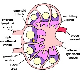
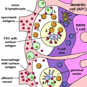
Lymph nodes (Figure \(\PageIndex{1}\)) contain many reticular fibers that support fixed macrophages and dendritic cells as well as ever changing populations of circulating B-lymphocytes and T-lymphocytes. When microorganisms and other antigens enter tissues, they are transported by tissue fluid into the lymph vessels. Lymph vessels, in turn, carry these antigens, now in the lymph, to regional lymph nodes. In addition, immature dendritic cells located under the surface epithelium of the skin and the surface epithelium of the mucous membranes of the respiratory tract, genitourinary tract, and the gastrointestinal tract capture antigens through pinocytosis and phagocytosis. The dendritic cells detach from their initial site, enter lymph vessels, and are carried to regional lymph nodes. Here the microbes and other antigens in the lymph encounter changing populations of B-lymphocytes, are filtered out and phagocytosed by the fixed macrophages and dendritic cells, and are presented to changing populations of T-lymphocytes (Figure \(\PageIndex{2}\)). Approximately 25 billion different lymphocytes migrate through each lymph node every day.
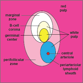
Like the lymph nodes, the spleen contains many reticular fibers that support fixed macrophages and dendritic cells as well as ever changing populations of circulating B-lymphocytes and T-lymphocytes. When microorganisms and other antigens enter the blood, they are transported by the blood vessels to the spleen. Most of the spleen is referred to as red pulp. This area is involved in the disposal of old red blood cells. Scattered throughout the spleen are isolated areas called the white pulp (Figure \(\PageIndex{3}\)). Here antigens in the blood encounter macrophages, dendritic cells, and ever-changing populations of B-lymphocytes and T-lymphocytes.
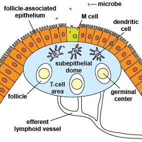
Mucosal surfaces within the body, the most common sites of microbial invasion, are protected by the mucosal immune system consisting of the mucosa-associated lymphoid tissue or MALT, an extensive diffuse system of small concentrations of lymphoid tissue found in various sites of the body such as the gastrointestinal tract, thyroid, breast, lung, salivary glands, eye, and skin. MALT is populated by loose clusters of T-lymphocytes, B-lymphocytes, plasma cells, activated TH cells, and macrophages. MALT can be subdivided into:
- GALT (gut-associated lymphoid tissue, such as the Peyer's patches (Figure \(\PageIndex{4}\)) in the lining of the small intestines, as well as the adenoids, tonsils, and appendix)
- BALT (bronchial-associated lymphoid tissue in the bronchi)
- SALT (skin-associated lymphoid tissue beneath the epidermis)
- NALT (nose-associated lymphoid tissue)
- LALT (larynx-associated lymphoid tissue)
- CALT (conjunctiva-associated lymphoid tissue in the eye)
As can be seen, no matter how microbes and other antigens enter the body, they will eventually encounter the lymphoid system to initiate adaptive immune responses.
Summary
- The body uses the lymphoid system to enable lymphocytes to encounter antigens and it is here that adaptive immune responses are initiated.
- The lymphoid system consists of primary lymphoid organs, secondary lymphoid organs, and lymphatic vessels.
- The bone marrow and the thymus constitute the primary lymphoid organs.
- While both B-lymphocytes and T-lymphocytes are produced from stem cells in the bone marrow, B-lymphocytes mature in the bone marrow and T-lymphocytes migrate to the thymus to mature.
- After maturation, both naive B-lymphocytes and naive T-lymphocytes circulate between the blood and the secondary lymphoid organs.
- Adaptive immune responses require antigen-presenting cells, such as macrophages and dendritic cells, and ever changing populations of B-lymphocytes and T- lymphocytes. These cells gather to detect and present antigens in secondary lymphoid organs.
- The secondary lymphoid organs include highly organized lymphoid organs such as lymph nodes and the spleen, as well as less organized accumulations of lymphoid organs scattered strategically throughout the body.
- Lymphatic vessels are responsible for flow of lymph within the lymphoid system and are a part of the body's fluid recirculation system. The lymph flows through regional lymph nodes and eventually enters the circulatory system at the heart to maintain the fluid volume of the circulation.