9.2: Medically Important Protozoa
- Page ID
- 3230
State a disease caused by each of the following protozoans and indicate their means of motility and how they are transmitted to humans: - Entamoeba histolytica
- Acanthamoeba
- Giardia lamblia
- Trichomonas vaginalis
- Trypanosoma brucei-gambiens
- Balantidium coli
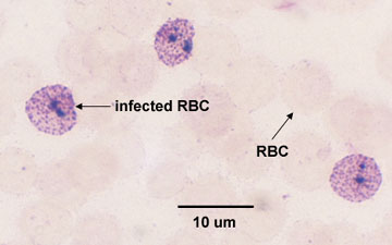
- Plasmodium species
- Toxoplasma gondii
- Cryptosporidium
The Sarcomastigophora (Amoeboflagellates)
The amoebas (subphylum Sarcodina) move by extending lobelike projections of their cytoplasm called pseudopodia .
- Photomicrograph of an amoeba.
Video YouTube movie an amoeba moving by forming pseudopodia (https://www.youtube.com/embed/7pR7TNzJ_pA).
a. Entamoeba histolytica (see photomicrograph) which causes a gastrointestinal infection called amoebic dysentery. The organism produces protective cysts which pass out of the intestines of the infected host and are ingested by the next host. It is transmitted by the fecal-oral route.
b. Acanthamoeba can cause rare, but severe infections of the eye, skin, and central nervous system. Acanthamoeba keratitis is an infection of the eye that typically occurs in healthy persons and can result in blindness or permanent visual impairment. Granulomatous amebic encephalitis (GAE) is an infection of the brain and spinal cord typically occurring in persons with a compromised immune system. Acanthamoeba is found in soil, dust, and a variety of water sources including lakes, tap water, swimming pools, and heating and air conditioning units. It typically enters the eyes and most cases are associated with contact lens use, but it can also enter cuts or wounds and be inhaled.
c. Naegleria fowleri (sometimes called the"brain-eating amoeba"), is another amoeba that can cause a rare but devastating infection of the brain called primary amebic meningoencephalitis (PAM). The amoeba is commonly found in warm freshwater rivers, lakes, rivers, and hot springs, as well as in the soil. It typically causes infections when contaminated water enters the body through the nose where it can subsequently travel to the brain.
| YouTube movie of Acanthamoeba |
The flagellates (subphylum Mastigophora) move by means of flagella. Some also have an undulating membrane .
a. Giardia lamblia (see photomicrograph) can cause a gastrointestinal infection called giardiasis. Cysts pass out of the intestines of the infected host and are ingested by the next host. It is transmitted by the fecal-oral route.
| YouTube animation illustrating giardiasis |
- Scanning electron micrograph of Giardia in the intestines; courtesy of Dennis Kunkel's Microscopy.
- Scanning electron micrograph of Giardia;courtesy of CDC.
b. Trichomonas vaginalis (see photomicrograph) infects the vagina and the male urinary tract causing an infection called genitourinary trichomoniasis. It does not produce a cysts stage and is usually transmitted by sexual contact.
| YouTube movie Trichomonas vaginalis. |
| YouTube movie showing motility of Trichomonas vaginalis. |
c. Trypanosoma brucei gambiens (see photomicrograph) causes African sleeping sickness and is transmitted by the bite of an infected Tsetse fly. The disease primarily involves the lymphatic and nervous systems of humans.
| YouTube movie of Trypanosoma |
The Ciliophora
The ciliates move by means of cilia.
- Scanning electron micrograph of Paramecium, a ciliated protozoan; courtesy of Dennis Kunkel's Microscopy.
YouTube movie showing motility of Balantidium coli.
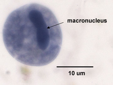
a. The only pathogenic ciliate is Balantidium coli (see photomicrograph) which causes a diarrhea-type infection called balantidiasis. Cysts pass out of the intestines of the infected host and are ingested by the next host. It is transmitted by the fecal-oral route.
Balantidium coli in a Fecal Smear
The Apicomplexans

Toxoplasma gondii is another intracellular apicomplexan and causes toxoplasmosis (see the AIDS pathology tutorial at the University of Utah). It can infect most mammals and is contracted by inhaling or ingesting cysts from the feces of infected domestic cats, where the protozoa reproduce both asexually and sexually, or by ingesting raw meat of an infected animal. Toxoplasmosis is usually mild in people with normal immune responses but can infect the brain, heart, or lungs of people who are immunosuppressed. It can also be transmitted congenitally and infect the nervous system of the infected child.
Cryptosporidium is an intracellular parasite that causes a gastrointestinal infection called cryptosporidiosis, although in people who are immunosuppressed it can also cause respiratory and gallbladder infections. It is transmitted by the fecal-oral route.
Virulence Factors that Promote Colonization of Protozoans
Virulence factors that promote protozoal colonization of the host include the ability to:
1. contact host cells;
2. adhere to host cells and resist physical removal;
3. invade host cells;
4. compete for nutrients;
5. resist innate immune defenses such as phagocytosis and complement; and
6. evade adaptive immune defenses.
Examples of virulence factors that promote protozoal colonization include:
- Some protozoa, such as Entamoeba histolytica,Trichomonas vaginalis, Giardia lamblia, and Balantidium coli use pseudopodia, flagella or cilia to swim through mucus and contact host cells.
- Protozoa use adhesins associated with their cytoplasmic membrane to adhere to host cells, colonize, and resist flushing.
- Some protozoa, such as the apicomplexans (Plasmodium (inf), Toxoplasma gondii (inf), and Cryptosporidium (inf)) possess a complex of organelles called apical complexes at their apex that contain enzymes used in penetrating host tissues and cells.
- Protozoans such as Trypanosoma brucei gambiens (inf) and Plasmodium species (inf) are able to change their surface antigens during their life cycle in the human. As the protozoa change the amino acid sequence and shape of their surface antigens, antibodies and cytotoxic T-lymphocytes made against a previous shape will no longer fit and the body has to start a new round of adaptive immunity against the new antigen shape.
- Some protozoa, such as Entamoeba histolytica (inf) shed their surface antigens so that antibodies made by the body against these surface antigens are tied up by the shed antigens.
To view a Quicktime movie of Cryptosporidium and electron micrographs of Giardia and Entamoeba, see the Parasites section of the CELL'S ALIVE web page.


