8.3: Molds
- Page ID
- 3225
Define: - mold
- hyphae
- mycelium
- vegetative mycelium
- aerial mycelium.
Briefly describe the following fungal asexual reproductive spores: - conidiospores
- macroconidia,
- microconidia
- sporangiospores
- arthrospores
Define dermatophyte, list 2 genera of dermatophytes, and name three dermatophytic infections. Describe what is meant by the term "dimorphic fungus", name two systemic infections caused by dimorphic fungi, and state how they are initially contracted.
- conidiospores
- macroconidia,
- microconidia
- sporangiospores
- arthrospores
Mold Morphology
Molds are multinucleated, filamentous fungi composed of hyphae. A hypha is a branching tubular structure approximately 2-10 µm in diameter which is usually divided into cell-like units by crosswalls called septa. The total mass of hyphae is termed a mycelium. The portion of the mycelium that anchors the mold and absorbs nutrients is called the vegetative mycelium , composed of vegetative hyphae; the portion that produces asexual reproductive spores is the aerial mycelium , composed of aerial hyphae (Figure \(\PageIndex{1}\)).
Molds have typical eukaryotic structures (Figure \(\PageIndex{2}\)) and have a cell wall usually composed of chitin, sometimes cellulose, and occasionally both. Furthermore, molds are obligate aerobes and grow by elongation at apical tips of their hyphae and thus are able to penetrate the surfaces on which they begin growing.
| For More Information: A Comparison of Prokaryotic and Eukaryotic Cells from Unit 1 |
Reproduction of Molds
1. Molds reproduce primarily by means of asexual reproductive spores (Figure \(\PageIndex{1}\)). These include the following.
a. conidiospores (conidia) See Figure \(\PageIndex{3}\).
Spores borne externally on an aerial hypha called a conidiophore ; see Figure \(\PageIndex{4}\) and Figure \(\PageIndex{5}\).
- Scanning electron micrographs of the conidiospores of Penicillium and of Aspergillus; courtesy of Dennis Kunkel's Microscopy.
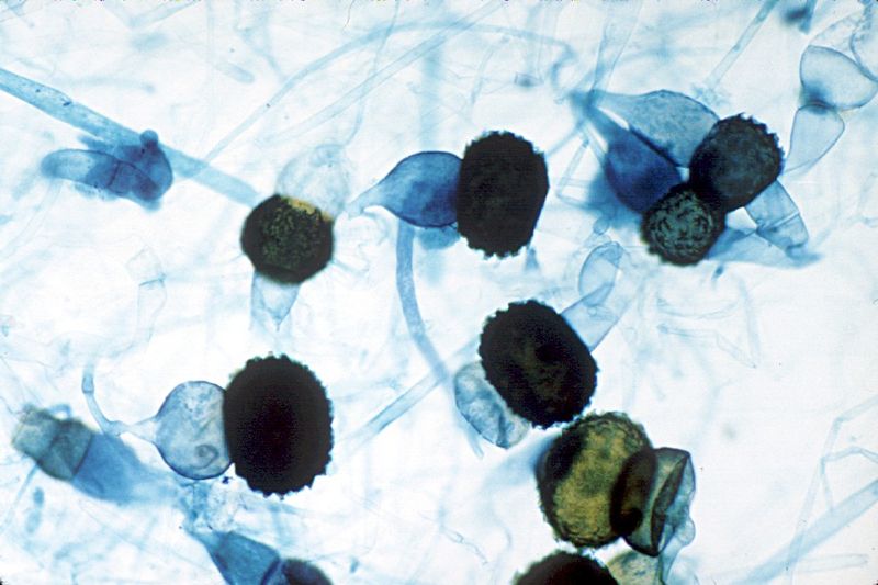
b. sporangiospores See Figure \(\PageIndex{6}\).
Spores borne in a sac or sporangium on an aerial hypha called a sporangiophore ; see Figure \(\PageIndex{7}\).
- Scanning electron micrograph of the conidiospores of Rhizopus; courtesy of Dennis Kunkel's Microscopy.
c. arthrospores See Figure \(\PageIndex{8}\).
spores produced by fragmentation of a vegetative hypha (Figure \(\PageIndex{9}\)).

Pathogenic Molds
Dermatophytes
The dermatophytes are a group of molds that cause superficial mycoses of the hair, skin, and nails and utilize the protein keratin, that is found in hair, skin, and nails, as a nitrogen and energy source. Infections are commonly referred to as ringworm or tinea infections and include:
- tinea capitis (infection of the skin of the scalp, eyebrows, and eyelashes)
- tinea barbae (infection of the bearded areas of the face and neck)
- tinea faciei (infection of the skin of the face)
- tinea corporis (infection of the skin regions other than the scalp, groin, palms, and soles)
- tinea cruris (infection of the groin; jock itch)
- tinea unguium (onchomycosis; infection of the fingernails and toenails)
- tinea pedis (athlete's foot; infection of the soles of the feet and between the toes).
The three most common dermatophytes are Microsporum, Trichophyton, and Epidermophyton. They produce characteristic asexual reproductive spores called macroconidia and microconidia (Figure \(\PageIndex{10}\) and Figure \(\PageIndex{11}\)).
- Scanning electron micrograph of the macroconidia of Epidermophyton; courtesy of Dennis Kunkel's Microscopy.
Another tinea infection of the skin is tinea versicolor caused by the yeast Malassezia globosa. Tinea versicolor appears as a hypopigmentation of the infected skin. M. globosa is also the most common cause of dandruff.
Dimorphic Fungi
Dimorphic fungi may exhibit two different growth forms. Outside the body they grow as a mold, producing hyphae and asexual reproductive spores, but in the body they grow in a non-mycelial yeast form. These infections appear as systemic mycoses and usually begin by inhaling spores from the mold form. After germination in the lungs, the fungus grows as a yeast. Factors such as body temperature, osmotic stress, oxidative stress, and certain human hormones activate a dimorphism-regulating histidine kinase enzyme in dimorphic molds, causing them to switch from their avirulent mold form to their more virulent yeast form.
For example:
a. Coccidioides immitis causes coccidioidomycosis (Figure \(\PageIndex{12}\)), a disease endemic to the southwestern United States. An estimated 100,000 infections occur annually in the United States, but one to two thirds of these cases are subclinical.
The mold form of the fungus grows in arid soil and produces thick-walled, barrel-shaped asexual spores called arthrospores (Figure \(\PageIndex{8}\)) by a fragmentation of its vegetative hyphae.
After inhalation, the arthrospores germinate and develop into endosporulating spherules (Figure \(\PageIndex{13}\)) in the terminal bronchioles of the lungs. The spherules reproduce by a process called endosporulation, where the spherule produces numerous endospores (yeast-like particles), ruptures, and releases viable endospores that develop into new spherules.
b. Histoplasma capsulatum (Figure \(\PageIndex{14}\))is a dimorphic fungus that causes histoplasmosis, a disease commonly found in the Great Lakes region and the Mississippi and Ohio River valleys. Approximately 250,000 people are thought to be infected annually in the US, but clinical symptoms of histoplasmosis occur in less than 5% of the population. Most individuals with histoplasmosis are asymptomatic. Those who develop clinical symptoms are typically either immunocompromised or are exposed to a large quantity of fungal spores.
The mold form of the fungus often grows in bird or bat droppings or soil contaminated with these droppings and produces large tuberculate macroconidia and small microconidia (Figure \(\PageIndex{15}\)). Although birds cannot be infected by the fungus and do not transmit the disease, bird excretions contaminate the soil and enrich it for mycelial growth. Bats, however, can become infected and transmit histoplasmosis through their droppings. After inhalation of the fungal spores and their germination in the lungs, the fungus grows as a budding, encapsulated yeast (Figure \(\PageIndex{16}\)).
- Chest X-ray of a person with histoplasmosis.
c. Blastomycosis, caused by Blastomyces dermatitidis, is common around the Great Lakes region and the Mississippi and Ohio River valleys.Infection can range from an asymptomatic, self-healing pulmonary infection to widely disseminated and potentially fatal disease. Pulmonary infection may be asymptomatic in nearly 50% of patients. Blastomyces dermatitidis can also sometimes infect the skin.
Blastomyces dermatitidis produces a mycelium with small conidiospores (Figure \(\PageIndex{17}\)) and grows actively in bird droppings and contaminated soil. When spores are inhaled or enter breaks in the skin, they germinate and the fungus grows as a yeast (Figure \(\PageIndex{18}\)).having a characteristic thick cell wall. It is diagnosed by culture and by biopsy examination.
These infections usually remains localized in the lungs, but in rare cases may spread throughout the body.
As mentioned earlier, the yeast Candida albicans can also exhibit dimorphism.
- To view additional photomicrographs of Coccidioides and Histoplasma, see the AIDS Pathology Tutorial at the University of Utah.
Opportunistic Molds
Certain molds once considered as non-pathogenic have recently become a fairly common cause of opportunistic lung and wound infections in the debilitated or immunosuppressed host. These include the common molds Aspergillus (Figure \(\PageIndex{4}\)) and Rhizopus (Figure \(\PageIndex{6}\)). Although generally harmless in most healthy individuals, Aspergillus species do cause allergic bronchopulmonary aspergillosis (ABPA), chronic necrotizing Aspergillus pneumonia (or chronic necrotizing pulmonary aspergillosis [CNPA]), aspergilloma (a mycetoma or fungus ball in a body cavity such as the lung), and invasive aspergillosis. In highly immunosuppressed individuals, however, Aspergillus may disseminate beyond the lung via the blood.
Mucormycoses are infections caused by fungi belonging to the order of Mucorales. Rhizopus species are the most common causative organisms. The most common infection is a severe infection of the facial sinuses, which may extend into the brain. Other mycoses include pulmonary, cutaneous, and gastrointestinal.


