Biology LibreTexts

5.9: The Endoplasmic Reticulum

  • Page ID
    68567
  • \( \newcommand{\vecs}[1]{\overset { \scriptstyle \rightharpoonup} {\mathbf{#1}} } \) \( \newcommand{\vecd}[1]{\overset{-\!-\!\rightharpoonup}{\vphantom{a}\smash {#1}}} \)\(\newcommand{\id}{\mathrm{id}}\) \( \newcommand{\Span}{\mathrm{span}}\) \( \newcommand{\kernel}{\mathrm{null}\,}\) \( \newcommand{\range}{\mathrm{range}\,}\) \( \newcommand{\RealPart}{\mathrm{Re}}\) \( \newcommand{\ImaginaryPart}{\mathrm{Im}}\) \( \newcommand{\Argument}{\mathrm{Arg}}\) \( \newcommand{\norm}[1]{\| #1 \|}\) \( \newcommand{\inner}[2]{\langle #1, #2 \rangle}\) \( \newcommand{\Span}{\mathrm{span}}\) \(\newcommand{\id}{\mathrm{id}}\) \( \newcommand{\Span}{\mathrm{span}}\) \( \newcommand{\kernel}{\mathrm{null}\,}\) \( \newcommand{\range}{\mathrm{range}\,}\) \( \newcommand{\RealPart}{\mathrm{Re}}\) \( \newcommand{\ImaginaryPart}{\mathrm{Im}}\) \( \newcommand{\Argument}{\mathrm{Arg}}\) \( \newcommand{\norm}[1]{\| #1 \|}\) \( \newcommand{\inner}[2]{\langle #1, #2 \rangle}\) \( \newcommand{\Span}{\mathrm{span}}\)\(\newcommand{\AA}{\unicode[.8,0]{x212B}}\)

    The endoplasmic reticulum (ER) is a series of interconnected membranous tubules that collectively modify proteins and synthesize lipids. However, these two functions are performed in separate areas of the endoplasmic reticulum: the rough endoplasmic reticulum and the smooth endoplasmic reticulum, respectively.

    A diagram showing the nucleus. The nuclear envelope is continuous with and becomes the endoplasmic reticulum; a webbing of membranes outside the nucleus. Regions of the endoplasmic reticulum with dots are labeled rough endoplasmic reticulum (RER) and regions without dots are labeled smooth endoplasmic reticulum (SER). The RER and SER are continuous with each other.
    Figure \(\PageIndex{1}\): The rough and smooth endoplasmic reticulum are part of the endomembrane system.

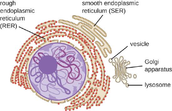
    The hollow portion of the ER tubules is called the lumen or cisternal space. The membrane of the ER, which is a phospholipid bilayer embedded with proteins, is continuous with the nuclear envelope (Figure \(\PageIndex{1}\)).

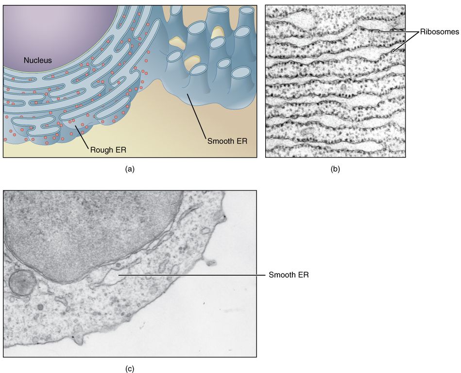
    The rough endoplasmic reticulum (RER) is so named because the ribosomes attached to its cytoplasmic surface give it a studded appearance when viewed through an electron microscope (Figure \(\PageIndex{2}\)). The ribosomes synthesize proteins while attached to the ER, resulting in transfer of their newly synthesized proteins into the lumen of the RER where they undergo modifications such as folding or addition of sugars. The RER also makes phospholipids for cell membranes.

    This figure shows structure of the endoplasmic reticulum. The diagram highlights the rough and smooth endoplasmic reticulum and the nucleus is labeled. Two micrographs show the structure of the endoplasmic reticulum in detail. The first micrograph shows the rough endoplasmic reticulum in a pancreatic cell and the second micrograph shows a smooth endoplasmic reticulum.
    Figure \(\PageIndex{2}\): (a) The ER is a winding network of thin membranous sacs found in close association with the cell nucleus. The smooth and rough endoplasmic reticula are very different in appearance and function (source: mouse tissue). (b) Rough ER is studded with numerous ribosomes, which are sites of protein synthesis (source: mouse tissue). EM × 110,000. (c) Smooth ER synthesizes phospholipids, steroid hormones, regulates the concentration of cellular Ca++, metabolizes some carbohydrates, and breaks down certain toxins (source: mouse tissue). EM × 110,510. (Micrographs provided by the Regents of University of Michigan Medical School © 2012). Figure from The Cytoplasm and Cellular Organelles; OpenStax.

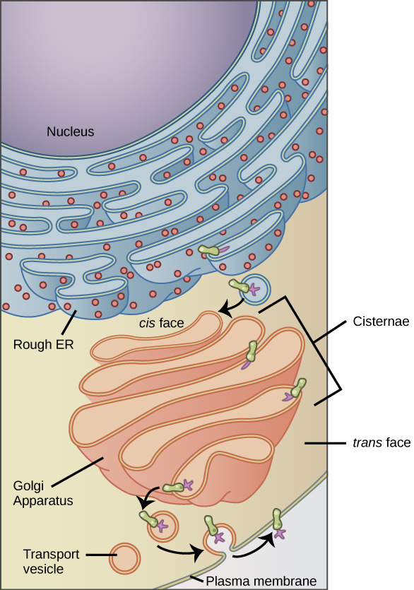
    If the phospholipids or modified proteins are not destined to stay in the RER, they will be packaged within vesicles and transported from the RER by budding from the membrane (Figure \(\PageIndex{3}\)). Since the RER is engaged in modifying proteins that will be secreted from the cell, it is abundant in cells that secrete proteins, such as the liver.

    The smooth endoplasmic reticulum (SER) is continuous with the RER but has few or no ribosomes on its cytoplasmic surface (see Figures \(\PageIndex{1}\)-\(\PageIndex{3}\)). The SER’s functions include synthesis of carbohydrates, lipids, and steroid hormones; detoxification of medications and poisons; alcohol metabolism; and storage of calcium ions.

    In muscle cells, a specialized SER called the sarcoplasmic reticulum is responsible for storage of the calcium ions that are needed to trigger the coordinated contractions of the muscle cells.

    This figure shows the nucleus, rough ER, Golgi apparatus, vesicles, and plasma membrane. The right side of the rough ER is shown with an integral membrane protein embedded in it. The part of the protein facing the inside of the ER has a carbohydrate attached to it. The protein is shown leaving the ER in a vesicle that fuses with the cis face of the Golgi apparatus. The Golgi apparatus consists of several layers of membranes, called cisternae. As the protein passes through the cisternae, it is further modified by the addition of more carbohydrates. Eventually, it leaves the trans face of the Golgi in a vesicle. The vesicle fuses with the cell membrane so that the carbohydrate that was on the inside of the vesicle faces the outside of the membrane. At the same time, the contents of the vesicle are released from the cell.
    Figure \(\PageIndex{3}\): The endomembrane system works to modify, package, and transport lipids and proteins. (credit: modification of work by Magnus Manske)

    References

    Unless otherwise noted, images on this page are licensed under CC-BY 4.0 by OpenStax.

    Text adapted from: OpenStax, Concepts of Biology. OpenStax CNX. May 18, 2016 http://cnx.org/contents/b3c1e1d2-839...9a8aafbdd@9.10


    5.9: The Endoplasmic Reticulum is shared under a CC BY license and was authored, remixed, and/or curated by LibreTexts.

    • Was this article helpful?