
2.6.3.2: Angiosperm Life Cycle
- Page ID
- 27725

\( \newcommand{\vecs}[1]{\overset { \scriptstyle \rightharpoonup} {\mathbf{#1}} } \)
\( \newcommand{\vecd}[1]{\overset{-\!-\!\rightharpoonup}{\vphantom{a}\smash {#1}}} \)
\( \newcommand{\dsum}{\displaystyle\sum\limits} \)
\( \newcommand{\dint}{\displaystyle\int\limits} \)
\( \newcommand{\dlim}{\displaystyle\lim\limits} \)
\( \newcommand{\id}{\mathrm{id}}\) \( \newcommand{\Span}{\mathrm{span}}\)
( \newcommand{\kernel}{\mathrm{null}\,}\) \( \newcommand{\range}{\mathrm{range}\,}\)
\( \newcommand{\RealPart}{\mathrm{Re}}\) \( \newcommand{\ImaginaryPart}{\mathrm{Im}}\)
\( \newcommand{\Argument}{\mathrm{Arg}}\) \( \newcommand{\norm}[1]{\| #1 \|}\)
\( \newcommand{\inner}[2]{\langle #1, #2 \rangle}\)
\( \newcommand{\Span}{\mathrm{span}}\)
\( \newcommand{\id}{\mathrm{id}}\)
\( \newcommand{\Span}{\mathrm{span}}\)
\( \newcommand{\kernel}{\mathrm{null}\,}\)
\( \newcommand{\range}{\mathrm{range}\,}\)
\( \newcommand{\RealPart}{\mathrm{Re}}\)
\( \newcommand{\ImaginaryPart}{\mathrm{Im}}\)
\( \newcommand{\Argument}{\mathrm{Arg}}\)
\( \newcommand{\norm}[1]{\| #1 \|}\)
\( \newcommand{\inner}[2]{\langle #1, #2 \rangle}\)
\( \newcommand{\Span}{\mathrm{span}}\) \( \newcommand{\AA}{\unicode[.8,0]{x212B}}\)
\( \newcommand{\vectorA}[1]{\vec{#1}} % arrow\)
\( \newcommand{\vectorAt}[1]{\vec{\text{#1}}} % arrow\)
\( \newcommand{\vectorB}[1]{\overset { \scriptstyle \rightharpoonup} {\mathbf{#1}} } \)
\( \newcommand{\vectorC}[1]{\textbf{#1}} \)
\( \newcommand{\vectorD}[1]{\overrightarrow{#1}} \)
\( \newcommand{\vectorDt}[1]{\overrightarrow{\text{#1}}} \)
\( \newcommand{\vectE}[1]{\overset{-\!-\!\rightharpoonup}{\vphantom{a}\smash{\mathbf {#1}}}} \)
\( \newcommand{\vecs}[1]{\overset { \scriptstyle \rightharpoonup} {\mathbf{#1}} } \)
\(\newcommand{\longvect}{\overrightarrow}\)
\( \newcommand{\vecd}[1]{\overset{-\!-\!\rightharpoonup}{\vphantom{a}\smash {#1}}} \)
\(\newcommand{\avec}{\mathbf a}\) \(\newcommand{\bvec}{\mathbf b}\) \(\newcommand{\cvec}{\mathbf c}\) \(\newcommand{\dvec}{\mathbf d}\) \(\newcommand{\dtil}{\widetilde{\mathbf d}}\) \(\newcommand{\evec}{\mathbf e}\) \(\newcommand{\fvec}{\mathbf f}\) \(\newcommand{\nvec}{\mathbf n}\) \(\newcommand{\pvec}{\mathbf p}\) \(\newcommand{\qvec}{\mathbf q}\) \(\newcommand{\svec}{\mathbf s}\) \(\newcommand{\tvec}{\mathbf t}\) \(\newcommand{\uvec}{\mathbf u}\) \(\newcommand{\vvec}{\mathbf v}\) \(\newcommand{\wvec}{\mathbf w}\) \(\newcommand{\xvec}{\mathbf x}\) \(\newcommand{\yvec}{\mathbf y}\) \(\newcommand{\zvec}{\mathbf z}\) \(\newcommand{\rvec}{\mathbf r}\) \(\newcommand{\mvec}{\mathbf m}\) \(\newcommand{\zerovec}{\mathbf 0}\) \(\newcommand{\onevec}{\mathbf 1}\) \(\newcommand{\real}{\mathbb R}\) \(\newcommand{\twovec}[2]{\left[\begin{array}{r}#1 \\ #2 \end{array}\right]}\) \(\newcommand{\ctwovec}[2]{\left[\begin{array}{c}#1 \\ #2 \end{array}\right]}\) \(\newcommand{\threevec}[3]{\left[\begin{array}{r}#1 \\ #2 \\ #3 \end{array}\right]}\) \(\newcommand{\cthreevec}[3]{\left[\begin{array}{c}#1 \\ #2 \\ #3 \end{array}\right]}\) \(\newcommand{\fourvec}[4]{\left[\begin{array}{r}#1 \\ #2 \\ #3 \\ #4 \end{array}\right]}\) \(\newcommand{\cfourvec}[4]{\left[\begin{array}{c}#1 \\ #2 \\ #3 \\ #4 \end{array}\right]}\) \(\newcommand{\fivevec}[5]{\left[\begin{array}{r}#1 \\ #2 \\ #3 \\ #4 \\ #5 \\ \end{array}\right]}\) \(\newcommand{\cfivevec}[5]{\left[\begin{array}{c}#1 \\ #2 \\ #3 \\ #4 \\ #5 \\ \end{array}\right]}\) \(\newcommand{\mattwo}[4]{\left[\begin{array}{rr}#1 \amp #2 \\ #3 \amp #4 \\ \end{array}\right]}\) \(\newcommand{\laspan}[1]{\text{Span}\{#1\}}\) \(\newcommand{\bcal}{\cal B}\) \(\newcommand{\ccal}{\cal C}\) \(\newcommand{\scal}{\cal S}\) \(\newcommand{\wcal}{\cal W}\) \(\newcommand{\ecal}{\cal E}\) \(\newcommand{\coords}[2]{\left\{#1\right\}_{#2}}\) \(\newcommand{\gray}[1]{\color{gray}{#1}}\) \(\newcommand{\lgray}[1]{\color{lightgray}{#1}}\) \(\newcommand{\rank}{\operatorname{rank}}\) \(\newcommand{\row}{\text{Row}}\) \(\newcommand{\col}{\text{Col}}\) \(\renewcommand{\row}{\text{Row}}\) \(\newcommand{\nul}{\text{Nul}}\) \(\newcommand{\var}{\text{Var}}\) \(\newcommand{\corr}{\text{corr}}\) \(\newcommand{\len}[1]{\left|#1\right|}\) \(\newcommand{\bbar}{\overline{\bvec}}\) \(\newcommand{\bhat}{\widehat{\bvec}}\) \(\newcommand{\bperp}{\bvec^\perp}\) \(\newcommand{\xhat}{\widehat{\xvec}}\) \(\newcommand{\vhat}{\widehat{\vvec}}\) \(\newcommand{\uhat}{\widehat{\uvec}}\) \(\newcommand{\what}{\widehat{\wvec}}\) \(\newcommand{\Sighat}{\widehat{\Sigma}}\) \(\newcommand{\lt}{<}\) \(\newcommand{\gt}{>}\) \(\newcommand{\amp}{&}\) \(\definecolor{fillinmathshade}{gray}{0.9}\)- Identify structures and phases in the angiosperm life cycle; know their ploidy.
- Explain how fertilization occurs within a flower.
- Label a developing ovary cross section.
Angiosperms have a complex life cycle. The gametophytes have been further reduced: antheridia were lost in the gymnosperms and archegonia were lost in the angiosperms. Both gametophytes are now housed within the flower, a structure composed of highly modified leaves specialized for pollination. From flowers, fruits are produced, a protective structure that (usually) develops from the ovary wall and is specialized for seed dispersal.
The Microgametophyte (AKA the Pollen Grain)
The microgametophyte develops and reaches maturity within the microsporangia (Figure \(\PageIndex{1}\)). The microsporangia, which are usually bi-lobed, are also called pollen sacs. These pollen sacs are found in the anther of the stamen, which is at the end of the filament.

Within the microsporangium (pollen sac), many microspore mother cells divide by meiosis to each give rise to four haploid microspores, each of which will ultimately form a pollen grain (Figure \(\PageIndex{2}\)). An inner layer of cells in the microsporangium, known as the tapetum, provides nutrition to the developing microspores and contributes key components to the pollen wall. Upon maturity, the microsporangia burst, releasing the pollen grains from the anther.
Each pollen grain has two coverings: the exine (thicker, outer layer) and the intine (Figure \(\PageIndex{2}\)). The exine contains sporopollenin, a complex waterproofing substance supplied by the tapetal cells. Sporopollenin allows the pollen to survive under unfavorable conditions and to be carried by wind, water, or biological agents without undergoing damage.

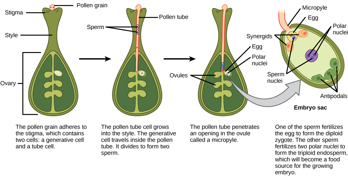
Mature pollen grains contain two cells: a generative cell and a pollen tube cell. The generative cell is contained within the larger pollen tube cell. When a pollen grain reaches the stigma, it germinates into a pollen tube. The generative cell migrates with the pollen tube to enter the ovary. During its transit inside the pollen tube, the generative cell divides to form two gametes (spermatia). These, along with the tube nucleus (also known as the vegetative nucleus), migrate down the pollen tube as it grows through the style, the micropyle, and into the ovule chamber.
In Arabidopsis, the pollen tube follows a gradient of increasing concentration of a small defensin-like protein secreted by the synergids (see The Megagametophyte).
The Megagametophyte
Within the ovary of the gynoecium, ovules are produced. Ovules consist of a double-layered integument with a small opening called the micropyle. The integument surrounds the megasporangium. Both the micropyle and megasporangium are diploid tissue of the sporophyte and are connected to the ovary wall by a region of tissue called the funiculus. The funiculus connects to a region of the ovary called the placenta, where nutritive support is provided the ovary wall and supplied to the developing ovule.
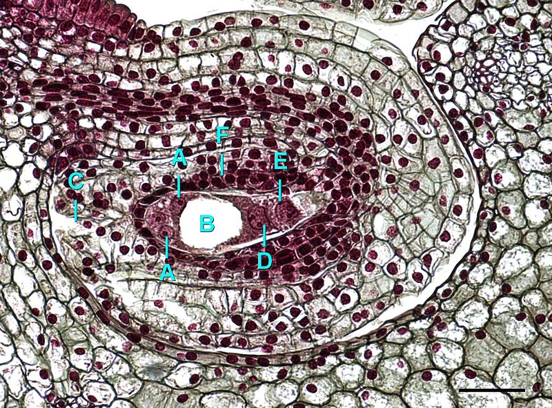
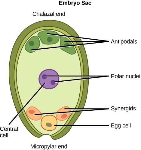
Within the megasporangium a single diploid megaspore mother cell divides by meiosis to produce four haploid megaspores. One of these will survive and three will disintegrate. The nucleus of the surviving megaspore undergoes 3 successive mitotic divisions. The 8 nuclei that result are distributed and partitioned off by cell walls to form the embryo sac (Figure \(\PageIndex{3}\)). The embryo sac is composed of 7 cells. The egg cell, located near the micropylar end, is flanked by 2 synergid cells. The large central cell contains 2 polar nuclei (a dikaryotic cell). The final three cells are the antipodal cells, located on the opposite side as the egg and synergids (Figure \(\PageIndex{4}\)).
The synergids help guide the pollen tube for successful fertilization, after which they disintegrate. One of the spermatia produced by the pollen's generative cell fuses with the egg to form a diploid zygote. This zygote will grow into the sporophyte. The second spermatium fuses with the polar nuclei to produce a triploid endosperm (Figure \(\PageIndex{5}\)). This event is called double fertilization. The endosperm will provide additional nutritive tissue for the growing embryo.
After fertilization, the integument will close the micropyle and develop into the seed coat, protecting the seed. The ovary wall will develop into the pericarp of the fruit.
Fruits
An unfertilized ovary, as shown in Figure \(\PageIndex{6}\), contains one or more developing ovules produced in compartments called locules. Each ovule is attached to a nutritional region of the ovary called the placenta by a strand of tissue called the funiculus. The sporophyte supports the developing ovule through this tissue pathway. Prior to fertilization, there is a small gap in the integument called the micropyle.

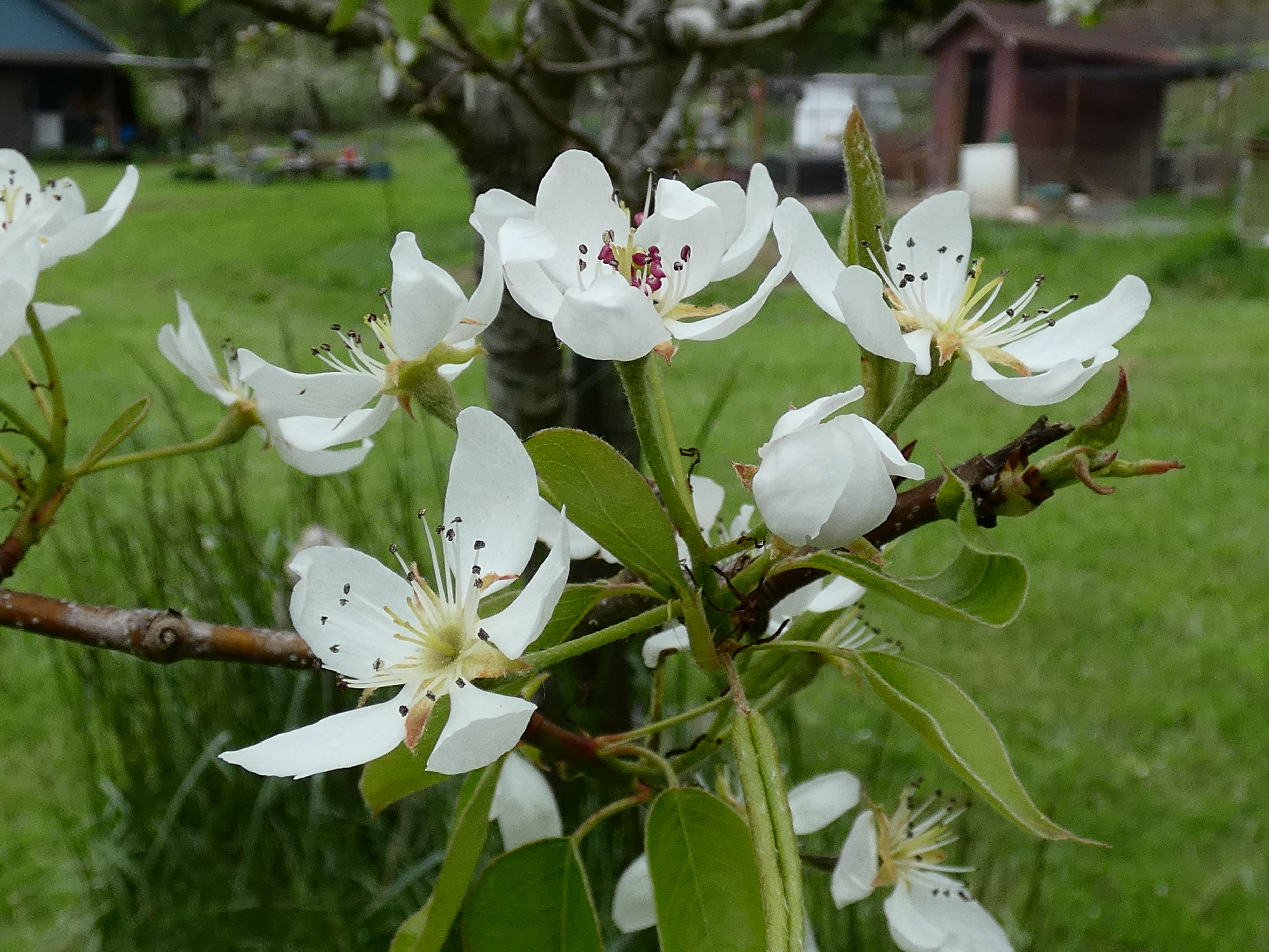
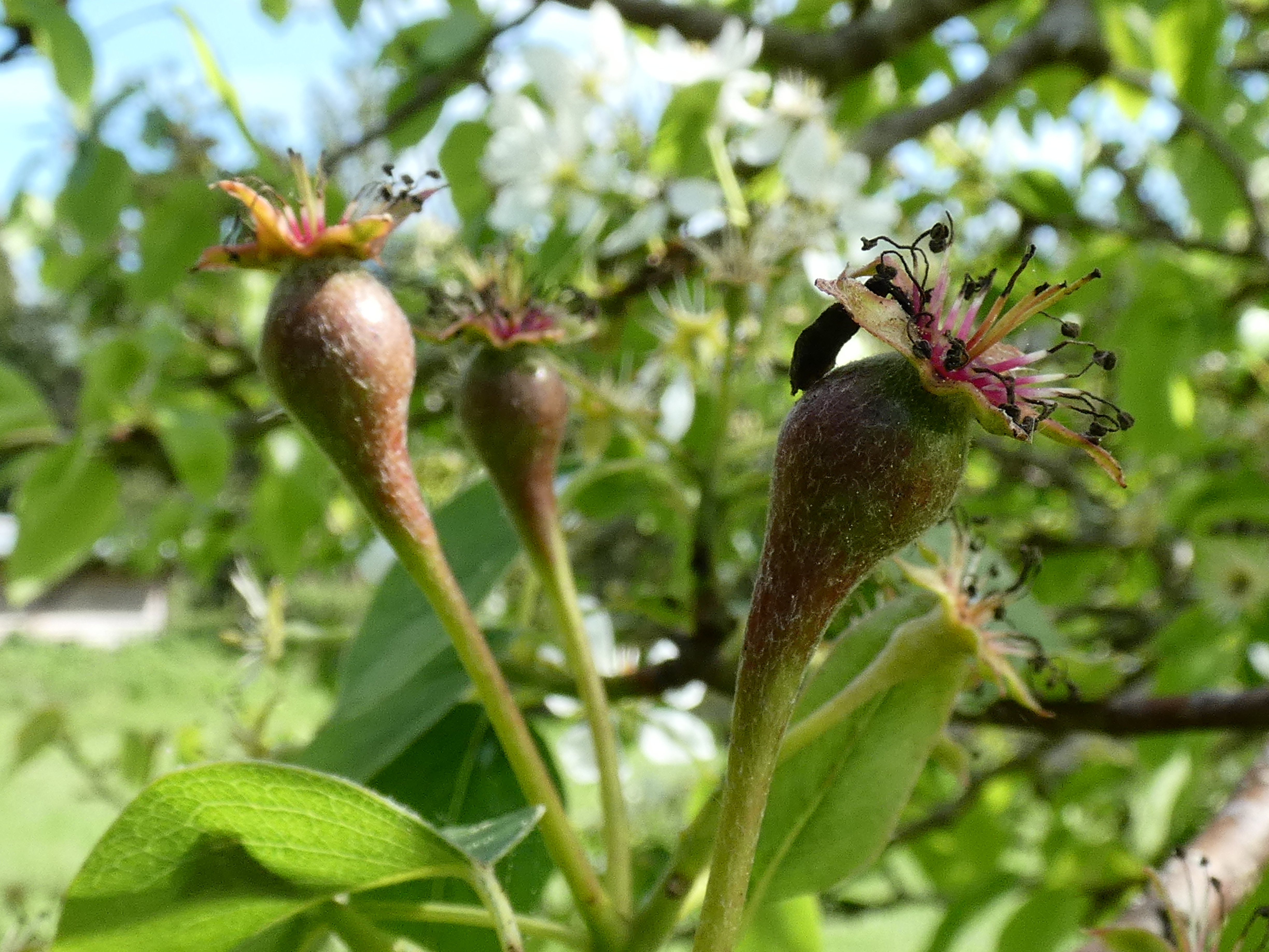
After fertilization, the ovary wall develops into the fruit, surrounding the seeds. In fleshy fruits that use animals for dispersal, like the pears shown in Figure \(\PageIndex{7}\), this might include a swelling of the cells, increased sugar production, and a change in pigmentation. These are the type of fruits we are familiar with. However, all flowers turn into fruits. The fruits might be dry, spiky, or in other ways completely unappetizing, and this is become many fruits do not use animal ingestion as their method of dispersal (more on this in Chapter 8.3 Fruits and Dispersal).
The Full Life Cycle
The angiosperm life cycle is shown in Figure \(\PageIndex{12}\) and Video \(\PageIndex{1}\).
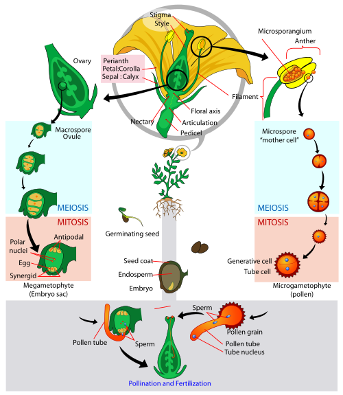
Watch Video \(\PageIndex{1}\) to help untangle this complex life cycle.
Video \(\PageIndex{1}\): A digital, narrated rendition of the angiosperm life cycle. Sourced from YouTube.
Attribution
Curated and authored by Maria Morrow, CC BY-NC, using the following sources:
- 32.1 Reproductive Development and Structure and 32.2 Pollination and Fertilization from Biology 2e by OpenStax (licensed CC-BY). Access for free at openstax.org.
- 8.1 Spermatophyta 2.0 from Introduction to Botany by Alexey Shipunov (public domain)