Biology LibreTexts

2.5.3.2: Polypodiopsida

  • Page ID
    37013
  • \( \newcommand{\vecs}[1]{\overset { \scriptstyle \rightharpoonup} {\mathbf{#1}} } \) \( \newcommand{\vecd}[1]{\overset{-\!-\!\rightharpoonup}{\vphantom{a}\smash {#1}}} \)\(\newcommand{\id}{\mathrm{id}}\) \( \newcommand{\Span}{\mathrm{span}}\) \( \newcommand{\kernel}{\mathrm{null}\,}\) \( \newcommand{\range}{\mathrm{range}\,}\) \( \newcommand{\RealPart}{\mathrm{Re}}\) \( \newcommand{\ImaginaryPart}{\mathrm{Im}}\) \( \newcommand{\Argument}{\mathrm{Arg}}\) \( \newcommand{\norm}[1]{\| #1 \|}\) \( \newcommand{\inner}[2]{\langle #1, #2 \rangle}\) \( \newcommand{\Span}{\mathrm{span}}\) \(\newcommand{\id}{\mathrm{id}}\) \( \newcommand{\Span}{\mathrm{span}}\) \( \newcommand{\kernel}{\mathrm{null}\,}\) \( \newcommand{\range}{\mathrm{range}\,}\) \( \newcommand{\RealPart}{\mathrm{Re}}\) \( \newcommand{\ImaginaryPart}{\mathrm{Im}}\) \( \newcommand{\Argument}{\mathrm{Arg}}\) \( \newcommand{\norm}[1]{\| #1 \|}\) \( \newcommand{\inner}[2]{\langle #1, #2 \rangle}\) \( \newcommand{\Span}{\mathrm{span}}\)\(\newcommand{\AA}{\unicode[.8,0]{x212B}}\)

    Learning Objectives
    • Differentiate between ferns, horsetails, and lycophytes.
    • Identify features of vegetative and reproductive shoots of Equisetum.
    • Identify features and phases of the fern life cycle; know their ploidy.
    • Label a fern gametophyte and sporophyte.

    The Polypodiopsida includes the horsetails and ferns. These plants produce leaves with branching veins of vascular tissue called megaphylls. Like the lycophytes, asexual reproduction of the sporophyte can be accomplished via rhizomes. Spores produced in this group develop into gametophytes that can produce both antheridia and archegonia.

    Characteristics

    • Megaphylls. Leaves have branching veins of vascular tissue. Megaphylls are thought to have evolved from branching stems. Webbing filled in the spaces between the branches, forming the flat blade of the leaf. The arrangement of the leaf veins (vascular bundles) reflects the original branching pattern of the stems.
    • Rhizomes. Asexual propogation of the sporophyte through underground stems.
    • Homosporous. Haploid spores grow into bisexual gametophytes that produce both antheridia and archegonia, or are capable of producing one or the other, dependent upon conditions.

    Equisteum (subclass Equisetidae)

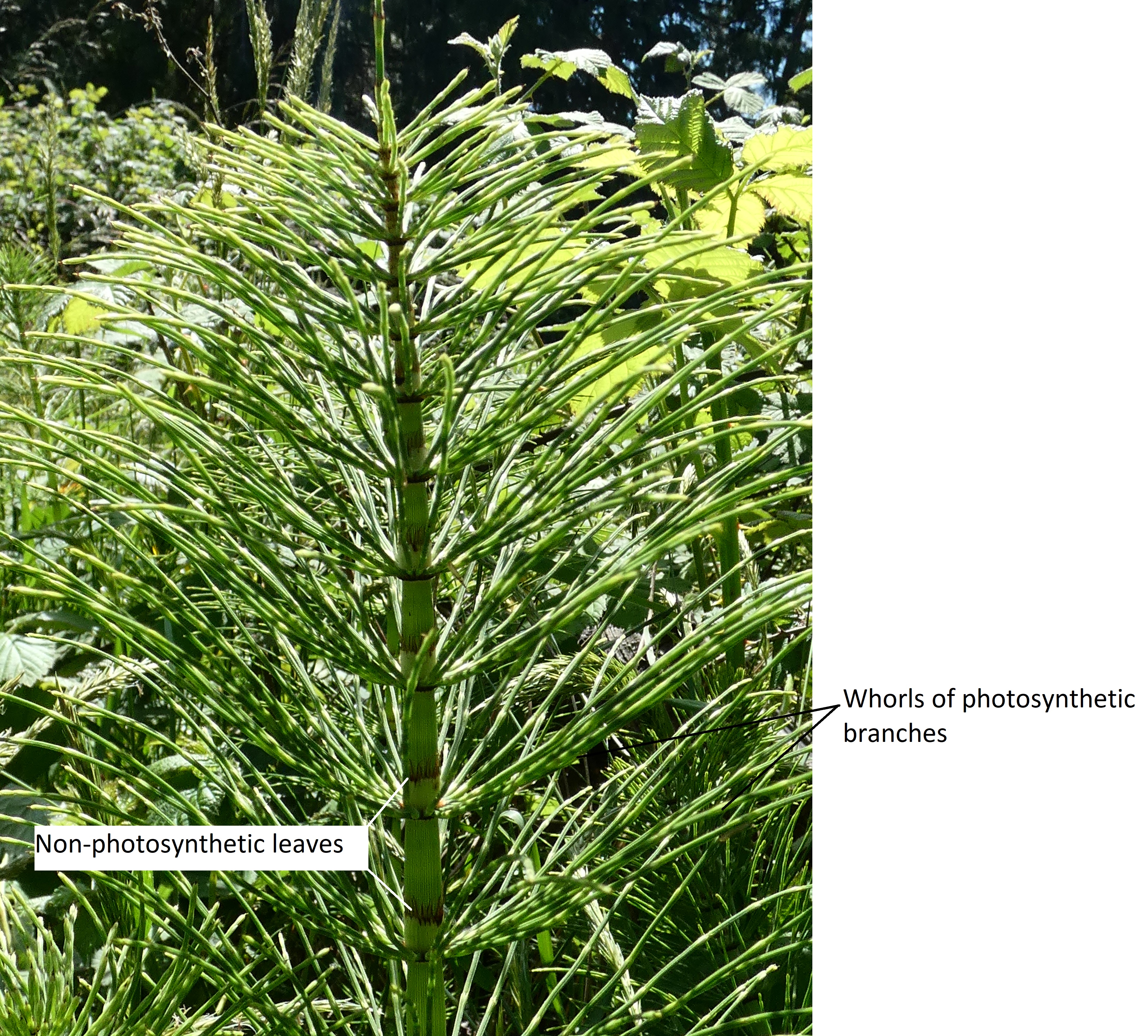
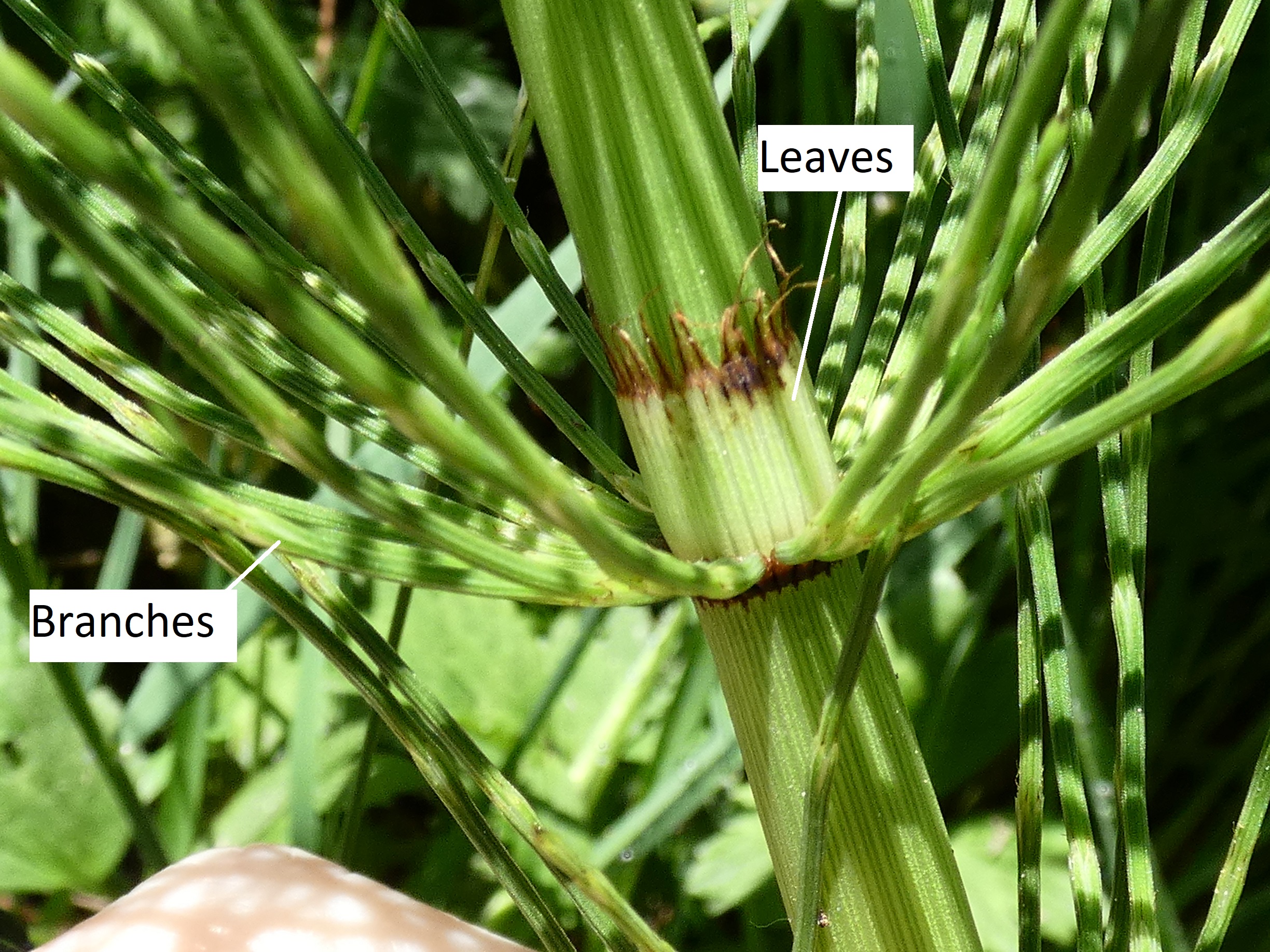
    Horsetails are a small group with a single extant genus, Equisetum, which has about 30 different herbaceous species that typically live in moist habitats. The common name comes from the characteristic pattern of branching: whorls or rings of branches arising from an above-ground shoot. The leaves of these plants have been reduced to scales, and instead the segmented stems are photosynthetic. If you look closely at the nodes of a green vegetative shoot, you will see that branches and leaves have not only switched roles, they have also switched places, with the photosynthetic branches emerging below the papery, non-photosynthetic leaves.

    Horsetails often grow in sandy places and incorporate silica in their stem epidermis, which gives it an abrasive surface. Because of this, American pioneers would use this plant to scour pots and pans. This is how it received the nickname “scouring rush.” The stem has multiple canals, an analogous characteristic to stems of grasses. The sporangia are associated with hexangular stalked sporangiophores produced on terminal strobili. Within the sporangia, there are elaters that are not separate cells but parts of the spore walls. Gametophytes are typically minute and dioecious, but the plants themselves are homosporous: smaller suppressed gametophytes develop only antheridia while larger gametophytes develop only archegonia.

    Gametophyte Morphology

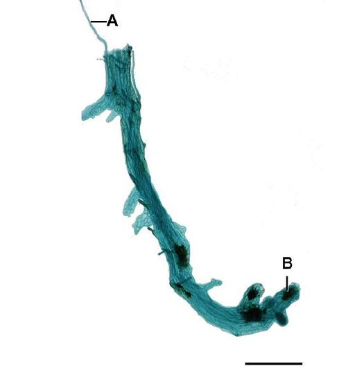
    Horsetail gametophytes are reduced and thalloid (figure \(\PageIndex{1}\)). Gametophytes grow from homospores and can produce both antheridia and archegonia.

    A long, thin thallus with a rhizoid emerging from one side and some darkened regions labeled as antheridia on the other side.
    Figure \(\PageIndex{1}\): Whole mount of an Equisetum gametophyte. A indicates a long, thin rhizoid. B indicates one of several antheridia, each positioned on an extending lobe from the long, linear thallus. Scale=0.6mm. Jon Houseman, CC BY-SA 3.0, via Wikimedia Commons.

    Sporophyte Morphology

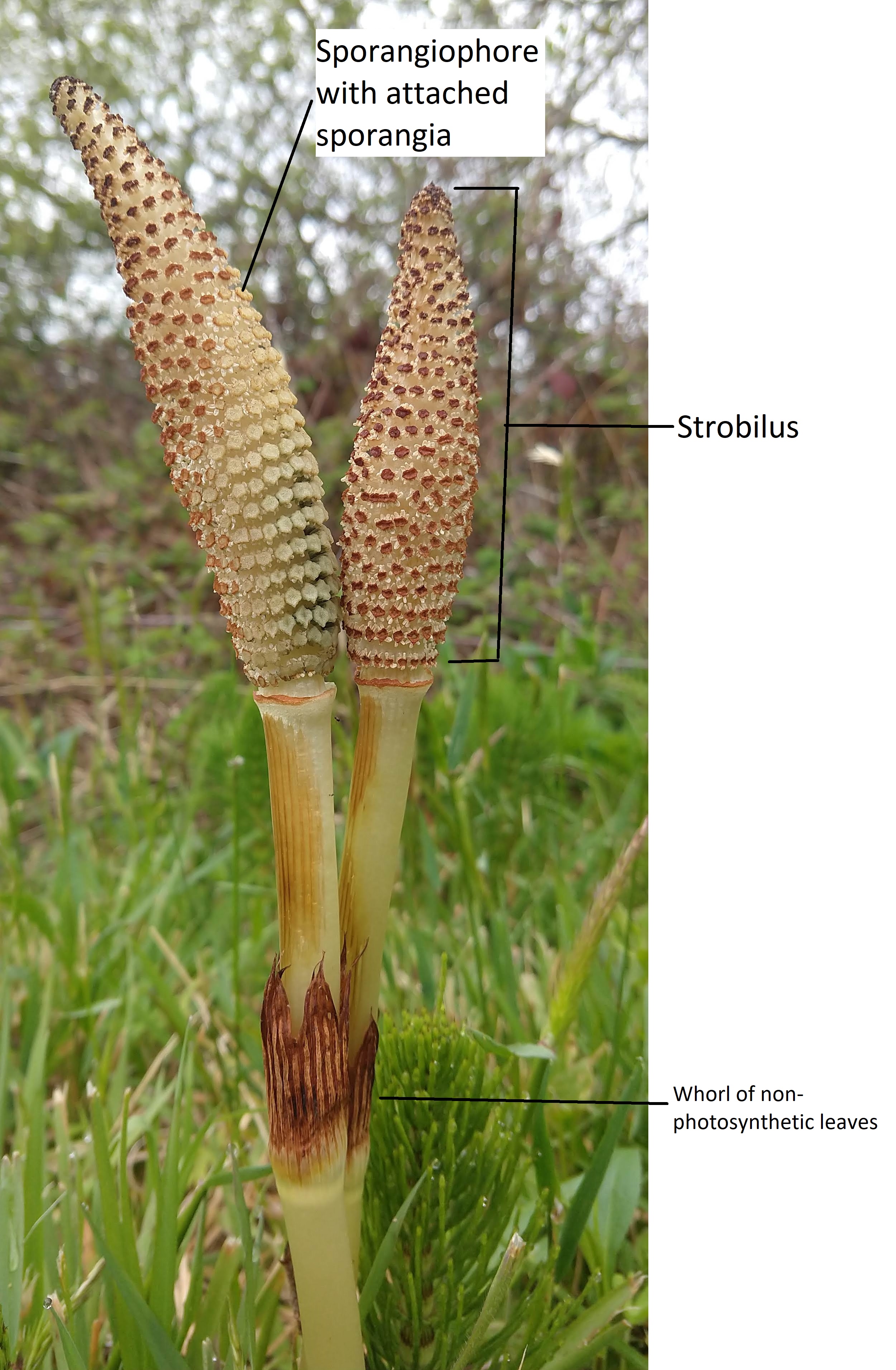
    In some Equisetum species, there are two different types of shoots produced by the sporophyte: vegetative shoots that perform photosynthesis and reproductive shoots that form strobili and undergo meiosis. In other species, the strobilus is formed at the apex of a photosynthetic shoot.

    Vegetative Shoots

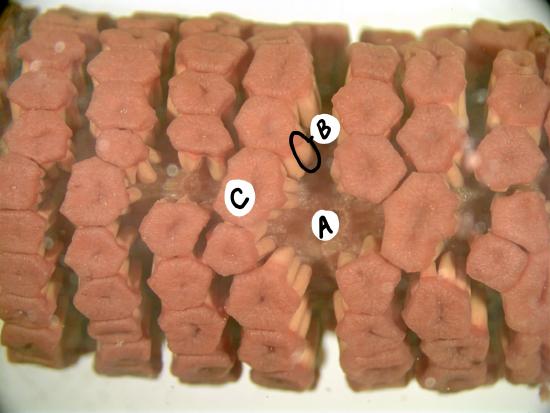
    On the vegetative shoot, the leaves are dark, papery and non-photosynthetic. Branches are photosynthetic and produced in whorls. Branches and leaves emerge at nodes, separated by regions of the main stem called internodes. Unlike most plants, the branches emerge below the leaves in the node (Figure \(\PageIndex{2-3}\)). The epidermis of the stems contain silica, which has an abrasive texture.

    A vegetative shoot of Equisetum
    Figure \(\PageIndex{2}\): A vegetative shoot of Equisetum. There are rings of long, thin, green branches emerging from each node. Above each whorl of branches, a ring of pointy brown leaves are pressed up against the main stem. Photo by Maria Morrow, CC-BY 4.0.
    Equisetum leaves and branches arranged in a whorl around a node
    Figure \(\PageIndex{3}\): A close up of the vegetative shoot of Equisetum. In this image, you get a better view of the leaves. They start out as a united tube surrounding the main stem, then turn dry and brown, separating into individual, wispy points. The whorl of leaves looks a bit like a grassy crown encircling the stem. Photo by Maria Morrow, CC-BY 4.0.

    Reproductive Shoots

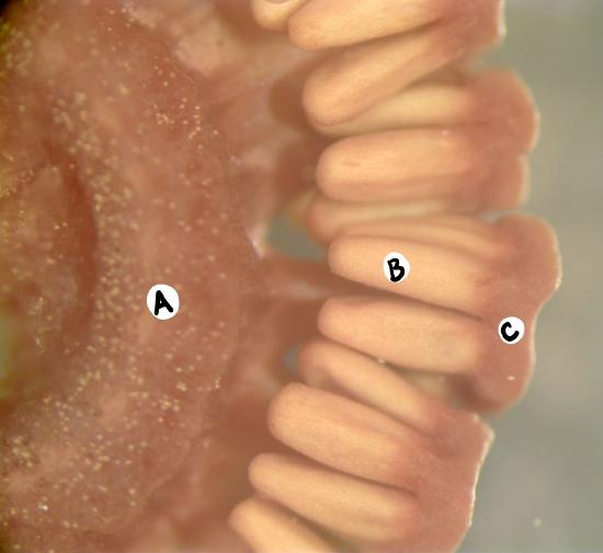
    Sporangia are produced in a terminal strobilus on the reproductive shoot (figure \(\PageIndex{4-5}\)). In some species, this reproductive shoot lacks chlorophyll and is instead fed through the rhizome of connected vegetative shoots. Spores are photosynthetic and have four hygroscopic arms called elaters.

    Two reproductive shoots of Equisetum
    Figure \(\PageIndex{4}\): Two reproductive shoots of Equisetum. These shoots are a very pale yellow, lacking chlorophyll and unable to do photosynthesis. The leaves on the reproductive shoots still occur in whorls and are larger than on the vegetative shoots shown above. At the top of each shoot, there is an elongated structure that comes to a point and looks like it is covered in polka dots. These are the strobili. Each polka dot is the top of a sporangiophore, each of which bear many sporangia, perhaps visible to you as little white frills surrounding the polka dots. Photo by Maria Morrow, CC-BY 4.0.
    An Equisetum strobilus on its side
    A cross section through the Equisetum strobilus shows the sporangia dangling from the T-shaped sporangiophore.
     
    Figure \(\PageIndex{5}\): These images show a preserved strobilus. In the first image, e are looking straight at the tops of the sporangiophores (C), but some sporangia (B) are visible underneath them. There is a section where you can see the cone axis (A). In both images, the labels represent the same structures: A) cone axis, B) sporangium, C) sporangiophore. Copyright by Curtis Clark, licensed as noted, CC BY-SA 2.5, via Wikimedia Commons with labels added by Maria Morrow.

    Video \(\PageIndex{1}\): This video shows how the elaters of Equisetum spores respond to changes in humidity. Retrieved from YouTube.

    Ferns (subclass Polypodiidae)

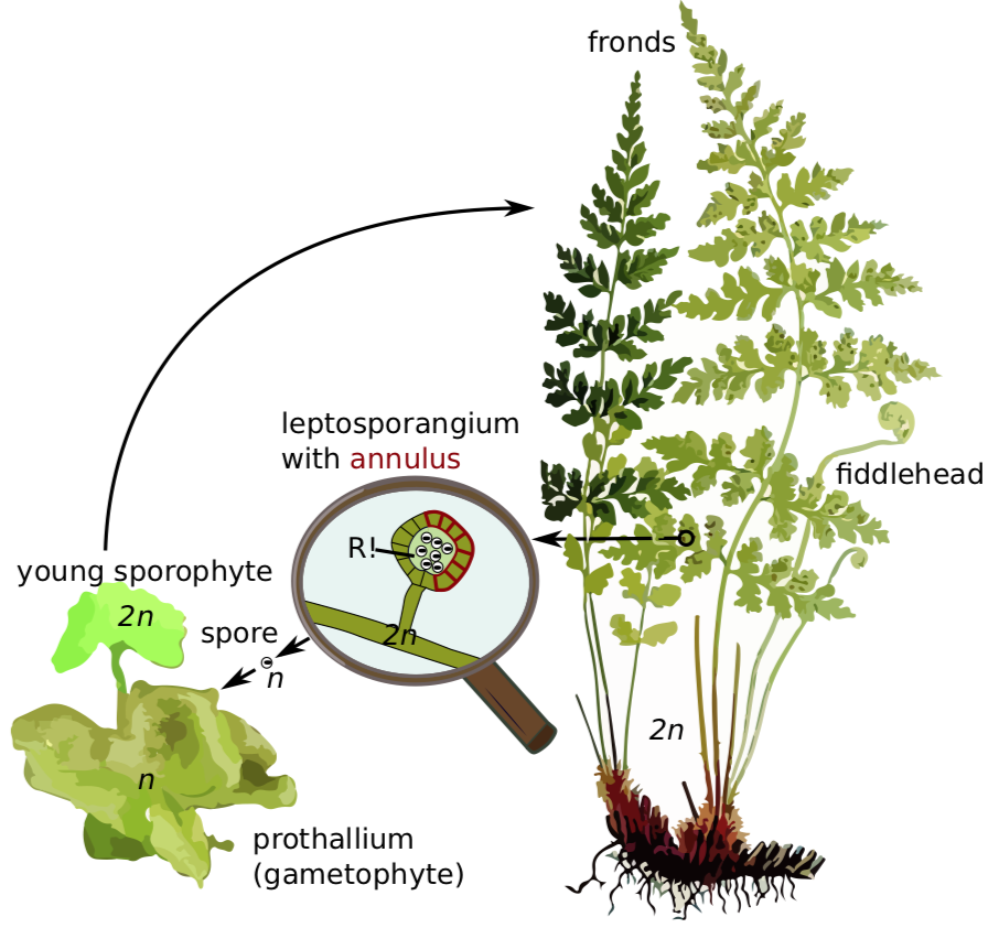
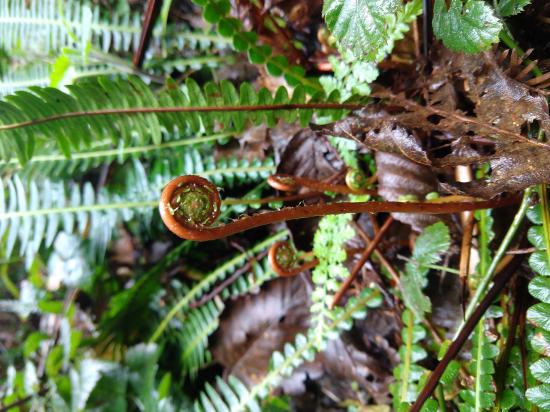
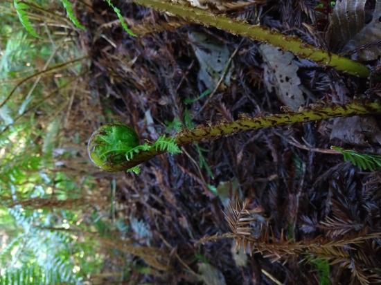
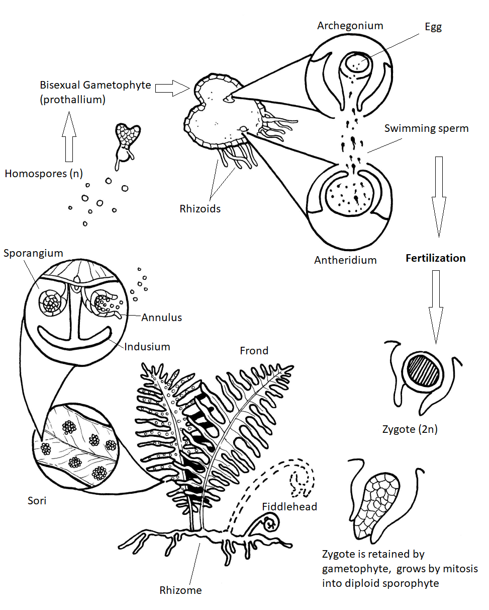
    Some 15,000 species of ferns live on earth today. Many of these are found in the tropics where some — the "tree ferns" — may grow to heights of 40 ft (13 m) or more. The ferns of temperate regions are smaller. They are usually found in damp, shady locations. They produce perennial rhizomes that can overwinter. Their leaves, called fronds due to apical growth, emerge from the rhizome each spring as coiled fiddleheads (Figure \(\PageIndex{6}\)). These fiddleheads open through a process called circinate vernation, where the growing tissues are protected at the center of the coil and emerge last.

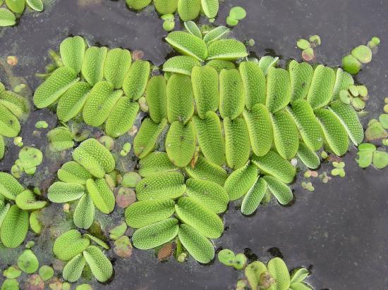
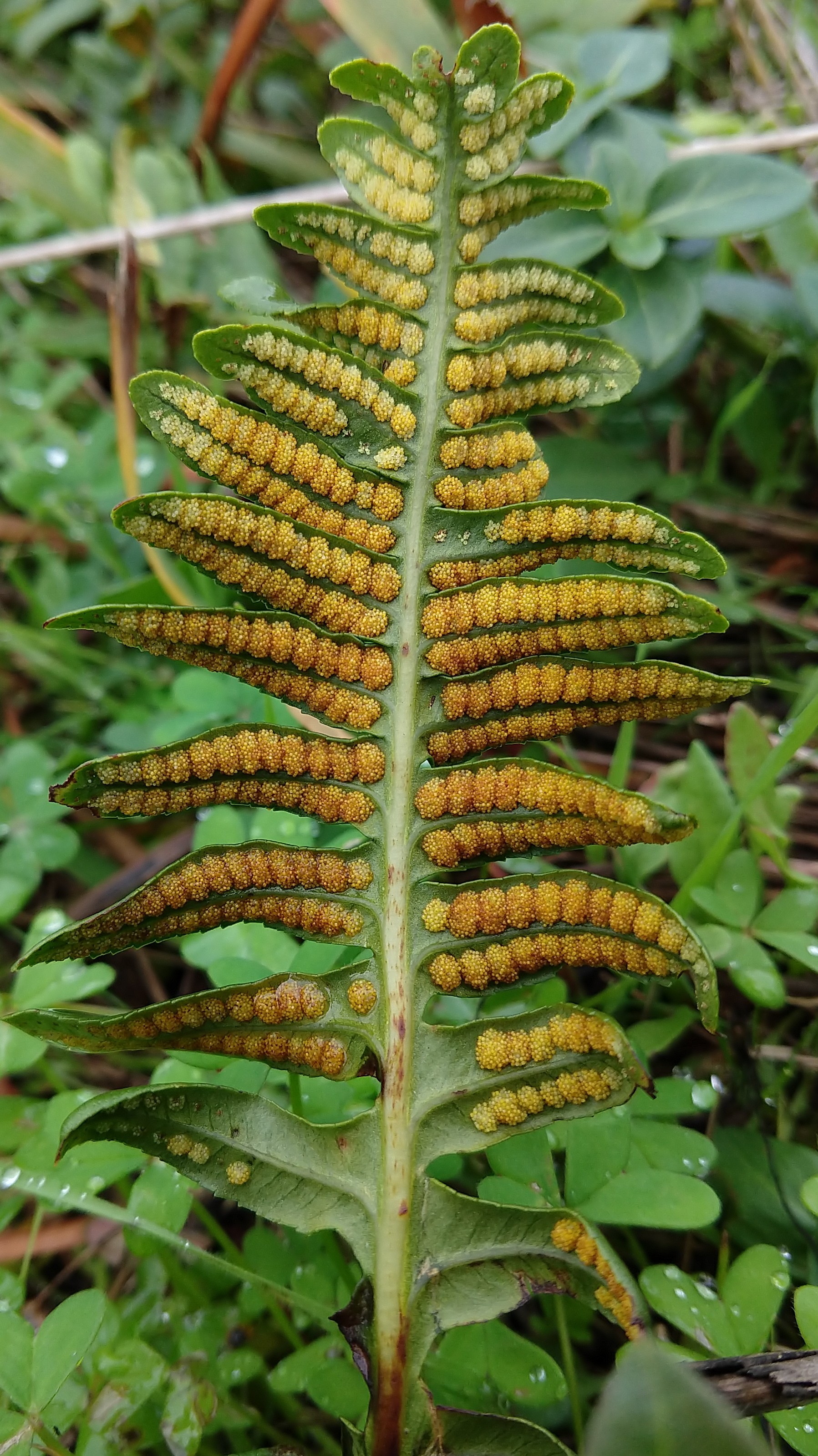
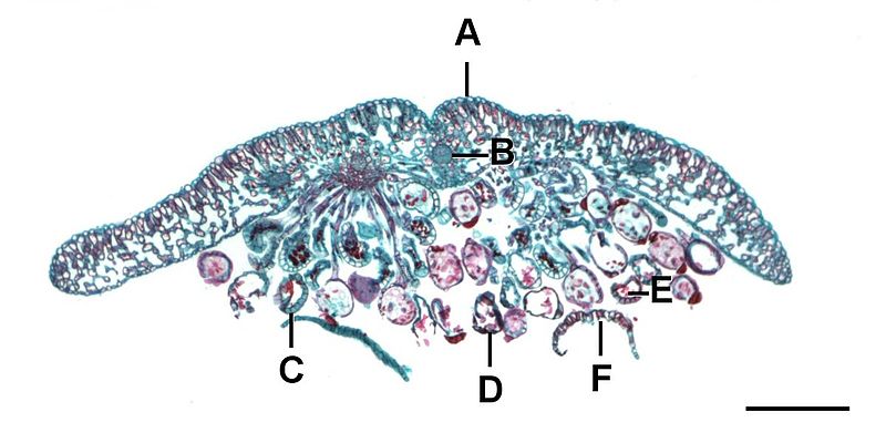
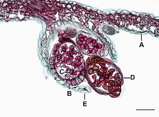
    True ferns are megaphyllous: their leaves originated from flattened branches and have branching veins of vascular tissue. True ferns have unique sporangia: leptosporangia (Figure \(\PageIndex{6}\)). Leptosporangia originate from a single cell in a leaf, they have long, thin stalks, and the wall of one cell layer. They also open actively: when sporangium matures (dries), a row of cells with thickened walls on the outside of the sporangium (called an annulus) will shrink slower than surrounding cells and finally would break and release all spores at once. Leptosporangia are grouped in clusters called sori which are often covered with umbrella- or pocket-like indusia. Gametophytes are minute and grow aboveground. While most ferns are homosporous, some genera of true ferns (like the water fern Azolla, water shamrock Marsilea and several others) are heterosporous (Figure \(\PageIndex{7}\)).

    Select stages of the fern life cycle
    Figure \(\PageIndex{6}\): Selected stages of Cystopteris life cycle, representative of Pteridopsida. In this life cycle, a leptosporangium is featured in the middle. It has a thickened row of cells along the outside of the sporangium (the annulus) that will split the sporangium open as it dries. This produces a haploid (n) spore that develops into a prothallium (gametophyte, n). From the prothallium, a diploid (2n) sporophyte grows out. This then develops into several mature fronds and a developing fiddlehead.
    Salvinia natans is shaped like tiny green tacos floating in the water. The inner surface of the leaves have hairs.
    Yasui Kono examining a flower
    Figure \(\PageIndex{7}\): Salvinia natans (left) is a heterosporous member of Polypodiidae. The hairs on the tops of the leaves repel water and help the leaves stay afloat. Kono Yasui (right), the first Japanese woman to earn her PhD, described the anatomy of this species in the Annals of Botany, making her the first Japanese woman to publish in a scientific journal centered outside of Japan. Over her career, she published 99 papers, including studies of plant cells (plant cytology), plant genetics, and even the formation of coal. Left image by Le.Loup.Gris, CC-BY-SA, and right image by 朝日新聞社 (public domain).

    True ferns are highly competitive even to angiosperms. In spite of their “primitive” life cycle, they have multiple advantages: abilities to photosynthesize in deep shade (they are not obliged to grow fast), to survive high humidity, and to make billions of reproductive units (spores). Ferns do not need to spend their resources on flowers and fruits, and are also less vulnerable to vertebrate herbivores and insect pests, probably because they do not employ them as pollinators and, therefore, can poison tissues against all animals.

    Gametophyte Morphology

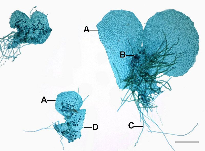
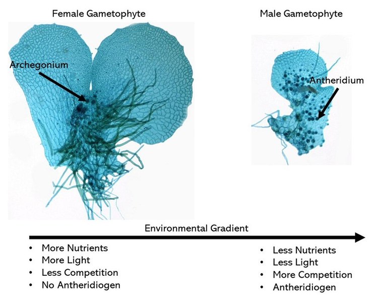
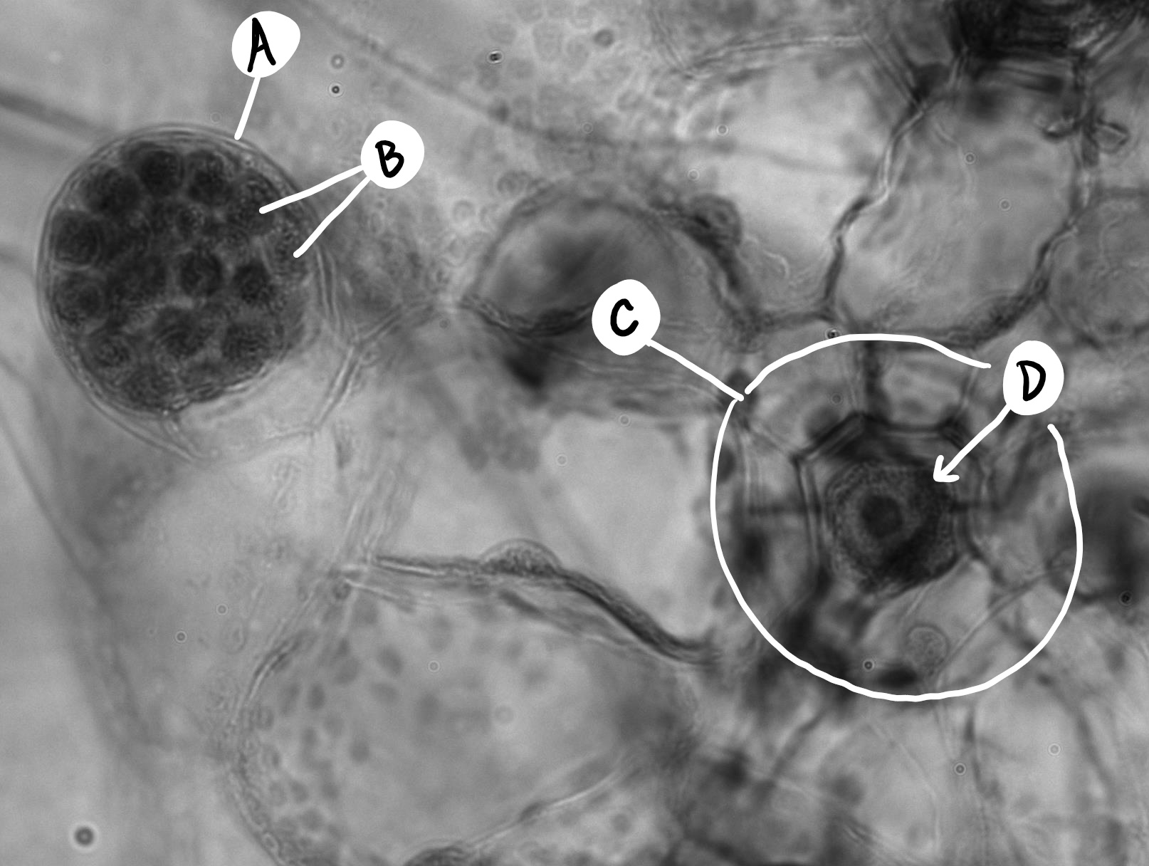
    Fern gametophytes are reduced, thalloid, and heart-shaped (Figure \(\PageIndex{8}\)). They are often referred to as a prothallus or prothallium. Rhizoids are produced from the underside of the thallus, just like in the bryophytes. Similar to the horsetails, whether a gametophyte produces antheridia or archegonia can be regulated by environmental cues (Figure \(\PageIndex{9}\)). Each antheridium produces many swimming sperm, but archegonia produce only a single egg (Figure \(\PageIndex{10}\)).

    Components of a fern gametophyte
    Figure \(\PageIndex{8}\): Three fern gametophytes that have been stained and so appear blue. The images are labeled as follows: A) Prothallus, B) Archegonium, C) Rhizoid, D) Antheridium. Scale=0.525mm. The rhizoids and archegonia emerge from the underside off the thallus. Descriptive text: A points to the flat, heart-shaped thallus, B points to structures with elongated necks close to the rhizoids, C points to long thin projections, D points to small dark structures spread across the thallus. Photo by Jon Houseman, CC BY-SA 4.0, via Wikimedia Commons.
    Fern gametophytes showing male and female structures and the environmental cues that select for each
    Figure \(\PageIndex{9}\): Fern gametophytes may develop either antheridia or archegonia based on environmental characteristics such as nutrients, competition, light, and antheridiogens. More nutrients, more light, less competition, and the absence of antheridiogen all select for the production of archegonia. Less nutrients, less light, more competition, and antheridiogen all select for the production of antheridia. Image by Jon Houseman, CC BY-SA 4.0, via Wikimedia Commons.
    A close up of the fern gametophyte, showing an antheridium and an archegonium
    Figure \(\PageIndex{10}\): A magnified view of a fern gametophyte that has both antheridia and archegonia simultaneously. The antheridium (A) has many sperm inside (B) and does not appear three dimensional. The archegonium (C) is surrounded by enlarged cells--we are looking straight down the neck. At the center of the archegonium, there is only a single egg (D). Photo by Maria Morrow, CC-BY 4.0.

    Sporophyte Morphology

    Fern sporophytes are composed of megaphylls, often pinnately compound fronds that emerging as coiled fiddleheads in the spring. Sporangia are produced in clusters called sori (sorus, singular) on the fronds (Figure \(\PageIndex{11}\)).

    A fern frond where each pinna has two rows of fuzzy-looking orange lumps (sori)
    Figure \(\PageIndex{11}\): The underside of Polypodium frond. Each fuzzy-look orange region is a developing sorus (a cluster of sporangia). These sori are not protected by an indusium. Photo by Maria Morrow, CC-BY 4.0.

    Circinate vernation is a term used to describe the development of the fern fiddlehead (Figure \(\PageIndex{12}\)) into a frond. Because plants grow apically, it is important to protect the apical meristems in growing organs (as we have seen in both axillary and terminal buds with the protective bud scales). The fiddlehead is essentially a structure that tucks away the growing tips of the fronds. As the frond develops, it gradually unfurls, releasing the tips last.

    The fiddlehead of a deer fern frond
    The fuzzy fiddlehead of a sword fern frond
    Figure \(\PageIndex{12}\): The two images above show fiddleheads of two different types of ferns, a deer fern in the first image and a sword fern in the second. Each has the same basic shape--a long stalk that transitions in a tightly coiled knot containing the developing leaf tissues. Photo by Maria Morrow, CC-BY 4.0.

    A sorus (plural, sori) is a cluster of sporangia, often protected by an umbrella-like structure called the indusium as the spores mature (Figure \(\PageIndex{13}\)). Some sori are protected by an extension of the leaf called a false indusium (Figure \(\PageIndex{14}\)), while others lack any protective covering. Each sporangium is lined by an inflated strip of cells called an annulus. When the spores have matured, the cells in the annulus begin to dry out, causing the cells to collapse and pull the sporangium open, releasing the spores.

    A cross section through a fern sorus with a small leaf.
    Figure \(\PageIndex{13}\): Cross section of a Polystichum sorus. The epidermis of the leaf (sporophyll) is labeled A. A large vascular bundle (B) is located in the center, as well as several others located along the length of the leaf. Under the leaf, a cluster of sporangia (D) are forming a sorus. Each sporangium is surrounded by a thickened layer of cells, the annulus (C). Within the sporangia, spores (E) are produced. The entire sorus was once covered by the residual structures labeled F, the indusium. Photo by Jon Houseman, CC BY-SA 3.0, via Wikimedia Commons.
    A cross section through a sorus with a false indusium where tissue from the leaf extends around it, rather than a central umbrella.
    Figure \(\PageIndex{14}\): Cross section of a Dicksonia sorus. In this fern, the cluster of sporangia is enveloped by an extension of the leaf (E), a false indusium. A=Epidermis, B=Annulus, C=Spore, D=Sporangium, E=False indusium. Scale=0.1mm. Photo by Jon Houseman, CC BY-SA 3.0, via Wikimedia Commons.

    Full Life Cycle Diagram

    Ferns rely on water for dispersal of the sperm, which must swim into an archegonium to fertilize an egg (Figure \(\PageIndex{15}\)). If moisture is plentiful, the sperm swim to archegonia - usually on another prothallus because the two kinds of sex organs generally do not mature at the same time on a single prothallus.

    Another method for promoting cross-fertilization: The first spores to germinate develop into prothallia with archegonia. These prothallia secrete a gibberellin into their surroundings. This is absorbed by younger prothallia and causes them to produce antheridia exclusively.

    Fertilization restores the diploid number and begins a new sporophyte generation. The embryo sporophyte develops a foot that penetrates the tissue of the prothallus and enables the sporophyte to secure nourishment until it becomes self-sufficient. Although it is tiny, the haploid fern prothallus is a fully-independent, autotrophic plant. Soon, the sporophyte is nutritionally independent. It is the larger, longer-lived stage of the life cycle. To reproduce, many sori are formed on the undersides of the fronds. Within each sporangium of the sorus, the spore mother cells undergo meiosis producing four haploid spores each.

    When the humidity drops, the thin-walled lip cells of each sporangium separate, the annulus slowly straightens out, then the annulus snaps forward expelling the spores. Each of these homospores can then grow into a gametophyte capable of producing antheridia and archegonia.

    Fern life cycle diagram
    Figure \(\PageIndex{15}\): The diagram above shows the life cycle of a Polypodium fern. The gametophyte generation (n) is shown in the top half of the diagram and the sporophyte generation (2n) in the bottom half. Starting from the release of haploid homospores, these spores grow by mitosis into bisexual gametophytes. The gametophyte is heart-shaped, thalloid, and produces root-like structures called rhizoids. The antheridium produces many swimming sperm that are dispersed by water into an archegonium. A sperm swims down the neck/venter of an archegonium and fertilizes the single egg produced there. This produces a diploid zygote, which is retained on the gametophyte. The zygote grows by mitosis within the archegonium, producing a sporophyll (frond). When fully developed, the sporophyte will likely have multiple fronds and a rhizome. Fronds start as fiddleheads and uncoil by circinate vernation. The frond on the left is producing sori on the underside of the leaflets. Each sorus is a cluster of sporangia, which is protected by an indusium. Meiosis happens within the sporangia. Each sporangium has an inflated annulus to help release the spores when conditions are right. One of these has torn open to release its haploid homospores, which brings us back to the beginning. Diagram by Nikki Harris CC-BY 4.0 with labels added.

    Attributions


    This page titled 2.5.3.2: Polypodiopsida is shared under a CC BY-SA 4.0 license and was authored, remixed, and/or curated by Melissa Ha, Maria Morrow, & Kammy Algiers (ASCCC Open Educational Resources Initiative) .