2.10: Nucleic Acids
- Page ID
- 24742
\( \newcommand{\vecs}[1]{\overset { \scriptstyle \rightharpoonup} {\mathbf{#1}} } \)
\( \newcommand{\vecd}[1]{\overset{-\!-\!\rightharpoonup}{\vphantom{a}\smash {#1}}} \)
\( \newcommand{\dsum}{\displaystyle\sum\limits} \)
\( \newcommand{\dint}{\displaystyle\int\limits} \)
\( \newcommand{\dlim}{\displaystyle\lim\limits} \)
\( \newcommand{\id}{\mathrm{id}}\) \( \newcommand{\Span}{\mathrm{span}}\)
( \newcommand{\kernel}{\mathrm{null}\,}\) \( \newcommand{\range}{\mathrm{range}\,}\)
\( \newcommand{\RealPart}{\mathrm{Re}}\) \( \newcommand{\ImaginaryPart}{\mathrm{Im}}\)
\( \newcommand{\Argument}{\mathrm{Arg}}\) \( \newcommand{\norm}[1]{\| #1 \|}\)
\( \newcommand{\inner}[2]{\langle #1, #2 \rangle}\)
\( \newcommand{\Span}{\mathrm{span}}\)
\( \newcommand{\id}{\mathrm{id}}\)
\( \newcommand{\Span}{\mathrm{span}}\)
\( \newcommand{\kernel}{\mathrm{null}\,}\)
\( \newcommand{\range}{\mathrm{range}\,}\)
\( \newcommand{\RealPart}{\mathrm{Re}}\)
\( \newcommand{\ImaginaryPart}{\mathrm{Im}}\)
\( \newcommand{\Argument}{\mathrm{Arg}}\)
\( \newcommand{\norm}[1]{\| #1 \|}\)
\( \newcommand{\inner}[2]{\langle #1, #2 \rangle}\)
\( \newcommand{\Span}{\mathrm{span}}\) \( \newcommand{\AA}{\unicode[.8,0]{x212B}}\)
\( \newcommand{\vectorA}[1]{\vec{#1}} % arrow\)
\( \newcommand{\vectorAt}[1]{\vec{\text{#1}}} % arrow\)
\( \newcommand{\vectorB}[1]{\overset { \scriptstyle \rightharpoonup} {\mathbf{#1}} } \)
\( \newcommand{\vectorC}[1]{\textbf{#1}} \)
\( \newcommand{\vectorD}[1]{\overrightarrow{#1}} \)
\( \newcommand{\vectorDt}[1]{\overrightarrow{\text{#1}}} \)
\( \newcommand{\vectE}[1]{\overset{-\!-\!\rightharpoonup}{\vphantom{a}\smash{\mathbf {#1}}}} \)
\( \newcommand{\vecs}[1]{\overset { \scriptstyle \rightharpoonup} {\mathbf{#1}} } \)
\(\newcommand{\longvect}{\overrightarrow}\)
\( \newcommand{\vecd}[1]{\overset{-\!-\!\rightharpoonup}{\vphantom{a}\smash {#1}}} \)
\(\newcommand{\avec}{\mathbf a}\) \(\newcommand{\bvec}{\mathbf b}\) \(\newcommand{\cvec}{\mathbf c}\) \(\newcommand{\dvec}{\mathbf d}\) \(\newcommand{\dtil}{\widetilde{\mathbf d}}\) \(\newcommand{\evec}{\mathbf e}\) \(\newcommand{\fvec}{\mathbf f}\) \(\newcommand{\nvec}{\mathbf n}\) \(\newcommand{\pvec}{\mathbf p}\) \(\newcommand{\qvec}{\mathbf q}\) \(\newcommand{\svec}{\mathbf s}\) \(\newcommand{\tvec}{\mathbf t}\) \(\newcommand{\uvec}{\mathbf u}\) \(\newcommand{\vvec}{\mathbf v}\) \(\newcommand{\wvec}{\mathbf w}\) \(\newcommand{\xvec}{\mathbf x}\) \(\newcommand{\yvec}{\mathbf y}\) \(\newcommand{\zvec}{\mathbf z}\) \(\newcommand{\rvec}{\mathbf r}\) \(\newcommand{\mvec}{\mathbf m}\) \(\newcommand{\zerovec}{\mathbf 0}\) \(\newcommand{\onevec}{\mathbf 1}\) \(\newcommand{\real}{\mathbb R}\) \(\newcommand{\twovec}[2]{\left[\begin{array}{r}#1 \\ #2 \end{array}\right]}\) \(\newcommand{\ctwovec}[2]{\left[\begin{array}{c}#1 \\ #2 \end{array}\right]}\) \(\newcommand{\threevec}[3]{\left[\begin{array}{r}#1 \\ #2 \\ #3 \end{array}\right]}\) \(\newcommand{\cthreevec}[3]{\left[\begin{array}{c}#1 \\ #2 \\ #3 \end{array}\right]}\) \(\newcommand{\fourvec}[4]{\left[\begin{array}{r}#1 \\ #2 \\ #3 \\ #4 \end{array}\right]}\) \(\newcommand{\cfourvec}[4]{\left[\begin{array}{c}#1 \\ #2 \\ #3 \\ #4 \end{array}\right]}\) \(\newcommand{\fivevec}[5]{\left[\begin{array}{r}#1 \\ #2 \\ #3 \\ #4 \\ #5 \\ \end{array}\right]}\) \(\newcommand{\cfivevec}[5]{\left[\begin{array}{c}#1 \\ #2 \\ #3 \\ #4 \\ #5 \\ \end{array}\right]}\) \(\newcommand{\mattwo}[4]{\left[\begin{array}{rr}#1 \amp #2 \\ #3 \amp #4 \\ \end{array}\right]}\) \(\newcommand{\laspan}[1]{\text{Span}\{#1\}}\) \(\newcommand{\bcal}{\cal B}\) \(\newcommand{\ccal}{\cal C}\) \(\newcommand{\scal}{\cal S}\) \(\newcommand{\wcal}{\cal W}\) \(\newcommand{\ecal}{\cal E}\) \(\newcommand{\coords}[2]{\left\{#1\right\}_{#2}}\) \(\newcommand{\gray}[1]{\color{gray}{#1}}\) \(\newcommand{\lgray}[1]{\color{lightgray}{#1}}\) \(\newcommand{\rank}{\operatorname{rank}}\) \(\newcommand{\row}{\text{Row}}\) \(\newcommand{\col}{\text{Col}}\) \(\renewcommand{\row}{\text{Row}}\) \(\newcommand{\nul}{\text{Nul}}\) \(\newcommand{\var}{\text{Var}}\) \(\newcommand{\corr}{\text{corr}}\) \(\newcommand{\len}[1]{\left|#1\right|}\) \(\newcommand{\bbar}{\overline{\bvec}}\) \(\newcommand{\bhat}{\widehat{\bvec}}\) \(\newcommand{\bperp}{\bvec^\perp}\) \(\newcommand{\xhat}{\widehat{\xvec}}\) \(\newcommand{\vhat}{\widehat{\vvec}}\) \(\newcommand{\uhat}{\widehat{\uvec}}\) \(\newcommand{\what}{\widehat{\wvec}}\) \(\newcommand{\Sighat}{\widehat{\Sigma}}\) \(\newcommand{\lt}{<}\) \(\newcommand{\gt}{>}\) \(\newcommand{\amp}{&}\) \(\definecolor{fillinmathshade}{gray}{0.9}\)Nucleic Acids
Nucleic acids are composed of linked nucleotides. DNA includes the sugar, deoxyribose, combined with phosphate groups and combinations of thymine, cytosine, guanine, and adenine. RNA includes the sugar, ribose with phosphate groups and combinations of uracil, cytosine, guanine, and adenine.
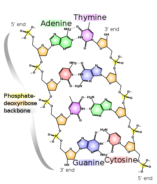
DNA and RNA are nucleic acids and make up the genetic instructions of an organism. Their monomers are called nucleotides, which are made up of individual subunits. Nucleotides consist of a 5-Carbon sugar (a pentose), a charged phosphate and a nitrogenous base (Adenine, Guanine, Thymine, Cytosine or Uracil). Each carbon of the pentose has a position designation from 1 through 5. One major difference between DNA and RNA is that DNA contains deoxyribose and RNA contains ribose. The discriminating feature between these pentoses is at the 2′ position where a hydroxyl group in ribose is substituted with a hydrogen.

DNA has a double-helical structure. Two antiparallel strands are bound by hydrogen bonds.
The following video illustrates the structure and properties of DNA.
DNA is a double helical molecule. Two antiparallel strands are bound together by hydrogen bonds. Adenine forms 2 H-bonds with Thymine. Guanine forms 3 H-bonds with Cytosine. This AT & GC matching is referred to as complementarity. While the nitrogenous bases are found on the interior of the double helix (like rungs on a ladder), the repeating backbone of pentose sugar and phosphate form the backbone of the molecule. Notice that phosphate has a negative charge. This makes DNA and RNA, overall negatively charged.

There are 10 bases for every complete turn in the double helix of DNA.
Nucleic Acids: DNA Extraction and Dische’s Diphenylamine Test (Activity)
Prelab Questions
- What are fruits?
- Where do they come from?
- What are they made of?
- Use phylogeny to classify plants (DKPCOFGS)
- Where is DNA located within the fruits? Where is it located in you?
- Why would you want to extract DNA from an organism?
- What class of molecule is DNA?
Extraction of DNA from fruit
single panel instructions can be found at https://github.com/jeremyseto/bio-oer/blob/master/figures/chemistry/DNA/fruitdnaisolation.svg
- Mash about 10g or 3cm of over-ripe banana OR 3 grapes OR 1 strawberry in zip-top bag.
- Over-ripe banana is best since the cell walls are already decomposing
- Physical mashing continues to break up the cell walls
2. Add 7ml of salt solution.
- The salt solution helps the DNA to aggregate (clump together).
3. Add 7ml of liquid detergent and mix.
- Dissolves the lipids in the cell and nuclear membranes
- Releases DNA into the salt solution
4. Place a coffee filter over a cup or beaker and fasten with an elastic band.
- Pour mash through the filter into a beaker
5. Pour about 5ml of filtrate into a test tube.
6. Slowly pour an EQUAL volume of cold ethanol down the side of the tube to form a layer on top of the fruit fluid.
- Carefully run the alcohol down the side to form a separate layer on top of the fruit solution.
- Do not mix the alcohol and banana solution.
- Ice-cold 100% ethanol works best.
7. Spool the DNA: use a plastic loop or glass rod to gently swirl at the interface of the two solutions.
- The interface is where the two solutions meet.
- DNA is not soluble in alcohol.
- Bubbles may form around a wooly substance (this is the DNA).
8. Transfer the DNA.
Dische Diphenylamine Test For DNA
DNA can be identified chemically with the Dische diphenylamine test. Acidic conditions convert deoxyribose to a molecule that binds with diphenylamine to form a blue complex. The intensity of the blue color is proportional to the concentration of DNA. The Dische’s Test will detect the deoxyribose of DNA and will not interact with the ribose in RNA. The amount of blue corresponds to the amount of DNA in solution.

The diphenylamine compound of the Dische’s test interacts with the deoxyribose of DNA to yield a blue coloration.
- Obtain 3 test tubes and number them 1-3.
- Suspend the spooled DNA in 3 ml of distilled water. MIX.
- Add to tubes:
- 2 ml of DNA solution
- 1 ml of DNA solution with 1 ml H2O
- 2 ml of H2O
- Add 2 ml of the Dische’s diphenylamine reagent to each tube and mix thoroughly.
- Place in a boiling water bath for 10 minutes.
- Evaluate your results. A clear tube indicates no nucleic acids. A blue color indicates the presence of DNA. A greenish color indicates the presence of RNA.


