10.2: Lipids Aggregates in Water - Micelles and Liposomes
- Page ID
- 14977
\( \newcommand{\vecs}[1]{\overset { \scriptstyle \rightharpoonup} {\mathbf{#1}} } \)
\( \newcommand{\vecd}[1]{\overset{-\!-\!\rightharpoonup}{\vphantom{a}\smash {#1}}} \)
\( \newcommand{\id}{\mathrm{id}}\) \( \newcommand{\Span}{\mathrm{span}}\)
( \newcommand{\kernel}{\mathrm{null}\,}\) \( \newcommand{\range}{\mathrm{range}\,}\)
\( \newcommand{\RealPart}{\mathrm{Re}}\) \( \newcommand{\ImaginaryPart}{\mathrm{Im}}\)
\( \newcommand{\Argument}{\mathrm{Arg}}\) \( \newcommand{\norm}[1]{\| #1 \|}\)
\( \newcommand{\inner}[2]{\langle #1, #2 \rangle}\)
\( \newcommand{\Span}{\mathrm{span}}\)
\( \newcommand{\id}{\mathrm{id}}\)
\( \newcommand{\Span}{\mathrm{span}}\)
\( \newcommand{\kernel}{\mathrm{null}\,}\)
\( \newcommand{\range}{\mathrm{range}\,}\)
\( \newcommand{\RealPart}{\mathrm{Re}}\)
\( \newcommand{\ImaginaryPart}{\mathrm{Im}}\)
\( \newcommand{\Argument}{\mathrm{Arg}}\)
\( \newcommand{\norm}[1]{\| #1 \|}\)
\( \newcommand{\inner}[2]{\langle #1, #2 \rangle}\)
\( \newcommand{\Span}{\mathrm{span}}\) \( \newcommand{\AA}{\unicode[.8,0]{x212B}}\)
\( \newcommand{\vectorA}[1]{\vec{#1}} % arrow\)
\( \newcommand{\vectorAt}[1]{\vec{\text{#1}}} % arrow\)
\( \newcommand{\vectorB}[1]{\overset { \scriptstyle \rightharpoonup} {\mathbf{#1}} } \)
\( \newcommand{\vectorC}[1]{\textbf{#1}} \)
\( \newcommand{\vectorD}[1]{\overrightarrow{#1}} \)
\( \newcommand{\vectorDt}[1]{\overrightarrow{\text{#1}}} \)
\( \newcommand{\vectE}[1]{\overset{-\!-\!\rightharpoonup}{\vphantom{a}\smash{\mathbf {#1}}}} \)
\( \newcommand{\vecs}[1]{\overset { \scriptstyle \rightharpoonup} {\mathbf{#1}} } \)
\( \newcommand{\vecd}[1]{\overset{-\!-\!\rightharpoonup}{\vphantom{a}\smash {#1}}} \)
\(\newcommand{\avec}{\mathbf a}\) \(\newcommand{\bvec}{\mathbf b}\) \(\newcommand{\cvec}{\mathbf c}\) \(\newcommand{\dvec}{\mathbf d}\) \(\newcommand{\dtil}{\widetilde{\mathbf d}}\) \(\newcommand{\evec}{\mathbf e}\) \(\newcommand{\fvec}{\mathbf f}\) \(\newcommand{\nvec}{\mathbf n}\) \(\newcommand{\pvec}{\mathbf p}\) \(\newcommand{\qvec}{\mathbf q}\) \(\newcommand{\svec}{\mathbf s}\) \(\newcommand{\tvec}{\mathbf t}\) \(\newcommand{\uvec}{\mathbf u}\) \(\newcommand{\vvec}{\mathbf v}\) \(\newcommand{\wvec}{\mathbf w}\) \(\newcommand{\xvec}{\mathbf x}\) \(\newcommand{\yvec}{\mathbf y}\) \(\newcommand{\zvec}{\mathbf z}\) \(\newcommand{\rvec}{\mathbf r}\) \(\newcommand{\mvec}{\mathbf m}\) \(\newcommand{\zerovec}{\mathbf 0}\) \(\newcommand{\onevec}{\mathbf 1}\) \(\newcommand{\real}{\mathbb R}\) \(\newcommand{\twovec}[2]{\left[\begin{array}{r}#1 \\ #2 \end{array}\right]}\) \(\newcommand{\ctwovec}[2]{\left[\begin{array}{c}#1 \\ #2 \end{array}\right]}\) \(\newcommand{\threevec}[3]{\left[\begin{array}{r}#1 \\ #2 \\ #3 \end{array}\right]}\) \(\newcommand{\cthreevec}[3]{\left[\begin{array}{c}#1 \\ #2 \\ #3 \end{array}\right]}\) \(\newcommand{\fourvec}[4]{\left[\begin{array}{r}#1 \\ #2 \\ #3 \\ #4 \end{array}\right]}\) \(\newcommand{\cfourvec}[4]{\left[\begin{array}{c}#1 \\ #2 \\ #3 \\ #4 \end{array}\right]}\) \(\newcommand{\fivevec}[5]{\left[\begin{array}{r}#1 \\ #2 \\ #3 \\ #4 \\ #5 \\ \end{array}\right]}\) \(\newcommand{\cfivevec}[5]{\left[\begin{array}{c}#1 \\ #2 \\ #3 \\ #4 \\ #5 \\ \end{array}\right]}\) \(\newcommand{\mattwo}[4]{\left[\begin{array}{rr}#1 \amp #2 \\ #3 \amp #4 \\ \end{array}\right]}\) \(\newcommand{\laspan}[1]{\text{Span}\{#1\}}\) \(\newcommand{\bcal}{\cal B}\) \(\newcommand{\ccal}{\cal C}\) \(\newcommand{\scal}{\cal S}\) \(\newcommand{\wcal}{\cal W}\) \(\newcommand{\ecal}{\cal E}\) \(\newcommand{\coords}[2]{\left\{#1\right\}_{#2}}\) \(\newcommand{\gray}[1]{\color{gray}{#1}}\) \(\newcommand{\lgray}[1]{\color{lightgray}{#1}}\) \(\newcommand{\rank}{\operatorname{rank}}\) \(\newcommand{\row}{\text{Row}}\) \(\newcommand{\col}{\text{Col}}\) \(\renewcommand{\row}{\text{Row}}\) \(\newcommand{\nul}{\text{Nul}}\) \(\newcommand{\var}{\text{Var}}\) \(\newcommand{\corr}{\text{corr}}\) \(\newcommand{\len}[1]{\left|#1\right|}\) \(\newcommand{\bbar}{\overline{\bvec}}\) \(\newcommand{\bhat}{\widehat{\bvec}}\) \(\newcommand{\bperp}{\bvec^\perp}\) \(\newcommand{\xhat}{\widehat{\xvec}}\) \(\newcommand{\vhat}{\widehat{\vvec}}\) \(\newcommand{\uhat}{\widehat{\uvec}}\) \(\newcommand{\what}{\widehat{\wvec}}\) \(\newcommand{\Sighat}{\widehat{\Sigma}}\) \(\newcommand{\lt}{<}\) \(\newcommand{\gt}{>}\) \(\newcommand{\amp}{&}\) \(\definecolor{fillinmathshade}{gray}{0.9}\)Single Chain Amphiphiles and Micelles
An understanding of lipids in simple solutions in the lab is incredibly helpful to understanding them in vivo. The same physical-chemical constraints would apply to the complex environment of the cell. What is different in the cell is that lipids are found in a cellular environment that is incredibly crowded with proteins that bind, synthesize, and breakdown lipids. Nevertheless, we can apply what we know from the test tube experiments to the cell.
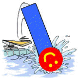
To understand how molecules might react, it helps a bit to pretend you are a molecule and ask yourself what would you do! We want to know how lipid molecules, specifically single and double-chain amphiphiles, interact with each other and solvent when they are added to water. Before you read the answer, look at the image below and ask yourself the question: What would I do if I were a single chain amphiphile and jumped into water as shown in Figure \(\PageIndex{1}\)?

Here is what they do. When added to water, some single-chain amphiphiles dissolve in water while others form a monolayer on the surface of the water. If enough enter the solution and exceed their net solubility, they self-aggregate to form micelles. These outcomes are shown in Figure \(\PageIndex{2}\).
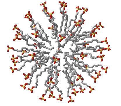
Figure \(\PageIndex{3}\) shows an interactive iCn3D model of an sodium dodecylsulfate (SDS) micelle
Figure \(\PageIndex{3}\): Sodium dodecylsulfate (SDS) micelle (Copyright; author via source).
Click the image for a popup or use this external link: not available
Double-chain amphiphiles, in contrast, form bilayers instead of micelles. (Note: single and double-chain amphiphiles can form other multimolecular aggregate structures as well, but micelles and bilayers are the most common and are the only ones we will consider.)
The micelle interior is completely nonpolar. Spherical bilayers that enclose an aqueous compartment are called vesicles or liposomes. Micelles and bilayers, formed from single and double-chain amphiphiles, respectively, represent noncovalent aggregates and hence are formed by an entirely physical process. No covalent steps are required.
Common single-chain amphiphiles that form micelles are detergents (like sodium dodecyl sulfate - SDS) as well as fatty acids, which themselves are detergents. Sodium hydroxide feels slippery on your skin since the base hydrolyses the fatty acids esterified to skin lipids. The free fatty acids then aggregate spontaneously to form micelles which act like detergents, and are also slippery.
Micelle/detergents in water are an example of an emulsion of two liquids that are generally immiscible in each other unless one is dispersed into small droplets into the other Fine oil drops can be dispersed in water and fine aqueous drops can be dispersed in a nonpolar liquid. Many vaccines are formulated as this later type of emulsion. Grease and oil in your clothes can be carried away by "diving" into the nonpolar part of the detergent micelle which is dispersed in water as an emulsion. Another example of an emulsion or more properly a colloid is a cloud, a dispersion of liquid water droplets in a solvent, the atmosphere.
The formation of these structures can be understood from the study of noncovalent interactions but also through thermodynamics. In a micelle, the buried acyl chains can interact and be stabilized by induced dipole-induced dipole forces as the nonpolar carbons and hydrogen are in van der Walls contact. They are sequestered from water. This view fits our simple axiom of "like-dissolves like". The polar head groups can be stabilized by ion-dipole bonds between charged head groups and water. Likewise, H-bonds between water and the head group stabilize the exposed head groups in water. Repulsive forces may also be involved. Head groups can repel each other through steric factors, or ion-ion repulsion from like-charged head groups. The attractive forces must be greater than the repulsive forces, which lead to these molecular aggregates.
From a thermodynamic approach, one problem arises with this simple explanation. For a micelle or bilayer to form, many monomers must aggregate to form a single micelle or vesicle, which is entropically disfavored! So let's delve into the thermodynamics of micelle formation.
ΔG, the free energy chance for a reaction, determines the spontaneity and extent of a chemical or physical reaction. The free energy of the system depends on 3 variables, temperature T, pressure P, and n, the number of moles of each substance. For the latter, think of solute X on two different sides of a permeable membrane. If the concentration of X is the same on each side, as shown in the system below, the system is in equilibrium as shown in Figure \(\PageIndex{4}\).
If the system is composed of two different parts, A and B, the system is at equilibrium (ΔG=0) if TA = TB, PA = PB, and the change in the absolute free energy per mole of A is ΔGA/Δn = ΔGB/Δn. More precisely, using simple calculus, we would discuss incremental changes in absolute free energy/mol, dGA/dn for A (often called the chemical potential of A, μA), and dGB/dn or μB)for B. At equilibrium dGA/dn = dGB/dn. (We will use the symbol G here instead of μ). G then is the absolute free energy/mol (again often called the chemical potential), where G=Go +RTln[A]. The equations you used in introductory chemistry can be written.
\begin{equation}
\begin{array}{l}
\Delta \mathrm{G}=\Delta \mathrm{G}^{0}+\mathrm{RTIn} \mathrm{Qr} \\
\Delta \mathrm{G}=\Delta \mathrm{H}-\mathrm{T} \Delta \mathrm{S} \\
\Delta \mathrm{G}^{0}=\Delta \mathrm{H}^{0}-\mathrm{T} \Delta \mathrm{S}^{0} \\
\Delta \mathrm{G}^{0}=-\mathrm{RTInK} \mathrm{eq}
\end{array}
\end{equation}
Now let's apply this to the chemical equation for micelle formation:
n SCA <==> 1 micelle
where SCA represents a single chain amphiphile. At first glance, we might suspect that:
- ΔH0 < 0 since the induced dipole-induced dipole interactions among the buried acyl chains in the micelle would be much more favorable than the water-acyl interactions for the monomeric amphiphile in solution. This notion is supported by our aphorism, "like dissolves like". Of course, we couldn't ignore polar interactions (H bonding for example) among the head groups and water, but we might expect these to be equally favorable in both the monomeric and micellular states.
- ΔS0 < 0 since we are forming a very ordered state (a single micelle) with much less entropy from a state (single chains amphiphiles dispersed in solution) with much more entropy.
Hence it would appear that micelle formation is enthalpically favored but entropically disfavored. Let's examine this issue more closely. First, we need to obtain a greater understanding of ΔGo which should give us a clue as to where a SCA would "want" to be in this mixture. Remember, ΔG0 is a constant that at a given T, P, and solvent conditions and depends only on the relative stability of a molecule in a given environment and not its concentration.
Traube, in 1891, noticed that single chain amphiphiles tend to migrate to the surface of the water and decrease its surface tension (ST.) He observed that the decrease in ST is directly proportional to the amount of amphiphile, added up until a certain point, at which added amphiphile has no additional effect. In other words, the response of ST saturates at some point.
We are more interested in what happens to amphiphiles in bulk water, not at the surface. As we showed in Figure 2 above, monomeric single chain amphiphiles are in equilibrium with single chain amphiphiles in micelles. Assume you have a way to measure monomeric single chain amphiphile in solution. What happens to its concentration as you add more and more SCA to the mixture? Turns out you observe the same effect that Traube noted with changes in surface tension. This explanation goes like this: as more amphiphile is added, more goes into bulk solution as monomers. At some point, there are enough amphiphiles added to form micelles. After this point, added amphiphiles form more micelles and no further increases in monomeric single chain amphiphiles are noted. The concentration of amphiphile at which this occurs is the critical micelle concentration (CMC). Figure \(\PageIndex{5}\) shows a graph of monomeric single chain amphiphile in solution versus the concentration added to the solution.
This saturation effect can be observed with other systems as well.
- Consider the amount of NaCl(aq) in the solution as more NaCl(s) is added to water. At some point, the water is saturated with dissolved NaCl, and no further increase in NaCl (aq) occurs.
- Consider the amount of a sparing soluble hydrocarbon (HC) in water. After saturation, phase separation occurs.
Now consider the addition of a drop of a slightly soluble hydrocarbon liquid (HCL) into water, as pictured in the diagram below. At t=0, the system is not at equilibrium and some of the HC will transfer from the pure liquid to water, so at time t=0, ΔGTOT < 0. This is illustrated in Figure \(\PageIndex{6}\).
The following equations can be derived.
\begin{equation}
\begin{array}{c}
\Delta \mathrm{G}_{\mathrm{TOT}}=\left(G_{\mathrm{HC}-\mathrm{W}}\right)-\left(G_{\mathrm{HC}-\mathrm{L}}\right)=\mathrm{G}_{\mathrm{HC}-\mathrm{W}}^{0}+R T \ln [\mathrm{HC}]_{\mathrm{W}}-\left(\mathrm{G}_{\mathrm{HC}-\mathrm{L}}^{0}+R T \ln [\mathrm{HC}]_{\mathrm{L}}\right)= \\
\Delta \mathrm{G}_{\mathrm{TOT}}=\left(\mathrm{G}_{\mathrm{HC}-\mathrm{W}}^{0}-\mathrm{G}_{\mathrm{HC}-\mathrm{L}}^{0}\right)+R T \ln \left([\mathrm{HC}]_{\mathrm{W}}-\ln [\mathrm{HC}]_{\mathrm{L}}\right)= \\
\Delta \mathrm{G}_{\mathrm{TOT}}=\Delta \mathrm{G}^{0}+R T \ln \frac{[\mathrm{HC}]_{\mathrm{W}}}{[\mathrm{HC}]_{\mathrm{L}}}
\end{array}
\end{equation}
Now add a bit more complexity to the last example. Add a hydrocarbon x, to a biphasic system of water and octanol and shake it vigorously as shown in Figure \(\PageIndex{7}\). At equilibrium, x would have "partitioned" between the two mostly immiscible phases.
A simple favorable reaction can be written for this system: x aq → x oct.
If X is a hydrocarbon, ΔG < 0. Also, ΔGo < 0, since this term is independent of concentration and depends only on the intrinsic stability of x in water in comparison to that of octanol. This simple equation holds:
\begin{equation}
\Delta \mathrm{G}_{\mathrm{TOT}}=\left(\mathrm{G}_{\mathrm{X}-\mathrm{oct}}^{0}-\mathrm{G}_{\mathrm{X}-\mathrm{w}}^{0}\right)+R T \ln \frac{[\mathrm{X}]_{\mathrm{oct}}}{[\mathrm{X}]_{\mathrm{w}}}=\Delta \mathrm{G}^{0}+R T \ln \frac{[\mathrm{X}]_{\mathrm{oct}}}{[\mathrm{X}]_{\mathrm{w}}}
\end{equation}
At equilibrium, ΔG0=0 and the equation can be rewritten as:
\begin{equation}
\Delta \mathrm{G}^{0}=-R T \ln \frac{[\mathrm{X}]_{\mathrm{oct}}}{[\mathrm{X}]_{\mathrm{w}}}=-\mathrm{RTlnK}_{\mathrm{part}}
\end{equation}
where Kpart is the equilibrium partition coefficient for X in octanol and water. This can readily be determined in the lab. Just shake a separatory flask with a biphasic system of octanol and water after injecting a bit of X. Then separate the layers and determine the concentration of x in each phase. Plug these numbers into the last equation. You should be able to predict the sign and relative magnitude of ΔGo since it does not depend on concentration, but only on the intrinsic stability of the molecules in the different environments. Kpaft values are often determined for drugs since they often must diffuse across cell membranes to move into the cytoplasm where they can act. Drugs hence must have a reasonable Kpart to pass through the membrane but not so high that they are insoluble.
Double Chain Amphiphiles and Bilayers
In contrast to single chain amphiphiles, double-chain amphiphiles added to water form monolayers and vesicles called liposomes, as shown in Figure \(\PageIndex{8}\).
They can be unilamellar, consisting of a single bilayer surrounding the internal aqueous compartment, or multilamellar, consisting of multiple bilayers surrounding the enclosed aqueous solution. You can image imagine that multilamellar vesicles resemble an onion with its multiple layers. Cartoons of unilamellar and multilamellar liposomes are shown in Figure \(\PageIndex{9}\), where each concentric circle represents a bilayer.

Liposomes vary in diameter. They can be generally categorized into small (S, diameter < 25 nm), intermediate (I, diameter around 100 nm), and large (L, diameter from 250-1000 nm). If these vesicles are unilamellar, they are abbreviated as SUV, IUV, and LUV
The chemical composition of liposomes made in the lab can be widely varied. Most contain neutral phospholipids like phosphatidylcholine phosphatidyl ethanolamine (PE), or sphingomyelin (SM), supplemented, if desired, with negatively charged phospholipids, like phosphatidyl serine (PS) and phosphatidyl glycerol (PG). In addition, single-chain amphiphiles like cholesterol (C) and detergents can be incorporated into the bilayer membrane, which modulates the fluidity and transition temperature (Tm) of the bilayer. If present in too great a concentration, single-chain amphiphiles like detergents, which form micelles, can disrupt the membrane so completely that the double-chain amphiphiles become incorporated into detergent micelles, now called mixed micelles, in a process that effectively destroys the membrane bilayer.
The properties of liposomes (charge density, membrane fluidity, and permeability) are determined by the lipid composition and size of the vesicle. The desired properties will be, in turn, determined by the use of the particular liposome. The vesicles offer wonderful, simple models to study the biochemistry and biophysics of natural membranes. Membrane proteins can be incorporated into the liposome bilayer using the exact method you will be using. But apart from these purposes, liposomes can be used to encapsulate water-soluble molecules such as nucleic acids, proteins, and toxic drugs. These liposomes can be targeted to specific cells if antibodies or other molecules which will bind specifically to the target cell can be incorporated into the bilayer of the vesicle. Intraliposomal material may then be transferred into the cell either by fusion of the vesicle with the cell or by endocytosis of the vesicle.
Liposomes could also be called lipid nanoparticles as they have sizes ranging up to 1000 nm. Liposomes are vesicular - with small aqueous-filled compartments. They can be made with encapsulated drugs for delivery to target cells through blood transport. They act as an emulsion in water.
Lipid nanoparticles can also be particulate (insoluble), which slowly degrade and release their contents slowly in situ. Most recently, particulate lipid nanoparticles have helped save the world by being used to encapsulate messenger RNA (mRNA) for the spike protein from the SARS-CoV-2 virus which causes the COVID-19 pandemic. These lipid nanoparticles are used in the vaccine against the coronavirus mRNA molecules which encode the spike protein are "encapsulated" in the lipid nanoparticle. The mRNA contains specially modified nucleotides to increase their stability. The lipid nanoparticles also contain positively-charged lipids which help stabilize the negatively charged mRNA from degradation. The nanoparticles are endocytosed into cells, where the mRNA can be translated into SARS-CoV-2 spike protein, required for whole virus entry into the cell. The spike protein is then recognized by the immune system.
The lipids used in the formulation of these nanoparticles include fatty acids, mono-, di- and triglycerols, glycerophospholipids, waxes (like cetyl palmitate), and other positively charged lipids including stearyl amine, benzalkonium chloride, cetrimide, cetyl pyridinium chloride, and dimethyldioctadecylammonium bromide. These are shown in Figure \(\PageIndex{10}\)
Figure \(\PageIndex{10}\): Amphiphiles used to make lipid nanoparticles
Why Micelles and Bilayers?
Micelles and liposomes form spontaneously - i.e. ΔG < 0. But why do single chain amphiphiles form micelles and double-chain amphiphiles form bilayers? Let's think about this from a thermodynamics and structural sense.
As the number of Cs in the nonpolar carbon (NC) chain increases, the ΔG for the transferring into a micelle, or by analogy, for a single chain amphiphile entering a micelle, becomes more and more negative (i.e more favored). The following equation seems to apply to the transfer of a single chain amphiphile into a micelle:
ΔGo = Go (mic) - Go (aq) = + number - 709 NC
\begin{equation}
\Delta \mathrm{G}^{\circ}=\mathrm{G}^{\circ} \text { (mic) }-\mathrm{G}^{\circ}(\mathrm{aq})=+\text { number }-709 \mathrm{NC}
\end{equation}
where NC is the number of carbon atoms in the chain. The first positive term depends on the nature of the head group, while the second negative term is independent of the head group. These + and - terms bring us back to the principle of opposing forces we discussed when we looked at the noncovalent interactions involved in micelle and bilayer formation.
There are attractive interactions, including induced dipole-induced dipole interactions among the chains and dipole-ion, and H-bond interactions with water and the head groups. There are likewise repulsive interactions arising from steric hindrance with bulky heady groups and ion-ion repulsions. Of course, there are also entropic considerations. Let us now consider these factors as we explore what might happen to a preformed micelle as we try to put more single chain amphiphiles (sca) into it.
As we increase the number of Cs in the SCA, the micelles would have a larger radius. For a given SCA with a fixed number of Cs, once a spherical micelle is formed, it can no longer retain its spherical shape if more SCAs are added. Imagine increasing the diameter of a spherical micelle 10x. A large part of the inside would have no atoms or be filled with water, which would not be favorable. Therefore, if the micelle is to grow, it can do so only by changing shape to something other than a sphere. By squeezing a tennis ball, one can imagine that the shape could distort to a circular cylinder with end caps. In this way, the acyl chains can still interact. The only problem is that head groups will now be closer than they were in the sphere. This is simplistically illustrated in Figure \(\PageIndex{11}\).
Imagine in a sphere the head groups radiating in a perpendicular direction from the sphere surface. As the sphere is distorted into a cylinder, the head groups would come closer together, and hence they will experience more steric interference. If a cylinder can be formed, however, it could continue to grow as long as needed with no further compression required. Imagine now that you further compressed the cylinder into a planar "bilayer" structure. The head groups would be even closer and experience even more repulsion. This bilayer will not form since growth can occur in the cylindrical phase without the added repulsion.
Now consider a double-chain amphiphile (DCA). In the case of a SCA, the number (N) of head groups (HG) = the number of acyl chains (CH). Hence the surface area per HG is equal to the area per HC. Or: As/N HG = As/ N CH. For a DCA, N HG = N CH/2, therefore As/N HG = 2As/NCH. There is twice the surface area available per head group compared to that of the SCA. Therefore the DCA can tolerate more compression. It can easily be compressed to a bilayer, which as we saw, has much less As/HG. The cylindrical form has too much space per head group since water can enter the structure. The extra closeness of head groups in the bilayer can be tolerated even more since the ΔGo for transfer of a DCA into a micelle is 60% more negative than that of a SCA. The As/HG for closed vesicles differs only slightly from that of a truly planar bilayer since the vesicles are so large compared to a micelle.
Once again, we have discovered that structure mediates function. We can account for the fact that SCA and DCA form micelles and bilayers, respectively, by understanding the structure of the monomers!
In reality, things are more complicated.
The general rule holds that single chain amphiphiles form micelles and double-chain amphiphiles form bilayers. However, under the right condition, single-chain fatty acids can form bilayers if the pH is low enough that the head group is protonated and uncharged. Why would that make a difference? Fatty acids like oleic acid would be a prime candidates for components of the membranes of protocells in the evolution of life from abiotic conditions. Likewise, short double-chain amphiphiles can make micelles. In addition, other lipids phases can be observed. Which aggregates or phases ultimately form depends on the structure of the lipid, the solvent conditions, and the temperature. These include the following phases:
- lamellar gel (Lb) and lamellar liquid crystalline (La) phases
- hexagonal HI (cylinders packed in the shape of a hexagon with polar heads facing out into the water
- hexagonal HII (cylinders packed in the shape of a hexagon with acyl chains pointing out as in reverse micelles, and
- micellar (M).
We will discuss them in more detail in the next section.


