

4.7: Fibrillar Proteins
- Page ID
- 62324


\( \newcommand{\vecs}[1]{\overset { \scriptstyle \rightharpoonup} {\mathbf{#1}} } \)
\( \newcommand{\vecd}[1]{\overset{-\!-\!\rightharpoonup}{\vphantom{a}\smash {#1}}} \)
\( \newcommand{\dsum}{\displaystyle\sum\limits} \)
\( \newcommand{\dint}{\displaystyle\int\limits} \)
\( \newcommand{\dlim}{\displaystyle\lim\limits} \)
\( \newcommand{\id}{\mathrm{id}}\) \( \newcommand{\Span}{\mathrm{span}}\)
( \newcommand{\kernel}{\mathrm{null}\,}\) \( \newcommand{\range}{\mathrm{range}\,}\)
\( \newcommand{\RealPart}{\mathrm{Re}}\) \( \newcommand{\ImaginaryPart}{\mathrm{Im}}\)
\( \newcommand{\Argument}{\mathrm{Arg}}\) \( \newcommand{\norm}[1]{\| #1 \|}\)
\( \newcommand{\inner}[2]{\langle #1, #2 \rangle}\)
\( \newcommand{\Span}{\mathrm{span}}\)
\( \newcommand{\id}{\mathrm{id}}\)
\( \newcommand{\Span}{\mathrm{span}}\)
\( \newcommand{\kernel}{\mathrm{null}\,}\)
\( \newcommand{\range}{\mathrm{range}\,}\)
\( \newcommand{\RealPart}{\mathrm{Re}}\)
\( \newcommand{\ImaginaryPart}{\mathrm{Im}}\)
\( \newcommand{\Argument}{\mathrm{Arg}}\)
\( \newcommand{\norm}[1]{\| #1 \|}\)
\( \newcommand{\inner}[2]{\langle #1, #2 \rangle}\)
\( \newcommand{\Span}{\mathrm{span}}\) \( \newcommand{\AA}{\unicode[.8,0]{x212B}}\)
\( \newcommand{\vectorA}[1]{\vec{#1}} % arrow\)
\( \newcommand{\vectorAt}[1]{\vec{\text{#1}}} % arrow\)
\( \newcommand{\vectorB}[1]{\overset { \scriptstyle \rightharpoonup} {\mathbf{#1}} } \)
\( \newcommand{\vectorC}[1]{\textbf{#1}} \)
\( \newcommand{\vectorD}[1]{\overrightarrow{#1}} \)
\( \newcommand{\vectorDt}[1]{\overrightarrow{\text{#1}}} \)
\( \newcommand{\vectE}[1]{\overset{-\!-\!\rightharpoonup}{\vphantom{a}\smash{\mathbf {#1}}}} \)
\( \newcommand{\vecs}[1]{\overset { \scriptstyle \rightharpoonup} {\mathbf{#1}} } \)
\(\newcommand{\longvect}{\overrightarrow}\)
\( \newcommand{\vecd}[1]{\overset{-\!-\!\rightharpoonup}{\vphantom{a}\smash {#1}}} \)
\(\newcommand{\ket}[1]{\left| #1 \right>}\)
\(\newcommand{\bra}[1]{\left< #1 \right|}\)
\(\newcommand{\braket}[2]{\left< #1 \vphantom{#2} \right| \left. #2 \vphantom{#1} \right>}\)
\(\newcommand{\braopket}[3]{\left< #1 \vphantom{#2}\vphantom{#3} \right| #2 \vphantom{#1}\vphantom{#3} \left| #3 \vphantom{#1}\vphantom{#2} \right>}\)
\(\newcommand{\qmvec}[1]{\mathbf{\vec{#1}}}\)
\(\newcommand{\op}[1]{\hat{\mathbf{#1}}}\)
\(\newcommand{\expect}[1]{\langle #1 \rangle}\)
\(\newcommand{\dfn}[1]{\emph{\textbf{#1}}}\)
(Learning goals written by Claude, Sonnet 4.6, Anthropic)
Collagen and α-Keratin: Structure and Assembly
- Describe the unusual primary structure of Type I collagen — (GXY)n repeats with glycine at every third position, ~17% proline, post-translational hydroxylation of Pro and Lys requiring vitamin C as cofactor, and glycosylation of hydroxylysine — and explain how each feature contributes to the formation and stability of the triple helix, including the role of interstrand hydrogen bonds and n→π* electron donation in protecting peptide bonds from hydrolysis.
- Explain the hierarchical assembly of α-keratin from Type I/Type II monomer coiled-coil dimers through staggered tetramers, protofilaments, and intermediate filaments, identify the noncovalent forces (hydrophobic interactions between apolar helical surfaces, complementary ion-ion interactions between acidic and basic monomers) that drive each level of assembly, and predict how vitamin C deficiency or keratin gene mutations would disrupt this hierarchy.
Elastic and Resilient Fibrous Proteins
- Compare elastin, resilin, and spider dragline silk in terms of amino acid composition (hydrophobic residues, Gly/Pro content), degree of intrinsic disorder, crosslinking chemistry (covalent vs. noncovalent), resilience values, and mechanical properties (strength, toughness, extensibility, Young's modulus), and explain at a molecular level why each protein's sequence and structural features produce its characteristic elastic behavior.
- Describe the three-state conformational pathway of spider dragline silk MaSp1 spidroins — soluble disordered monomer → liquid-liquid phase separation (LLPS) condensate → β-sheet crystalline fiber — explaining how phosphate-mediated ion exchange promotes Arg–Tyr cation–π interactions, displaces Arg from Ala contacts, and nucleates poly-Ala β-sheet formation, and connect this to the broader principle of LLPS/liquid-liquid demixing discussed earlier in the course.
Fibrinogen, Myosin, and the Relationship Between Fibrous Structure and Function
- Describe how thrombin-mediated proteolytic removal of fibrinopeptides A and B from fibrinogen's central E region exposes "knob" sequences (Gly-Pro-Arg and Gly-His-Arg) that bind noncovalently to complementary "holes" on adjacent fibrinogen molecules, and explain how fibrinogen's elongated, flexible α/β/γ hexameric architecture with intrinsically disordered αC regions enables fibrin clot assembly.
- Explain how myosin heavy chain alternates between the extended 6S filament-forming conformation (active in actin-mediated ATP hydrolysis and force generation) and the compact folded 10S conformation (in which head-head and head-tail interactions inhibit all aspects of contraction), and connect the 28-residue heptad repeat of the coiled-coil tail to the structural basis of thick filament assembly.
Introduction
Most proteins have a roughly spherical or "globular" tertiary structure. However, many proteins form elongated fibrils with properties such as elasticity: they deform when a force is applied and return to their original state. Elastic molecules must store energy (go to a higher energy state) when the elongating force is applied, and the energy must be released on return to the equilibrium resting structure. Structures that can store and release energy when subjected to a force have resiliency. Proteins that stretch with an applied force include elastin (in blood vessels, lungs, and skin where elasticity is required), resilin in insects (which stretches on wing beating), silk (found in spider webs and whose partial structure we showed in 4.2), and fibrillin (found in most connective tissues and cartilage). Some proteins have high resiliency (90% in elastin and resilin), while others are only partially resilient (35% in silk, which has a tensile strength approaching that of stainless steel).
In contrast to rubber, which has an amorphous structure that imparts elasticity, these proteins, although they have dissimilar amino acid sequences, appear to share a common structure inferred from their DNA sequences. In some proteins (such as fibrillin), a folded beta-sheet domain unfolds like a stretched accordion. Others, like elastin and spider silk, have a beta-sheet domain and other secondary structures (alpha-helices and beta turns) along with Pro and Ala repetitions. Scientists are studying these structures to help synthesize new elastic and resilient products.
Elongated structures characterize fibrous proteins. These proteins often aggregate into filaments or bundles, forming structural scaffolds in biological systems. Within animals, the two most abundant fibrous protein families are collagen and α-keratin. Let's start our exploration of fibrillar proteins with these.
Collagen
Collagen is the most abundant protein in mammals, making up 25% to 35% of the whole-body protein content. It is predominantly found in the extracellular space of various connective tissues. Collagen has a unique quaternary structure: three protein strands wound together to form a triple helix. It is mostly found in fibrous tissues such as tendons, ligaments, and skin.
Depending upon the degree of mineralization, collagen tissues may be rigid (bone), compliant (tendon), or have a gradient from rigid to compliant (cartilage). It is also abundant in corneas, blood vessels, the gut, intervertebral discs, and dentin. In muscle tissue, it serves as a major component of the endomysium. Collagen constitutes one to two percent of muscle tissue and accounts for 6% of the weight of strong, tendinous muscles. Fibroblasts are the most common cells that produce collagen. Gelatin, used in food and industry, is irreversibly hydrolyzed collagen. Partially and fully hydrolyzed collagen powders are also used as dietary supplements. Collagen also participates in many binding interactions with target proteins, in addition to its structural role.
The name collagen comes from the Greek (kólla), meaning "glue," and the suffix -gen, denoting "producing." This refers to the compound's early use in boiling the skin and tendons of horses and other animals to make glue.
Over 90% of the collagen in the human body is type I. However, as of 2011, researchers had identified and described 28 types of collagen, divided into several groups based on the structures they form. The five most common types are:
- Type I: skin, tendon, vasculature, organs, bone (the main component of the organic part of bone)
- Type II: cartilage (the main collagenous component of cartilage)
- Type III: reticulate (the main component of reticular fibers), commonly found alongside Type I
- Type IV: forms basal lamina, the epithelium-secreted layer of the basement membrane
- Type V: cell surfaces, hair, and placenta
Let's focus on Type I collagen, which has an unusual amino acid composition and sequence:
- Glycine occurs at almost every third residue.
- Proline makes up about 17% of collagen.
- Collagen contains many hydroxyprolines and hydroxylysines, which are formed by post-translational modifications catalyzed by different enzymes, both of which require vitamin C as a cofactor.
- Some hydroxylysines are glycosylated, mostly with disaccharides.
Figure \(\PageIndex{1}\) shows the post-translational hydroxylations of lysine and proline.

Vitamin C deficiency causes scurvy, a serious and painful disease in which defective collagen prevents the formation of strong connective tissue. Gums deteriorate and bleed, with loss of teeth; skin discolors, and wounds do not heal. Before the 18th century, this condition was notorious among long-duration military expeditions, particularly naval ones. Participants were deprived of foods containing vitamin C. Many bacteria and viruses secrete virulence factors, such as the enzyme collagenase, which destroys collagen or interferes with its production.
Collagen has many (GXY)n repeats, where G is glycine (Gly), and X and Y are frequently proline (Pro) and hydroxyproline (Hyp). Three strands of collagen self-associate to form a triple-stranded helix with 10 GXY triplets in 3 complete turns of the helix. Others suggest seven triplet units in 2 turns of the strands. Note that the helix of each strand in the triple helix is not an alpha helix and has different phi/psi angles. Each strand is "frameshifted" by one amino acid, resulting in a staggered arrangement of the individual strands and helices. The glycines are buried along the central axis, so no essential hydrophobic core exists. The X and Y amino acids are solvent-exposed. All the other side chains, both hydrophobic and hydrophilic, are likewise exposed to solvent. Hydrogen bonding occurs between the amide hydrogen of the peptide bond of Gly and the carbonyl O of an X amino acid in another chain.
Figure \(\PageIndex{2}\) shows an interactive iCn3D model of a triple helical collagen-like peptide (4Z1R). The main chain atoms, shown in CPK colors, form hydrogen bonds with neighboring chains. The side chains are colored by chain (blue, brown, and magenta). Two sets of Pro-HPro-Gly repeats are labeled.

 Figure \(\PageIndex{2}\): Triple helical collagen-like peptide (4Z1R). (Copyright; author via source).
Click the image for a popup or use this external link: https://structure.ncbi.nlm.nih.gov/icn3d/share.html?TCrji1wPhekypJtc6
Given its widespread nature and its role in the structural stability of organisms, collagen is a very stable biomolecule. It is so stable that traces of it have even been found in the fossil remains of dinosaurs like T. rex from 80 million years ago and a Lufengosaurus from 195 million years ago. The peptide bond is thermodynamically unstable in an aqueous solution, so collagen, like other proteins, would be expected to hydrolyze over a period of hundreds of years. Its unique repetitive sequence, aggregation into a triple helix, and biological environment (such as in bone) help protect it from degradation, but not for millions of years. Another previously overlooked structural feature must inhibit hydrolysis at the electrophilic carbonyl of its peptide bond.
To investigate this extreme stability, the resistance of small nitrophenyl esters of N-acylated prolines in both cis and trans conformations to hydrolysis was studied in an aqueous solution using NMR. Figure \(\PageIndex{3}\) below shows the structure of these cis and trans isomers.

Figure \(\PageIndex{3}\): Hydrolysis of N-acylated-Pro-p-nitrophenol esters
The equilibrium constant (Kcis/trans) for the cis ↔ trans isomers increases with R group size. Remember, for X-Y peptide bonds in dipeptides, the Kcis/trans is about 1000 (trans favored), but for X-Pro, it's around 4. However, in collagen, all X-Pro bonds are trans to enable triple helix formation.
The kinetics of spontaneous hydrolysis in solution (Tris-HCl buffer, pH 8, 0.1 M NaCl, and 1% DMSO to increase solubility) were studied by measuring A400 nm during the release of the nitrophenolate anion. There is a remarkable resistance to hydrolysis in the trans isomer that can best be accounted for by an interaction between a nonbonded (n) electron pair on the acyl group (corresponding to the X C=O in an XY dipeptide sequence) and the C=O of the ester. This likely results from some stabilizing electron donation from the n orbital of the O to the pi* antibonding C=O orbital. In hydrolysis, water or OH- would donate a lone pair. This becomes disallowed in the sterically restricted trans form since only two electrons can occupy an orbital (in this case, the pi* antibonding orbital). The authors describe this as an example of the Pauli Exclusion principle (only 2 electrons per orbital), which you encountered in introductory chemistry classes.
Figure \(\PageIndex{4}\) below shows representations of the interactions and an energy diagram from Chapter 2.4 (Solubility in an aqueous world - noncovalent interactions in depth) to illustrate these points.
 |
 |
Figure \(\PageIndex{4}\): Left, Interactions between a lone pair of electrons (n) on an adjacent group and the pi* MO of an adjacent C=O. Right, Relative energy levels of molecular orbitals (not to scale). https://chem.libretexts.org/Bookshel...c_Spectroscopy
In the triple collagen helix, the n→ pi* interactions are on the same strand, while stabilizing hydrogen bonds are between donors and acceptors on adjacent strands, as illustrated in Figure \(\PageIndex{5}\) below.

Figure \(\PageIndex{5}\): Interactions in a collagen triple helix.
(A) Crystal structure of a (Pro-Hyp-Gly)n triple helix. The three strands are white, gray, or black. In each strand, peptide-bond carbons are yellow (Pro-Hyp), orange (Hyp-Gly), or red (Gly-Pro). The image was generated with PyMOL using PDB entry 1v7h.59.
(B) n→π* Interactions and hydrogen bonds within the main chain of a collagen triple helix. Carbons are colored as in panel A. Interstrand hydrogen bonds polarize the shaded peptide bonds. Jinyi Yang, Volga Kojasoy, Gerard J. Porter, and Ronald T. Raines. ACS Central Science Article ASAP. DOI: 10.1021/acscentsci.4c00971. Sept 2024. DOI: 10.1021/acscentsci.4c00971. CC-BY 4.0
α-Keratin
α-keratin is the key structural element of hair, nails, horns, claws, hooves, and the outer layer of skin. Because of its tightly wound structure, it can function as one of the strongest biological materials and has many uses in mammals, from predatory claws to hair for warmth.
Hanukoglu and Fuchs determined the first α-keratin sequences. These sequences revealed two distinct but homologous keratin families: Type I and Type II keratins. There are 54 keratin genes in humans, 28 of which code for type I and 26 for type II. Type I proteins are enriched in Asp and Glu amino acids, while type II proteins contain more basic amino acids, such as lysine. This differentiation is especially important in α-keratins because, in forming a keratin dimer, the coiled-coil requires that one protein coil be type I and the other type II. Some type I and type II pairs are particularly complementary within each organism. For example, in human skin, K5, a type II α-keratin, pairs primarily with K14, a type I α-keratin, to form the α-keratin complex of the epidermis layer of cells in the skin.
Coiled-coil dimers then assemble into a tetramer of two staggered coiled-coil dimers. Two tetramers can then pack together to form an elongated protofilament, a very stable, left-handed superhelical structure, as shown in the figure below. The keratin filaments stay associated through hydrophobic interactions between apolar residues along the keratin's helical segments. This is illustrated in Figure \(\PageIndex{6}\).
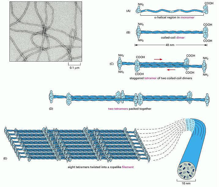
Figure \(\PageIndex{6}\): Assembly of Keratin Fibers. Wiki lectures. https://www.wikilectures.eu/w/Indivi...e_and_function
Initially, two keratin monomers (A) form a coiled-coil dimer structure (B). Two coiled-coil dimers join to form a staggered tetramer (C); next, the tetramers start to join together (D), ultimately forming a sheet of eight tetramers (E). The sheet of eight tetramers is then twisted into a left-handed helix, forming the final intermediate filament (E). An electron micrograph of the intermediate filament is shown in the upper left-hand corner.
Figure \(\PageIndex{7}\) shows an interactive iCn3D model of a dimer of Type I alpha-keratin (magenta backbone) and Type II (blue backbone) (6JFV).

Figure \(\PageIndex{7}\): Dimer of a Type I and Type II alpha-keratin backbones (6JFV). (Copyright; author via source).
Click the image for a popup or use this external link: https://structure.ncbi.nlm.nih.gov/i...g3L3sQh9dj5pw8
Acidic (red) side chains (Asp and Glu) and basic (blue) side chains (Lys) project away from the dimer. Both A and B chains have negative and positive side chains. This dimer's A (more acidic) chain has 5 Lys, 7 Arg, 1 Asp, and 16 Glu side chains, for a net charge of +12 -17 = -5. The dimer's B (more basic) chain has 8 Lys, 9 Arg, 5 Asp, and 12 Glu side chains for a net charge of +17 -17 = 0. It is more basic, with 17 lysine and arginine side chains, than the A chain, with 12. Depending on their 3D orientation, they could present a more positive face to the more negative monomer in the dimer.
Figure \(\PageIndex{8}\) shows an interactive iCn3D model of a spacefill model of the dimer. Note that a significant fraction of the nonpolar side chains are pointed inward between the two monomers and are much less exposed to solvent.

Figure \(\PageIndex{8}\): Dimer of a Type I and Type II alpha-keratin backbones in spacefill (6JFV). (Copyright; author via source).
Click the image for a popup or use this external link: https://structure.ncbi.nlm.nih.gov/i...uyJrw28mZ6x5h7
Elastin
As its name implies, the protein confers elasticity in target structures such as connective tissue and blood vessels. It has low-complexity hydrophobic domains, and the protein is cross-linked to form larger structures. It contains repeating hydrophobic amino acid sequences, mostly valine, proline, glycine, and alanine, and mimetics of the repeating structure (LGGVG)6 have been studied. This protein also displays significant disorder.
Resilin
The following description of resilin is taken directly from an article under Creative Commons Attribution 4.0 International License available at http://creativecommons.org/licenses/by/4.0/. Balu, R., Dutta, N.K., Dutta, A.K. et al. Resilin-mimetics as a smart biomaterial platform for biomedical applications. Nat Commun 12, 149 (2021). https://doi.org/10.1038/s41467-020-20375-x
Native elastomeric proteins are biomaterials that natural selection has perfected over billions of years to act as molecular springs in a wide range of biological systems, driving unique functions. Among native proteins, resilin is considered one of the most efficient elastic proteins. It is a structural protein mainly found in insect exoskeletons and exhibits outstanding resilience and fatigue life. Resilin was first described in the 1960s as a rubber-like protein found in the locust wing hinge and the dragonfly tendon. Early studies on resilin composition and structure revealed that the protein contains about 66% hydrophobic residues (much lower than in elastin), with about 45% proline and glycine combined. In their native state, resilins exist as di- and tri-tyrosine-crosslinked hydrogels and exhibit highly amorphous structures, as observed by X-ray diffraction and electron microscopy. During biosynthesis, pro-resilins (uncrosslinked) are secreted from the apical surface of the epidermal cells into the subcuticular space, where an enzyme-mediated process crosslinks them to form hydrogels. Over the next three decades, researchers also identified resilin in many other insects and arthropods, including copepods, reduviidae, and moths. In arthropods, resilin is largely involved in several different functions, including the flexibility and deformability of the membranous cuticle and joint systems, the storage of elastic energy in locomotion (jumping, flying, etc.) and catapulting systems, the adaptability to surface topography by multiple contact attachment, and prey catching systems, and the reduction of fatigue and damage in feeding and traumatic reproductive systems.
Researchers first identified the amino acid sequence of resilin in the early 2000s from the CG15920 gene segment of the fruit fly Drosophila melanogaster. This opened up new opportunities for the synthesis and development of biomimetic resilins. The CG15920 gene comprises N-terminal (exon-1), C-terminal (exon-3), and the middle chitin-binding (exon-2) domains, where exon-1 and exon-3 consist of 18 and 11 copies of consensus amino acid sequences: GGRPSDSYGAPGGGN and GYSGGRPGGQDLG, respectively. The first recombinant pro-resilin or resilin-like polypeptide (RLP), namely Rec1-resilin (encoded from the exon-1 of CG15920 gene) was synthesized in mid-2000s as a water soluble polypeptide expressed in the bacteria Escherichia coli. The synthesized pro-resilin was photo-crosslinked (dityrosine) using a ruthenium-persulfate crosslinking system to form hydrogels, which exhibited 97% resilience, outperforming native resilin dissected from dragonfly tendon (92%), natural elastin (90%), and synthetic polybutadiene rubber (80%)
The synthesized RLPs have several advantages over other elastomeric polypeptides, such as elastin-like polypeptides (ELPs), silk-like polypeptides (SLPs), and collagen-like polypeptides (CLPs). These include:
- Unique sequences rich in uncharged, polar amino acids and devoid of canonical hydrophobic residues, and containing high proportions of glycine- and proline-rich segments.
- Average negative hydropathy index.
- Intrinsically disordered protein structure with a rapidly interchangeable conformational ensemble in physiological conditions.
- Multi-stimuli (pH, temperature, ions, mechanical stress, other molecules, etc.) responsiveness, including dual-phase transition behavior (existence of both upper critical solution temperature, UCST, and lower critical solution temperature, LCST).
- Low stiffness, high extensibility, outstanding resilience, and excellent fatigue life.
- No inflammatory response.
Figure \(\PageIndex{9}\) shows possible transitions in the resilin protein that demonstrate such resiliency.
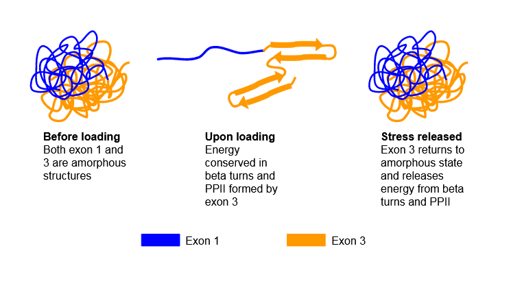
Given its disordered nature, no PDB structures of resilin are available.
Fibrinogen
This very large molecule is a hexamer of three monomers (Aα, Bβ, and γ), each present in two copies (α2β2γ2). Disulfide bonds connect one structural unit (αβγ) with another. When two fibrinopeptides (FpA and FpB) are cleaved from the amino ends of the α and β chains by the clotting enzyme thrombin, small structural "knobs" form that bind to "holes" on another fibrinogen, causing the formation of large fibrils of fibrin clots. Figure \(\PageIndex{10}\) shows an interactive iCn3D model of human fibrinogen (3ghg). The "floppy" parts of the alpha chain (αC region) and FpA and FpB peptides are not shown as they were not resolved (due to their disorder) in the crystal structure.

Figure \(\PageIndex{10}\): Human fibrinogen (3ghg). (Copyright; author via source).
Click the image for a popup or use this external link: https://structure.ncbi.nlm.nih.gov/i...S3dXzzsrkujKZ8
It is a very long, flexible molecule. The alpha chains are magenta and light green, the beta chains are dark blue and gray, and the gamma chains are brown and orange. Note that the helical chains are alpha-helical for this molecule.
Figure \(\PageIndex{11}\) shows the domain structure and hints at the flexibility of this long molecule, which is required to form a fibrous mesh clot as well as to be accessible to an enzyme, plasmin, which cleaves fibrin clots, facilitating their removal. The Aα, Bβ, and γ are shown in blue, red, and green, respectively. Carbohydrates are shown in orange in the space-filling model (b).
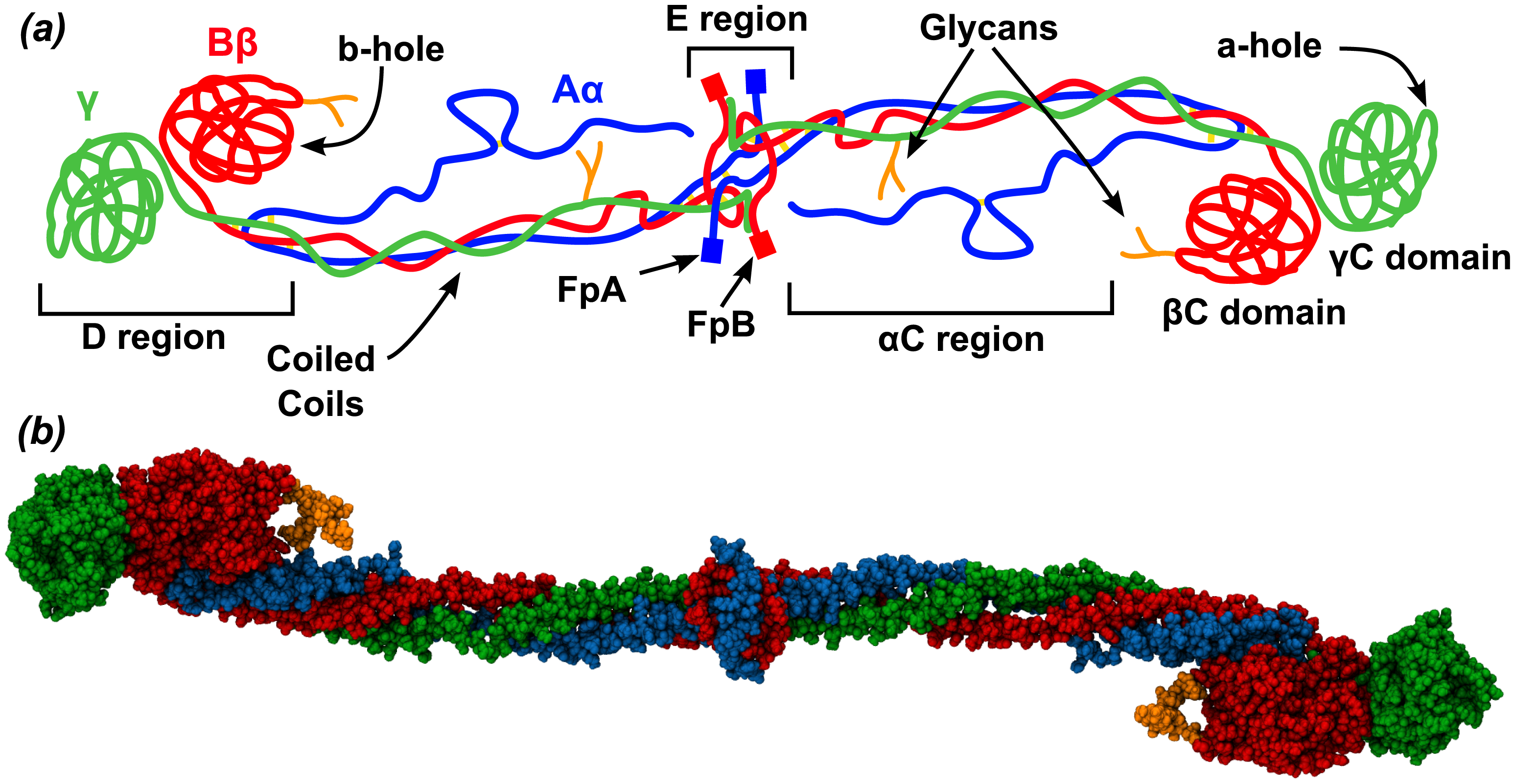
The coiled coils are mostly alpha-helical. The central E region is where the N-terminal ends of all of the fibrinogen chains are located and where the fibrinopeptides A (FpA, 16 residues) and B (FpB, 14 residues) are located, which will be cleaved by thrombin in clot formation. The C-termini of the chains are in the distal D region, which contains two C domains. After cleavage of the fibrinopeptides, conformational changes occur in the central E region to produce the "knobs" A (with starting sequence Gly-Pro-Arg) and B (Gly-His-Arg). These knobs bind through noncovalent interactions corresponding to "holes" a and b at the distal D regions of another fibrinogen to form a dimer and, subsequently, a fibrin clot.
Myosin Heavy Chain
Myosin is a chief component of muscles and, in complex with actin and other proteins, allows muscle contraction. It has two distinct domains and an elongated, rod-like tail with 28-residue repeats of 4 heptapeptides, characteristic of alpha-helical proteins that form coiled-coil quaternary structures. Figure \(\PageIndex{12}\) shows the domain structure of human myosin heavy chain 1. The orange represents the motor domain of the protein, which binds and hydrolyzes ATP, providing the free energy that powers muscle contraction.

Figure \(\PageIndex{13}\) shows a cartoon of myosin heavy chains (blue) associated with myosin light chains and how they interact with actin in the actinomyosin complex in striated muscle cells. The motor domain also binds actin filaments. Simply put, thick myosin filaments slide back and forth during muscle contraction and extension.

Myosin can exist in two major conformations. One is the "6S" (extended tail) form that assembles into myosin filaments, which interact with actin as shown in Figure \(\PageIndex{10}\) to transduce the chemical energy from ATP hydrolysis into mechanical forces and filament sliding. The other is the "10S" conformation, which is folded on itself. The heads interact with each other and the tail. All necessary steps (ATP cleavage, filament assembly, actin activation) required for actin/myosin-mediated contraction are inhibited in this compact form.
Figure \(\PageIndex{14}\) shows an interactive iCn3D model of an inactive (nonextended) conformation of myosin heavy chain (6xe9) from smooth muscle. The long cyan and green chains are smooth muscle myosin (II) heavy chains.

Figure \(\PageIndex{14}\): Inactive (nonextended) conformation of myosin heavy chain (6xe9). (Copyright; author via source).
Click the image for a popup or use this external link: https://structure.ncbi.nlm.nih.gov/i...thwKbyQQ5ezB96
Recent Updates: February 2026
Spider Silk - an unusual fibrillar protein
Silk from both spiders and silkworms is a fibrillar protein. In its final state, silk has long, aligned protein chains that form extensive β-sheets, which in turn form fibrils, which in turn form insoluble, extremely strong fibers. However, before the spider spins its web, the protein exists in a soluble form called a spidroin (250–400 kD), which forms a family. They aggregate together into a liquid condensate in which the spidroins are intrinsically disordered (a topic we discussed in Chapter 4.6: Intrinsically Disordered Proteins) and are stored in the silk gland. Liquid condensate formation is another example of liquid-liquid phase separation (LLPS), or liquid-liquid demixing, a topic we covered in previous sections (1.1: Cellular Foundations and 4.10: Protein Aggregates - Amyloids, Prions and Intracellular Granules). Potassium phosphate promotes LLPS. LLPS, along with changes in pH and ion exchange in the LLPS, leads to the formation of β-sheet fibrils, which self-associate into macroscopic fibers. This drastic conformational change during spinning is an amazing and unusual example of a rapid conversion of an IDP into a fibril-forming protein with extensive organization.
The structure of a typical spider web is shown below in Figure \(\PageIndex{15}\). The dragline is the strongest.

Figure \(\PageIndex{15}\): Schematic representation of different types of spider silk and their role in spider webs (compiled according to Vollrath and Knight 2001). Ramezaniaghdam Maryam, Nahdi Nadia D. , Reski Ralf. Recombinant Spider Silk: Promises and Bottlenecks. Frontiers in Bioengineering and Biotechnology, 10, 2022. https://www.frontiersin.org/journals...oe.2022.835637. Creative Commons Attribution License (CC BY).
The mechanical properties of different types of spider silk and some commonly used fibers are shown in Table \(\PageIndex{1}\) below.
| Materials | Strength [MPa] | Strain [%] | Toughness [MJ m−3] | Young's modulus [GPa] |
|---|---|---|---|---|
| MA silk (L. venusta) | 1469 | 23.3 | 151 | 10.6 |
| B. mori silk | 500–600 | 15.0–18.0 | 70–80 | 7 |
| Elastin | 2 | 150 | 1.6 | 0.0011 |
| Tendon collagen | 150 | 12 | 7.5 | 1.5 |
| Kevlar 49 | 3600 | 2.7 | 50 | 130 |
| Nylon 66 | 750–950 | – | 80 | 2–3.6 |
| Carbon fiber | 4000 | 1.3 | 25 | 300 |
| High‐tensile steel | 1500 | 0.8 | 6 | 200 |
Table \(\PageIndex{1}\): The mechanical properties of different types of spider silk and some commonly used fibers. Li J, Li S, Huang J, Khan AQ, An B, Zhou X, Liu Z, Zhu M. Spider Silk-Inspired Artificial Fibers. Adv Sci (Weinh). 2022 Feb;9(5):e2103965. doi: 10.1002/advs.202103965. Epub 2021 Dec 19. PMID: 34927397; PMCID: PMC8844500. CC BY 4.0
Here is the meaning of the terms:
- strength: How much pulling force the fiber can withstand before it breaks
- strain: How much the fiber stretches before breaking
- toughness: How much energy the fiber can absorb before breaking
- Young's modulus: How stiff the fiber is (higher value stiffer, lower value more flexible)
We'll explore dragline silk from the black widow spider. The dragline (the strongest type of silk fiber) is predominantly composed of two types of spidroins called major ampullate silk protein (MaSP). The two types are MaSp1 (most abundant) and MaSp2. These proteins have stretches of repeating poly-alanine residues that form crystalline beta-sheet fibers. They have many glycine-rich repeats of the sequence GGX, where X is Ala, Gln, and, to a lesser degree, Tyr, Arg, and Ser. These are between the poly-Ala regions. Tyr occurs in all glycine-rich regions. The motif RGG is found in every other Gly-rich region in MaSP1, while RQQ is found in the MaSP2 spidroin repeats.
Detailed structural characterization of the protein before and after spinning is complicated. Structural information from NMR spectroscopy of MaSP1 before spinning indicates it is intrinsically disordered, with little secondary structure. Molecular structure predictions show that a MaSP forms a micelle-like structure with a buried hydrophobic core enriched in poly-Ala, with the more hydrophilic side chains (such as Arg and the hydroxyl group of Tyr) on the surface and solvent-exposed.
In the spun fiber, the poly-Ala tract forms a β-sheet crystalline structure. The Gly-rich regions are generally disordered. Given their high Gly content, they likely form reverse turns (for GPGXX turns in MaSp2) and tight 310-helical structures (for GGX).
In general, the spidroin proteins can exist in a variety of states and undergo many transitions from the soluble monomer to the insoluble β-sheet crystalline fiber. These are described in Table \(\PageIndex{2}\) below.
| Protein State | aggregation state | changes in transition to next state |
| 1. Soluble spidroin "dope" in silk gland reservoir |
monomer, "micelle" like in that poly-Ala generally buried; Gly, Arg, Tyr mostly exposed; mainly disordered; neutral pH, NaCl elevated, high protein concentration |
increase phosphate (H2PO4-); ion exchange with NaCl; phosphate interacts with Arg+, displacing H2O; increase Arg–Tyr cation–π interactions |
| 2. Liquid-liquid phase separation (LLPS) | Proteins are very condensed but still hydrated with enhanced Arg-Tyr noncovalent interactions; little β-sheet, mostly disordered. Many weak side-chain interactions between monomers (multivalency) | LLPS moves through the spinning duct, leading to pH drop, shear forces, removal of water, chain alignment, β-sheet nucleation, and β-sheet formation |
| 3. Fiber | Poly(Ala) hydrogen bonding drives β-sheet nanocrystals forming rigid domains; glycine-rich is more flexible and elastic; Arg, Try are at β-sheet interfaces |
Table \(\PageIndex{2}\): Structures and transition in the spidron protein on fiber formation.
Recent structural work using NMR spectroscopy and structural predictions using Google ColabFold (v1.5.5 - AlphaFold2 using MMseqs2) for the prespun silk and AlphaFold3 for the spun silk has been performed on shorter fragments and multimers (hexamers and trimers) of the fragments of Latrodectus hesperus (Western Black Widow spider) MaSp1. The structures described in the Table above are illustrated below.
Figure \(\PageIndex{14}\) below shows the sequence of the 116-residue MaSp1 fragment from Latrodectus hesperus (Western Black Widow spider) used in structural predictions and molecular dynamics simulations, with poly(Ala) (red), Arg (purple), and Tyr (blue) highlighted. It represents a typical repeat within the protein sequence.
1AAAAAAAAGGAGQGGQGGYGQGGYGQGGAGQGGAAAAAAAAGGAGQGGYGRGGAGQGG58
59AAAAAGAGQGGYGGQGAGQGGAGAAAAAAAAGGAGQGGQGGYGRGGYGQGGAGQGGAG116
Figure \(\PageIndex{14}\): sequence of the 116-residue MaSp1 fragment from Latrodectus hesperus (Western Blackwidow spider)
First, let's look at the simulated/predicted structure of the soluble (before spinning) disordered MaSp1 116-mer fragment in NaCl. It's shown in Figure \(\PageIndex{14}\) below (left panel). It looks similarly globular in KH2PO4.
 |
 |
Figure \(\PageIndex{14}\): Left: Representative structures from simulations run with NaCl. Right: Example of a phosphate-bridged Arg–Tyr interaction observed in KH2PO4. H.R. Johnson, K. Chalek, N. Elathram, A.T. Chau, A.R. Domingo, J.E. Aldana, H. Nguyen, A. de Loera, B.A. Duarte, L. Shapakidze, D. Onofrei, G.T. Debelouchina, C.D. Lorenz, & G.P. Holland, Arg–Tyr cation–π interactions drive phase separation and β-sheet assembly in native spider dragline silk, Proc. Natl. Acad. Sci. U.S.A. 122 (52) e2523198122, https://doi.org/10.1073/pnas.2523198122 (2025). Creative Commons Attribution License 4.0 (CC BY).
Google CoLabFold seed structure: Figure \(\PageIndex{14}\)

Figure \(\PageIndex{14}\): Google CoLabFold seed structure
To view the AlphaFold3 model below in iCn3D:
1. Download this PNG appendable file. This might download as an image. If so, then resave the png file again using right-click on PC. 2. Open iCn3D. 3. File, Open File, iCn3D png appendable, and select the downloaded file on your computer (long load time)
The right panel shows a key interaction between the phosphate anion and proximal Arg and Tyr side chains. Molecular dynamics simulations reveal that phosphate promotes Arg–Tyr interactions and disrupts Arg–Ala contacts, freeing Ala for eventual β-sheet fiber formation.
Figure \(\PageIndex{15}\) shows that AlphaFold3 models with β-sheet fiber architecture of a hexamer of the 116mer with Arg–Tyr cation–π interactions at structured–unstructured interfaces.
 |
To view the AlphaFold3 model below in iCn3D: 1. Download this PNG appendable file. This might download as an image. If so, then resave the png file again using right-click on PC. 2. Open iCn3D. 3. File, Open File, iCn3D png appendable, and select the downloaded file on your computer (long load time)
|

Figure \(\PageIndex{15}\): Left - (A) Hexamer AlphaFold3 model of a 116-residue MaSp1 hexamer repeat predicts highly ordered β-sheet architecture, with poly(Ala) regions forming the β-sheet core (blue), and Arg (orange) and Tyr (purple) side chains positioned near β-sheet interfaces. H.R. Johnson et al., ibid.
Right: iCn3D AlphaFold3 model of a hexamer of 116-residue MaSP1. Two monomers are shown in sticks to illustrate the interchain hydrogen bonds (dotted lines). The Tyr and Arg side chains are both shown in cyan.
Figure \(\PageIndex{16}\) below shows AlphaFold3 models of the β-sheet fiber architecture of a trimer of the 43mer with Arg–Tyr cation–π interactions at structured–unstructured interfaces.
 |
To view the AlphaFold3 model below in iCn3D: 1. Download this PNG appendable file. This might download as an image. If so, then resave the png file again using right-click on PC. 2. Open iCn3D. 3. File, Open File, iCn3D png appendable, and select the downloaded file on your computer (long load time)
|


Figure \(\PageIndex{16}\): H.R. Johnson, et al., ibid.
Panel B: Trimer model of a 43-residue sequence containing Arg–Tyr motifs exhibits short β-sheets in poly(Ala) regions (purple). Arg (red) and Tyr (green) residues are located at β-sheet interfaces, with Arg side chains positioned near Tyr aromatic rings. The average distance between the Tyr Y19 ring (center-of-mass) and Arg R21 Nη atoms is ~5.7 Å.
Panel C: All-atom CHARMM36m MD simulation (200 ns) preserves poly(Ala) β-sheet structure in the trimer assembly, but reveals noteworthy sidechain disorder in interfacial Arg and Tyr residues.
Right Panel: iCn3D AlphaFold3 Model of the 43-mer. Tyrosine and arginine are shown in sticks and labeled. Two gold dotted lines show the ion-pi interactions between the adjacent Arg and Tyr on the same chain. The red dotted line between adjacent aromatic rings of the tyrosines indicates pi-pi interactions. If you open the iCn3D model and measure the distance between the Arg and Tyr side chains, it is too long (around 8 Å) for an actual ion-pi interaction. The AlphaFold3 program used to make the model returned the structure shown. The manuscript shows that the distance is closer to 5.7 Å, and in MD simulations, sometimes closer.
We will discuss another type of protein, β-amyloid proteins (Chapter 4.10: Protein Aggregates - Amyloids, Prions and Intracellular Granules), which form insoluble aggregates enriched in beta-sheets. They differ from silk, for example, in many ways. Amyloid protein monomers are soluble but typically enriched in alpha-structure. Upon misfolding (which can be caused by mutations), the initial form is metastable. The monomers, induced to misfold, aggregate to form fibrils with an interchain beta-sheet structure.
Summary
(Summary written by Claude, Sonnet 4.6, Anthropic)
This chapter surveys the structural and mechanical diversity of fibrous and elastic proteins, tracing how recurring structural motifs — coiled coils, triple helices, β-sheets, and disordered sequences — are organized at multiple hierarchical levels to produce materials with precisely tuned mechanical properties essential for biological function.
Collagen, the most abundant mammalian protein (~25–35% of total body protein), owes its exceptional mechanical properties to a unique primary structure and quaternary organization. Nearly every third residue is glycine, whose small side chain is absolutely required to fit at the central axis of the triple helix. Proline (~17%) and post-translationally modified hydroxyproline restrict backbone flexibility and stabilize the helix through stereoelectronic effects; hydroxylysine provides glycosylation sites and participates in intermolecular crosslinking. Both hydroxylation reactions require vitamin C as a cofactor, explaining why its deficiency causes scurvy — a failure of connective tissue integrity. Three collagen strands wind together in a left-handed superhelix with a pitch of approximately 10 GXY triplets per three turns; the strands are staggered by one residue, placing glycines along the central axis and exposing X and Y side chains to solvent. Interstrand hydrogen bonds form between glycine amide H donors and X-position carbonyl O acceptors on adjacent chains. A recently described mechanism for the extraordinarily long-term stability of collagen — including its preservation in dinosaur fossils — involves n→π* interactions between lone pairs on the acyl oxygen of trans X-Pro peptide bonds and the adjacent carbonyl π* antibonding orbital. In the trans isomer, steric restrictions invoke a Pauli exclusion-like effect: the lone pair on water (the hydrolytic nucleophile) cannot donate into the already partially occupied π* orbital, retarding hydrolysis. This structural feature, operating alongside the biological environment of bone matrix, contributes to collagen's remarkable persistence over geological timescales.
α-Keratin is the structural protein in hair, nails, horns, and skin. Two keratin monomer types — acidic (Type I, enriched in Asp/Glu) and basic (Type II, enriched in Lys) — form obligate heterodimeric coiled coils in which hydrophobic residues at heptad-repeat positions a and d pack into a knob-into-hole interface between the two wound α-helices, while complementary charged residues stabilize the dimer through ion-ion interactions across the helix interface. Coiled-coil dimers then assemble hierarchically into staggered antiparallel tetramers, which pack laterally into protofilaments, and ultimately into left-handed helical intermediate filaments. The result is one of the strongest biological materials, with tensile properties approaching those of stainless steel in some keratins.
Elastic and resilient proteins — elastin, resilin, spider silk, and fibrillin — span a spectrum of mechanical properties despite sharing architectural features including high glycine and proline content, intrinsic disorder, and modular repeat sequences. Elastin and resilin store and release elastic energy with efficiencies of 90% and 97%, respectively, enabled by their disordered, hydrated hydrogel-like structures crosslinked through lysine (elastin) or dityrosine (resilin) bonds. Their disorder is not incidental but essential: conformational entropy in the extended state provides the restoring force upon load release. Spider dragline silk (MaSp1/MaSp2 spidroins) is arguably the most remarkable biological material, combining a tensile strength approaching Kevlar with toughness exceeding all synthetic fibers. Before spinning, spidroins are stored as intrinsically disordered liquid condensates in the silk gland, maintained in solution by LLPS stabilized by multivalent Arg–Tyr cation–π interactions and elevated NaCl. During spinning, phosphate ions (H₂PO₄⁻) displace Na⁺, interact with positively charged Arg side chains through a bridging mechanism, and promote enhanced Arg–Tyr cation–π contacts that free poly-Ala sequences for β-sheet nucleation. Shear forces, pH drop, and water removal along the spinning duct then drive rapid, irreversible conversion of the disordered condensate into stacked β-sheet nanocrystalline domains (from poly-Ala tracts) embedded in a flexible, glycine-rich disordered matrix (from GGX and GPGXX regions) — a composite architecture that simultaneously maximizes both stiffness and toughness.
Fibrinogen exemplifies an elongated, flexible fibrous protein whose function depends on proteolytic activation. This hexameric glycoprotein (α₂β₂γ₂) features a long coiled-coil central domain flanked by globular D regions, with intrinsically disordered αC regions that are not resolved in crystal structures. Thrombin cleaves fibrinopeptides A and B from the N-termini of the α and β chains, exposing Gly-Pro-Arg and Gly-His-Arg "knob" sequences that bind noncovalently to complementary "holes" in the D regions of adjacent fibrinogen molecules, driving end-to-end and lateral association into the fibrin clot mesh. The same flexibility that enables clot formation also allows access by plasmin, the fibrinolytic enzyme that dissolves clots. Myosin heavy chain exemplifies a fibrous motor protein with two functionally essential conformational states: the extended 6S form, in which the coiled-coil tail assembles into thick filaments and the globular motor domains bind actin and hydrolyze ATP to generate force through a power stroke, and the folded 10S form, in which autoinhibitory head-head and head-tail interactions suppress all motor activity. The 28-residue heptad repeats of the coiled-coil tail region are the structural basis for thick filament assembly through hydrophobic packing, analogous in principle to the α-keratin coiled coil.
References
OpenStax, Proteins. OpenStax CNX. Sep 30, 2016 http://cnx.org/contents/bf17f4df-605c-4388-88c2-25b0f000b0ed@2.
File:Chirality with hands.jpg. (2017, September 16). Wikimedia Commons, the free media repository. Retrieved 17:34, July 10, 2019 from commons.wikimedia.org/w/index.php?title=File:Chirality_with_hands.jpg&oldid=258750003.
Wikipedia contributors. (2019, July 6). Zwitterion. In Wikipedia, The Free Encyclopedia. Retrieved 21:48, July 10, 2019, from en.Wikipedia.org/w/index.php?title=Zwitterion&oldid=905089721
Wikipedia contributors. (2019, July 8). Absolute configuration. In Wikipedia, The Free Encyclopedia. Retrieved 15:28, July 14, 2019, from en.Wikipedia.org/w/index.php?title=Absolute_configuration&oldid=905412423
Structural Biochemistry/Enzyme/Active Site. (2019, July 1). Wikibooks, The Free Textbook Project. Retrieved 16:55, July 16, 2019 from en.wikibooks.org/w/index.php?title=Structural_Biochemistry/Enzyme/Active_Site&oldid=3555410.
Structural Biochemistry/Proteins. (2019, March 24). Wikibooks, The Free Textbook Project. Retrieved 19:16, July 18, 2019 from en.wikibooks.org/w/index.php?title=Structural_Biochemistry/Proteins&oldid=3529061.
Fujiwara, K., Toda, H., and Ikeguchi, M. (2012) Dependence of a α-helical and β-sheet amino acid propensities on teh overall protein fold type. BMC Structural Biology 12:18. Available at: https://bmcstructbiol.biomedcentral.com/track/pdf/10.1186/1472-6807-12-18
Wikipedia contributors. (2019, July 16). Keratin. In Wikipedia, The Free Encyclopedia. Retrieved 17:50, July 19, 2019, from en.Wikipedia.org/w/index.php?title=Keratin&oldid=906578340
Wikipedia contributors. (2019, July 13). Alpha-keratin. In Wikipedia, The Free Encyclopedia. Retrieved 18:17, July 19, 2019, from en.Wikipedia.org/w/index.php?title=Alpha-keratin&oldid=906117410
Open Learning Initiative. (2019) Integumentary Levels of Organization. Carnegie Mellon University. In Anatomy & Physiology. Available at: https://oli.cmu.edu/jcourse/webui/syllabus/module.do?context=4348901580020ca6010f804da8baf7ba.
Wikipedia contributors. (2019, July 16). Collagen. In Wikipedia, The Free Encyclopedia. Retrieved 03:42, July 20, 2019, from en.Wikipedia.org/w/index.php?title=Collagen&oldid=906509954
Wikipedia contributors. (2019, July 2). Rossmann fold. In Wikipedia, The Free Encyclopedia. Retrieved 16:01, July 20, 2019, from https://en.Wikipedia.org/w/index.php?title=Rossmann_fold&oldid=904468788
Wikipedia contributors. (2019, May 30). TIM barrel. In Wikipedia, The Free Encyclopedia. Retrieved 16:46, July 20, 2019, from en.Wikipedia.org/w/index.php?title=TIM_barrel&oldid=899459569
Wikipedia contributors. (2019, July 16). Protein folding. In Wikipedia, The Free Encyclopedia. Retrieved 18:30, July 20, 2019, from https://en.Wikipedia.org/w/index.php?title=Protein_folding&oldid=906604145
Wikipedia contributors. (2019, June 11). Globular protein. In Wikipedia, The Free Encyclopedia. Retrieved 18:49, July 20, 2019, from en.Wikipedia.org/w/index.php?title=Globular_protein&oldid=901360467
Wikipedia contributors. (2019, July 11). Intrinsically disordered proteins. In Wikipedia, The Free Encyclopedia. Retrieved 19:52, July 20, 2019, from en.Wikipedia.org/w/index.php?title=Intrinsically_disordered_proteins&oldid=905782287
Comprehensive Database for Protein Analysis - Biozon
SCOP: Structural Characterization of Proteins - Database showing folds, superfamiles, families, and domains