Reading: Seedless Plants
- Page ID
- 7683
\( \newcommand{\vecs}[1]{\overset { \scriptstyle \rightharpoonup} {\mathbf{#1}} } \)
\( \newcommand{\vecd}[1]{\overset{-\!-\!\rightharpoonup}{\vphantom{a}\smash {#1}}} \)
\( \newcommand{\dsum}{\displaystyle\sum\limits} \)
\( \newcommand{\dint}{\displaystyle\int\limits} \)
\( \newcommand{\dlim}{\displaystyle\lim\limits} \)
\( \newcommand{\id}{\mathrm{id}}\) \( \newcommand{\Span}{\mathrm{span}}\)
( \newcommand{\kernel}{\mathrm{null}\,}\) \( \newcommand{\range}{\mathrm{range}\,}\)
\( \newcommand{\RealPart}{\mathrm{Re}}\) \( \newcommand{\ImaginaryPart}{\mathrm{Im}}\)
\( \newcommand{\Argument}{\mathrm{Arg}}\) \( \newcommand{\norm}[1]{\| #1 \|}\)
\( \newcommand{\inner}[2]{\langle #1, #2 \rangle}\)
\( \newcommand{\Span}{\mathrm{span}}\)
\( \newcommand{\id}{\mathrm{id}}\)
\( \newcommand{\Span}{\mathrm{span}}\)
\( \newcommand{\kernel}{\mathrm{null}\,}\)
\( \newcommand{\range}{\mathrm{range}\,}\)
\( \newcommand{\RealPart}{\mathrm{Re}}\)
\( \newcommand{\ImaginaryPart}{\mathrm{Im}}\)
\( \newcommand{\Argument}{\mathrm{Arg}}\)
\( \newcommand{\norm}[1]{\| #1 \|}\)
\( \newcommand{\inner}[2]{\langle #1, #2 \rangle}\)
\( \newcommand{\Span}{\mathrm{span}}\) \( \newcommand{\AA}{\unicode[.8,0]{x212B}}\)
\( \newcommand{\vectorA}[1]{\vec{#1}} % arrow\)
\( \newcommand{\vectorAt}[1]{\vec{\text{#1}}} % arrow\)
\( \newcommand{\vectorB}[1]{\overset { \scriptstyle \rightharpoonup} {\mathbf{#1}} } \)
\( \newcommand{\vectorC}[1]{\textbf{#1}} \)
\( \newcommand{\vectorD}[1]{\overrightarrow{#1}} \)
\( \newcommand{\vectorDt}[1]{\overrightarrow{\text{#1}}} \)
\( \newcommand{\vectE}[1]{\overset{-\!-\!\rightharpoonup}{\vphantom{a}\smash{\mathbf {#1}}}} \)
\( \newcommand{\vecs}[1]{\overset { \scriptstyle \rightharpoonup} {\mathbf{#1}} } \)
\(\newcommand{\longvect}{\overrightarrow}\)
\( \newcommand{\vecd}[1]{\overset{-\!-\!\rightharpoonup}{\vphantom{a}\smash {#1}}} \)
\(\newcommand{\avec}{\mathbf a}\) \(\newcommand{\bvec}{\mathbf b}\) \(\newcommand{\cvec}{\mathbf c}\) \(\newcommand{\dvec}{\mathbf d}\) \(\newcommand{\dtil}{\widetilde{\mathbf d}}\) \(\newcommand{\evec}{\mathbf e}\) \(\newcommand{\fvec}{\mathbf f}\) \(\newcommand{\nvec}{\mathbf n}\) \(\newcommand{\pvec}{\mathbf p}\) \(\newcommand{\qvec}{\mathbf q}\) \(\newcommand{\svec}{\mathbf s}\) \(\newcommand{\tvec}{\mathbf t}\) \(\newcommand{\uvec}{\mathbf u}\) \(\newcommand{\vvec}{\mathbf v}\) \(\newcommand{\wvec}{\mathbf w}\) \(\newcommand{\xvec}{\mathbf x}\) \(\newcommand{\yvec}{\mathbf y}\) \(\newcommand{\zvec}{\mathbf z}\) \(\newcommand{\rvec}{\mathbf r}\) \(\newcommand{\mvec}{\mathbf m}\) \(\newcommand{\zerovec}{\mathbf 0}\) \(\newcommand{\onevec}{\mathbf 1}\) \(\newcommand{\real}{\mathbb R}\) \(\newcommand{\twovec}[2]{\left[\begin{array}{r}#1 \\ #2 \end{array}\right]}\) \(\newcommand{\ctwovec}[2]{\left[\begin{array}{c}#1 \\ #2 \end{array}\right]}\) \(\newcommand{\threevec}[3]{\left[\begin{array}{r}#1 \\ #2 \\ #3 \end{array}\right]}\) \(\newcommand{\cthreevec}[3]{\left[\begin{array}{c}#1 \\ #2 \\ #3 \end{array}\right]}\) \(\newcommand{\fourvec}[4]{\left[\begin{array}{r}#1 \\ #2 \\ #3 \\ #4 \end{array}\right]}\) \(\newcommand{\cfourvec}[4]{\left[\begin{array}{c}#1 \\ #2 \\ #3 \\ #4 \end{array}\right]}\) \(\newcommand{\fivevec}[5]{\left[\begin{array}{r}#1 \\ #2 \\ #3 \\ #4 \\ #5 \\ \end{array}\right]}\) \(\newcommand{\cfivevec}[5]{\left[\begin{array}{c}#1 \\ #2 \\ #3 \\ #4 \\ #5 \\ \end{array}\right]}\) \(\newcommand{\mattwo}[4]{\left[\begin{array}{rr}#1 \amp #2 \\ #3 \amp #4 \\ \end{array}\right]}\) \(\newcommand{\laspan}[1]{\text{Span}\{#1\}}\) \(\newcommand{\bcal}{\cal B}\) \(\newcommand{\ccal}{\cal C}\) \(\newcommand{\scal}{\cal S}\) \(\newcommand{\wcal}{\cal W}\) \(\newcommand{\ecal}{\cal E}\) \(\newcommand{\coords}[2]{\left\{#1\right\}_{#2}}\) \(\newcommand{\gray}[1]{\color{gray}{#1}}\) \(\newcommand{\lgray}[1]{\color{lightgray}{#1}}\) \(\newcommand{\rank}{\operatorname{rank}}\) \(\newcommand{\row}{\text{Row}}\) \(\newcommand{\col}{\text{Col}}\) \(\renewcommand{\row}{\text{Row}}\) \(\newcommand{\nul}{\text{Nul}}\) \(\newcommand{\var}{\text{Var}}\) \(\newcommand{\corr}{\text{corr}}\) \(\newcommand{\len}[1]{\left|#1\right|}\) \(\newcommand{\bbar}{\overline{\bvec}}\) \(\newcommand{\bhat}{\widehat{\bvec}}\) \(\newcommand{\bperp}{\bvec^\perp}\) \(\newcommand{\xhat}{\widehat{\xvec}}\) \(\newcommand{\vhat}{\widehat{\vvec}}\) \(\newcommand{\uhat}{\widehat{\uvec}}\) \(\newcommand{\what}{\widehat{\wvec}}\) \(\newcommand{\Sighat}{\widehat{\Sigma}}\) \(\newcommand{\lt}{<}\) \(\newcommand{\gt}{>}\) \(\newcommand{\amp}{&}\) \(\definecolor{fillinmathshade}{gray}{0.9}\)Introduction
Plants (kingdom Plantae) are autotrophs; they make their own organic nutrients. The term “organic” refers to compounds that contain carbon. Organic nutrients such as sugars are made by photosynthesis.
Plants are adapted to living on land. For example, the above-ground parts of most plants are covered by a waxy layer called a cuticle to prevent water loss. Aquatic plants are secondarily adapted to living in water.
Some evidence that suggests that plants evolved from the green algae is:
- they both use chlorophyll a, chlorophyll b, and carotenoid pigments during photosynthesis.
- the primary food reserve of both is starch.
- they both have cellulose cell walls.
Genetic and morphological evidence indicates that plants evolved from a group of green algae called charophyceans. Many charophyceans inhabit shallow freshwater environments. Natural selection may have favored individuals capable of surviving occasional drying in these environments and this gave rise to land plants.
These traits occur in plants but not charophyceans. Some evolved independently in other algae.
- Apical meristems
- Alternation of generations
- Spores with protective walls
- Spores produced in sporangia
- Gametes are produced in multicellular structures called gametangia; Antheridia produce sperm; Archegonia produce eggs
- Multicellular dependent embryos
- Many have a cuticle that waterproofs and offers some protection
Alternation of Generations
The basic alternation of generations life cycle is illustrated below.

The diploid plant that produces spores is called a sporophyte. The haploid plant that produces gametes is called a gametophyte.
Some protists also have an alternation of generations life cycle but the structures that produce gametes in protists are usually single cells. Plants produce gametes in multicullar structures that have an outer protective layer. Sperm are produced in structures called antheridia (sing. antheridium), eggs are produced in archegonia (sing. archegonium),. As in protists and fungi, spores of plants are produced insporangia (sing. sporangium).
A dependent sporophyte is a sporophyte that is small and grows attached to the gametophyte. It obtains nutrients from the gametophyte. An independent sporophyte grows separately from the gametophyte. Similarly, a dependent gametophyte is small and grows attached to the sporophyte while an independent gametophyte grows separately from the sporophyte.
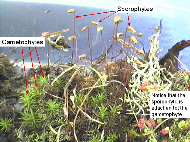
The evolutionary trend in plants has been from plants with a dominant gametophyte and reduced, dependent sporophyte (ex. Mosses) to plants with a dominant, independent sporophyte and a reduced, dependent gametophyte (ex. Seed plants).
Classification
Evolutionary relationships among the plants are shown below.

We will study the following phyla of plants.
| Characteristics | Classification |
| Bryophytes (no vascular tissue) |
Liverworts (Phylum Hepatophyta) Mosses (Phylum Bryophyta) Hornworts (Phylum Anthocerophyta |
| Seedless vascular plants | Club mosses, Spike Mosses, Quillworts (Phylum Lycophyta)Horsetails, Whisk Ferns, Ferns (Phylum Pterophyta) |
| Gymnosperms (vascular, naked seeds) |
Conifers (Phylum Coniferophyta) Cycads (Phylum Cycadophyta) Ginkgos (Phylum Ginkgophyta) Gnetophytes (Phylum Gnetophyta) |
| Angiosperms (vascular, protected seeds) |
Flowering Plants (Phylum Anthophyta)Monocots Eudicots |
Bryophytes
Phylum: Bryophyta (Mosses)
- Observe different kinds of moss on display and note the body form of the gametophyte.
- Obtain live sporulating moss and identify the sporophyte and gametophyte generations.
- Draw the life cycle of a typical bryophyte such as moss. Your drawing should contain the following terms:
- 2N
- N
- sporophyte
- sporangium
- meiosis
- spores
- protonema
- gametophyte
- antheridium
- sperm
- archegonium
- egg
- fertilization
- Observe a slide showing the antheridial head of Mnium (a moss). Begin using the scanning (4X) objective and then switch to the low power objective (10X).
- What is produced in this structure (the antheridium)?
- Show where the antheridium occurs on the live moss plant. Indicate where this structure occurs in the life cycle diagram that you prepared (above).
Figure 3. Mnium (a moss) antheridial head
Figure 4. Mnium (a moss) antheridial head x40
Figure 5. Mnium (a moss) antheridial head x100
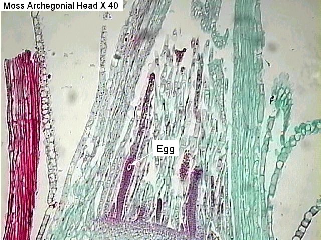
- Observe a slide showing the archegonial head of Mnium (a moss). Begin using the scanning (4X) objective and then switch to the low power objective (10X).
- What is produced in this structure?
- Show where the archegonium occurs on the live moss plant. Indicate where this structure occurs in the life cycle diagram that you prepared (above).
Figure 6. Moss archegonial head x 40
Figure 7. Moss archegonial head x 100
- After the egg is fertilized, it grows and produces a sporophyte. A capsule containing a sporangium is found at the tip of the mature sporophyte. Refer back to figure 2 to view a sporophyte.
- Use a dissecting microscope to view a longitudinal section (cut lengthwise).
- What is produced within this structure (the capsule)?
- Be sure that you can identify the sporophyte and the sporangium on the live moss plant. Indicate where these structures occur in the life cycle diagram that you prepared (above).
Figure 8. Moss capsule containing spores
Figure 9. Moss capsule x40
- How are moss spores dispersed to new locations?
Seedless Vascular Plants
Phylum: Pterophyta
Ferns
Figure 11. Fern gametophyte
- Draw the life cycle of a fern. Your drawing should contain the following terms:
- 2N
- N
- sporophyte
- sorus
- sporangium
- meiosis
- spores
- gametophyte
- antheridium
- sperm
- archegonium
- egg
- fertilization
- Observe sori on the underside of a fern leaf. Are sporangia visible? Indicate where this structure occurs in the life cycle diagram that you prepared (above).


Figure 13. Fern sorus x40
- View a slide of a fern gametophyte showing antheridia. What reproductive cells are produced by gametophytes? Indicate where the gametophyte occurs in the life cycle diagram that you prepared.
Phylum: Lycophyta
Members of this phylum have horizontal stems, upright stems, and small, spike-shaped leaves called microphylls.
Club Mosses
Observe a specimen of live club mosses such as Lycopodium. Find rhizomes. Identify microphylls. Do the specimens have any strobili? (Be sure to look up these words if you do not understand them.)
Figure 17. Club moss (lycopodium)
Spike Mosses
Observe a specimen of a spike moss such as Selaginella. Note the structure of the microphylls.
Figure 18. Spike moss (selaginella)
LICENSES AND ATTRIBUTIONS
CC LICENSED CONTENT, ORIGINAL
- Seedless Plants (Kingdom: Plantae), Biology 102. Authored by: Michael J. Gregory, Ph.D.. Provided by: LibreTexts. Located at: http://bio.libretexts.org/Under_Construction/BioStuff/BIO_102/Laboratory_Exercises/Seedless_Plants. Project: The Biology Web. License: CC BY-NC-SA: Attribution-NonCommercial-ShareAlike


