
3.4.2: Kingdoms Bacteria and Archaea
- Page ID
- 85710

Unit 3.4.2 - Kingdoms Bacteria and Archaea
- Please read and watch the following Learning Resources
- Reading the material for understanding, and taking notes during videos, will take approximately 1.5 hours.
- Optional Activities are embedded.
- Bolded terms are located at the end of the unit in the Glossary. There is also a Unit Summary at the end of the Unit.
- To navigate to Unit 3.4.3, use the Contents menu at the top of the page OR the right arrow on the side of the page.
- If on a mobile device, use the Contents menu at the top of the page OR the links at the bottom of the page.
Learning Objectives
- Describe the basic structure of a typical prokaryote
- Describe important differences in structure between Archaea and Bacteria
- Identify the macronutrients needed by prokaryotes, and explain their importance
- Describe the ways in which prokaryotes get energy and carbon for life processes
- Describe the roles of prokaryotes in the carbon and nitrogen cycles
Introduction
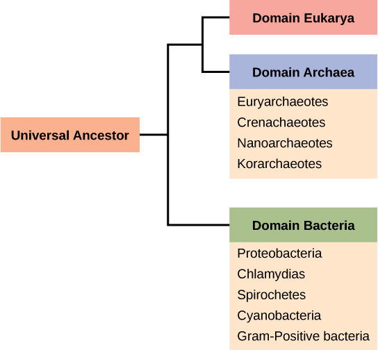
The domain Bacteria comprises all organisms in the kingdom Bacteria, the domain Archaea comprises the rest of the prokaryotes, and the domain Eukarya comprises all eukaryotes—including organisms in the kingdoms Animalia, Plantae, Fungi, and Protista (Figure \(\PageIndex{1}\)).
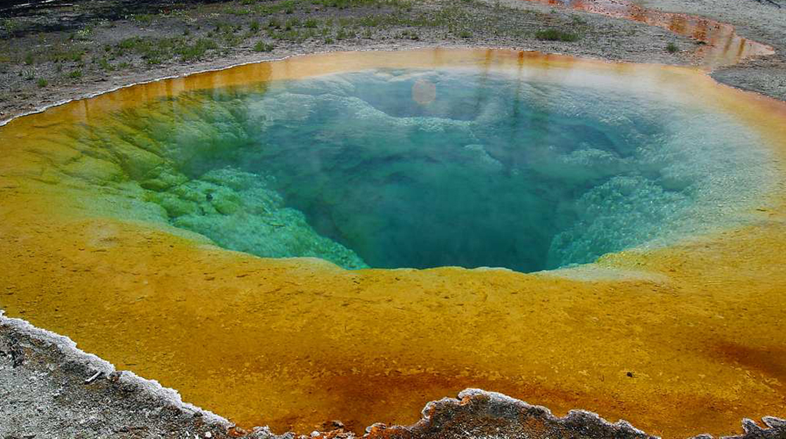
Unit 3.3 explored the diversity of prokaryotes, their formation, and the role they played in the early Earth. Two of the three domains—Bacteria and Archaea—are prokaryotic. Prokaryotes were the first inhabitants on Earth, appearing 3.5 to 3.8 billion years ago. These organisms are abundant and ubiquitous; that is, they are found everywhere on the planet. In addition to inhabiting moderate environments, they are found in extreme conditions: from boiling springs to permanently frozen environments in Antarctica; from salty environments like the Dead Sea to environments under tremendous pressure, such as the depths of the ocean; and from areas without oxygen, such as a waste management plant, to radioactively contaminated regions (Figure \(\PageIndex{2}\)). In addition, the evolution from prokaryotic to eukaryotic life was explored.
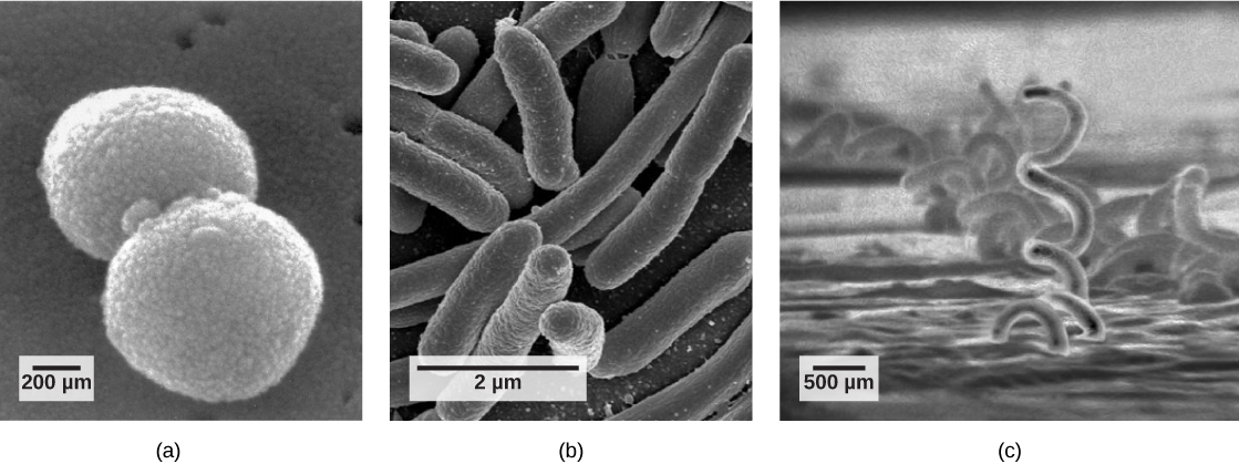
In this section, the focus will be on prokaryotes beginning with the definition of what they are. There are many differences between prokaryotic and eukaryotic cells. However, all cells have four common structures: the plasma membrane, which functions as a barrier for the cell and separates the cell from its environment; the cytoplasm, a jelly-like substance inside the cell; nucleic acids, the genetic material of the cell; and ribosomes, where protein synthesis takes place. Prokaryotes come in many shapes, but most fall into three categories: cocci (spherical), bacilli (rod-shaped), and spirilli (spiral-shaped) (Figure \(\PageIndex{3}\)).
This 7-minute video describes the prokaryotic cells that play an important role in human disease and health. They can cause disease but are also part of the human microbiota and live on our skin, body and on everyday objects in our environment.
Question after watching: In Section 3.3.3, you were asked, in the section called "Current Model", to draw a Venn Diagram showing the similarities and differences between prokaryotes and eukaryotes. While watching this video, add to this Venn Diagram with new information.
Evolution Connection: The Evolution of Prokaryotes
How do scientists answer questions about the evolution of prokaryotes? Unlike animals, prokaryotes have few fossils to offer clues. Fossils of ancient prokaryotes look like tiny bubbles in rock. Scientists turn to genetics and to the principle of the molecular clock to piece together the evolution of prokaryotes. The principle of the molecular clock holds that the more recently two species have diverged, the more similar their genes (and thus proteins) will be. Conversely, species that diverged long ago will have independently accumulated more mutations and will have more dissimilar genes.
Scientists at the NASA Astrobiology Institute and at the European Molecular Biology Laboratory collaborated to analyze the molecular evolution of 32 specific proteins common to 72 species of prokaryotes.1 The model they derived from their data indicates that three important groups of bacteria—Actinobacteria, Deinococcus, and Cyanobacteria (which the authors call Terrabacteria)—were the first to colonize land. (Deinococcus is a genus of prokaryote that is highly resistant to ionizing radiation.) Cyanobacteria are photosynthesizers, while Actinobacteria are a group of common bacteria that include species important in the decomposition of organic wastes.
The timelines of divergence suggest that Bacteria diverged from common ancestral species between 2.5 and 3.2 billion years ago, whereas Archaea diverged earlier: between 3.1 and 4.1 billion years ago. Eukarya later diverged off the Archaean line. The work further suggests that stromatolites that formed prior to the advent of cyanobacteria (about 2.6 billion years ago) photosynthesized in an anoxic environment and that because of the modifications of the Terrabacteria for land (which includes resistance to drying and the possession of compounds that protect the organism from excess light), photosynthesis producing oxygen may be closely linked to adaptations to survive on land.
Which of the following consist of prokaryotic cells?
- Bacteria and Fungi
- Archaea and Fungi
- Protista and Animalia
- Bacteria and Archaea
- Answer
- D. Bacteria and Archaea.
The Prokaryotic Cell
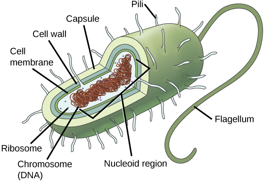
Recall that prokaryotes (Figure \(\PageIndex{4}\)) are unicellular organisms that lack organelles or internal membrane-bound structures. Therefore, they do not have a nucleus. They generally have a single chromosome—a piece of circular, double-stranded DNA located in an area of the cell called the nucleoid. Most prokaryotes have a cell wall outside the plasma membrane.
This quick 2-minute video describes the similarities and differences between the two types of prokaryotes: Bacteria and Archaea.
Question after watching: What are three similarities and three differences between Bacteria and Archaea?
The Cell Wall
The cytoplasm of prokaryotic cells has a high concentration of dissolved solutes that the cell needs to function. Therefore, the osmotic pressure within the cell is relatively high. The prokaryotic cell wall is a protective layer that surrounds some cells and gives them shape and rigidity. It is located outside the cell membrane and prevents osmotic lysis (bursting due to increasing volume). The chemical composition of the cell walls varies between Archaea and Bacteria and between bacterial species. The composition of their cell walls is also different from the eukaryotic cell walls found in plants (cellulose) or fungi and insects (chitin).
Bacterial cell walls contain peptidoglycan, composed of polysaccharide chains that are cross-linked by unusual peptides. Many of our antibiotics work by mimicking portions of these chains and therefore have specific effects on bacterial cell wall synthesis. There are more than 100 different forms of peptidoglycan. Surface layer proteins are also present on the outside of cell walls of both Archaea and Bacteria.
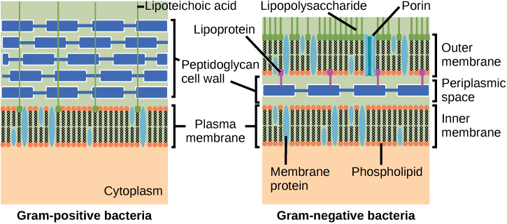
Some bacteria have a capsule outside the cell wall. Other structures are present in some prokaryotic species, but not in others (Table \(\PageIndex{1}\)). Up to 90 percent of the cell wall in bacteria without a capsule is composed of peptidoglycan. Most of the rest is composed of acidic substances called teichoic acids. Teichoic acids may be covalently linked to lipids in the plasma membrane to form lipoteichoic acids. Lipoteichoic acids anchor the cell wall to the cell membrane.
Bacterial cells with a capsule have a relatively thin cell wall composed of a few layers of peptidoglycan (only 10 percent of the total cell wall), surrounded by an outer envelope containing lipopolysaccharides (LPS) and lipoproteins. This outer envelope is sometimes referred to as a second lipid bilayer. The chemistry of this outer envelope is very different, however, from that of the typical lipid bilayer that forms plasma membranes.
Researchers and clinicians like to categorize bacteria as belonging to either Gram-positive or Gram-negative. This grouping is based on the result of a quick and easy-to-administer laboratory test that can help researchers and clinicians identify a bacterial species known as Gram staining. Under the Domain Bacteria and the Kingdom Eubacteria, Gram-positive bacteria belong to one phylum; bacteria that are Gram-negative are spread over several phyla.
The different bacterial responses to the staining procedure are ultimately due to cell wall structure. The different bacterial responses to the staining procedure are ultimately due to cell wall structure. Gram-positive organisms typically lack a capsule. Gram-negative organisms have a capsule outside of their cell wall (Figure \(\PageIndex{8}\)).
The Plasma Membrane
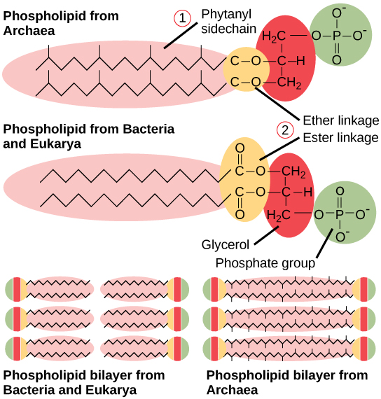
As with all living organisms, Bacteria and Archaea have a thin lipid bilayer, the plasma membrane, that surrounds the cell and separates the inside from the outside. Its selectively permeable nature keeps ions, proteins, and other molecules within the cell and prevents them from diffusing into the extracellular environment, while other molecules (usually small, non-polar ones) may move through the membrane. Recall that the general structure of a cell membrane is a phospholipid bilayer composed of two layers of lipid molecules. In archaeal cell membranes, glycerol is linked to isoprene (phytanyl) chains instead of fatty acids chains as in cell membranes of Bacteria and Eukarya. Some archaeal membranes are lipid monolayers instead of bilayers (Figure \(\PageIndex{5}\) and Table \(\PageIndex{1}\)).
This 9-minute video highlights the similarities and differences between Gram-positive and Gram-negative bacteria.
Question after watching: How does the process of Gram staining differentiate between the two types of bacteria?
Archaean cell walls do not have peptidoglycan (Table \(\PageIndex{1}\)). There are four different types of Archaean cell walls. One type is composed of pseudopeptidoglycan, which is similar to peptidoglycan in morphology but contains different sugars in the polysaccharide chain. The other three types of cell walls are composed of polysaccharides, glycoproteins, or pure protein.
| Structural Characteristic | Bacteria | Archaea |
|---|---|---|
| Cell type | Prokaryotic | Prokaryotic |
| Cell morphology | Variable | Variable |
| Cell wall | Contains peptidoglycan | Does not contain peptidoglycan |
| Cell membrane type | Lipid bilayer | Lipid bilayer or lipid monolayer |
| Plasma membrane lipids | Fatty acids | Phytanyl groups (contains the lipid isoprene) |
Prokaryotes stain as Gram-positive or Gram-negative because of differences in the cell _______.
- wall
- cytoplasm
- nucleus
- chromosome
- Answer
-
A. wall
Identify three differences between Bacteria and Archaea.
- Answer
-
Responses will vary. A possible answer is: Bacteria contain peptidoglycans in the cell wall; Archaea do not. The cell membrane in Bacteria is a lipid bilayer; in Archaea, it can be a lipid bilayer or a monolayer. Bacteria contain fatty acids on the cell membrane, whereas Archaea contain phytanyl (the lipid isoprene).
Prokaryotic Reproduction
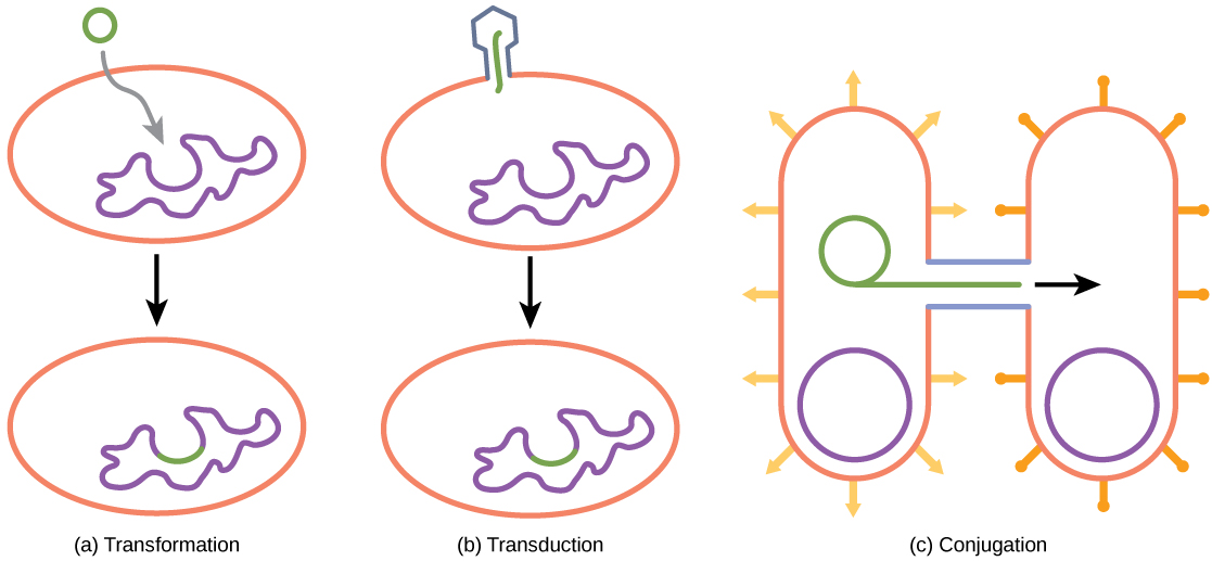
Reproduction in prokaryotes is asexual and usually takes place by binary fission. Recall that the DNA of a prokaryote exists as a single, circular chromosome. Prokaryotes do not undergo mitosis. Rather the chromosome is replicated and the two resulting copies separate from one another, due to the growth of the cell. The prokaryote, now enlarged, is pinched inward at its equator, and the two resulting cells, which are clones, separate. Binary fission does not provide an opportunity for genetic recombination or the generation of genetic diversity, but prokaryotes can share genes by three other mechanisms: transformation, transduction, and conjugation. All three depend on the presence of plasmids. Plasmids, which are extra-chromosomal DNA, are also present in many species of Bacteria and Archaea.
In transformation, the prokaryote takes in DNA found in its environment that is shed by other prokaryotes. If a nonpathogenic bacterium takes up DNA for a toxin gene from a pathogen and incorporates the new DNA into its own chromosome, it also may become pathogenic.
In transduction, bacteriophages, the viruses that infect bacteria, move short pieces of chromosomal DNA from one bacterium to another. Transduction results in a recombinant organism. Archaea are not affected by bacteriophages but instead have their own viruses that translocate genetic material from one individual to another.
In conjugation, DNA is transferred from one prokaryote to another by means of a pilus, which brings the organisms into contact with one another. The DNA transferred can be in the form of a plasmid or as a hybrid, containing both plasmid and chromosomal DNA. These three processes of DNA exchange are shown in Figure \(\PageIndex{7}\).
Reproduction can be very rapid: a few minutes for some species. This short generation time coupled with mechanisms of genetic recombination and high rates of mutation results in the rapid evolution of prokaryotes, allowing them to respond to environmental changes (such as the introduction of an antibiotic) very quickly.
These two videos (7.5-minutes and 6-minutes, respectively) summarize the two Domains and Kingdoms of prokaryotes: Bacteria and Archaea
Question after watching: In what ways are prokaryotes problematic and helpful for humans?
Prokaryotic Metabolism
The diverse environments and ecosystems on Earth have a wide range of conditions in terms of temperature, available nutrients, acidity, salinity, and energy sources. Prokaryotes are very well equipped to make their living out of a vast array of nutrients and conditions. To live, prokaryotes need a source of energy, a source of carbon, and some additional nutrients.
Energy and Carbon Sources
Prokaryotes can use different sources of energy to assemble macromolecules from smaller molecules. Photoautotrophs (or phototrophic organisms) obtain their energy from sunlight. Chemoautotrophs (or chemosynthetic organisms) obtain their energy from chemical compounds. Chemoautotrophs that can use organic compounds as energy sources are called chemoorganotrophs. Those that can also use inorganic compounds as energy sources are called chemolitotrophs.
Prokaryotes not only can use different sources of energy but also different sources of carbon compounds. Organisms that can fix inorganic carbon into organic molecules are called autotrophs. Autotrophic prokaryotes synthesize organic molecules from carbon dioxide. In contrast, heterotrophic prokaryotes obtain carbon from organic compounds.
To make the picture more complex, the terms that describe how prokaryotes obtain energy and carbon can be combined. Thus, photoautotrophs use energy from sunlight, and carbon from carbon dioxide and water, whereas chemoheterotrophs obtain energy and carbon from an organic chemical source. Chemolitoautotrophs obtain their energy from inorganic compounds, and they build their complex molecules from carbon dioxide. Table \(\PageIndex{2}\) summarizes carbon and energy sources in prokaryotes.
| Energy Source | ||
| Carbon Source | Sunlight | Chemicals (such as sulfur dioxide, iron, etc.) |
| Carbon Dioxide (from the atmosphere or water) | Photoautotrophs | Chemoautotrophs |
| Organic Compounds (from other organic matter) | Photoheterotrophs | Chemoheterotrophs |
Prokaryotes that obtain their energy from chemical compounds are called _____.
- phototrophs
- auxotrophs
- chemotrophs
- lithotrophs
- Answer
-
C. chemotrophs
Prokaryote Niches and Habitats
Prokaryotes are ubiquitous: There is no niche or ecosystem in which they are not present. Prokaryotes play many roles in the environments they occupy. The roles they play in the carbon and nitrogen cycles are vital to life on Earth.
Prokaryotes and the Carbon Cycle
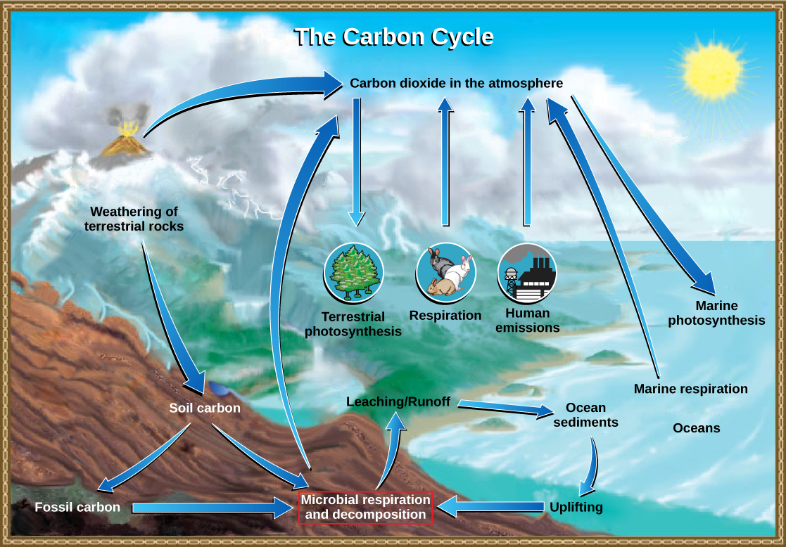
Carbon is one of the most important macronutrients, and prokaryotes play an important role in the carbon cycle (Figure \(\PageIndex{8}\)). Carbon is cycled through Earth’s major reservoirs: land, the atmosphere, aquatic environments, sediments and rocks, and biomass. The movement of carbon is via carbon dioxide, which is removed from the atmosphere by land plants and marine prokaryotes in photosynthesis and is returned to the atmosphere via the respiration of chemoorganotrophic organisms, including prokaryotes, fungi, and animals. Although the largest carbon reservoir in terrestrial ecosystems is in rocks and sediments, that carbon is not readily available.
A large amount of available carbon is found in land plants. Plants, which are producers, use carbon dioxide from the air to synthesize carbon compounds (i.e. organic molecules). Related to this, one very significant source of carbon compounds is humus, which is a mixture of prokaryotes and organic materials from dead plants that have resisted decomposition. Consumers such as animals use organic compounds generated by producers and release carbon dioxide into the atmosphere when they respire. Then, bacteria and fungi, collectively called decomposers, carry out the breakdown (decomposition) of plants and animals and their organic compounds. The most important contributor of carbon dioxide to the atmosphere is microbial decomposition of dead material (dead animals, plants, and humus) through respiration.
In aqueous environments and their anoxic sediments, there is another carbon cycle taking place. In this case, the cycle is based on one-carbon compounds. In anoxic sediments, prokaryotes, mostly archaea, produce methane (CH4). This methane moves into the zone above the sediment, which is richer in oxygen and supports bacteria called methane oxidizers that oxidize methane to carbon dioxide as a way to fuel their energy needs. The carbon dioxide produced then returns to the atmosphere.
Prokaryotes and the Nitrogen Cycle
Nitrogen is a very important element for life because it is part of proteins and nucleic acids. It is a macronutrient, and in nature, it is recycled from organic compounds to ammonia (NH3), ammonium ions (NH4+), nitrate (NO-3), nitrite (NO-2), and nitrogen gas (N2) by myriad processes, many of which are carried out only by prokaryotes. As illustrated in Figure \(\PageIndex{9}\), prokaryotes are key to the nitrogen cycle. The largest pool of nitrogen available in the terrestrial ecosystem is gaseous nitrogen from the air, but this nitrogen is not usable by plants, which are primary producers.
Gaseous nitrogen is transformed, or “fixed” into more readily available forms such as ammonia through the process of nitrogen fixation. Bacteria in the soil or living on or in plant roots perform this function, making nitrogen available to plants. Ammonia can be used by plants or converted to other forms.
Another source of ammonia is ammonification, the process by which ammonia is released during the decomposition of nitrogen-containing organic compounds. The majority of the nitrogen produced during ammonification is N2. The rest is as NH4+ and nitrous oxides (NxO). Ammonia is catabolized anaerobically, meaning no oxygen needs to be present. This process can therefore take place deep in the soil by some prokaryotes.
Nitrification is the conversion of ammonium to nitrite and nitrate. Nitrification in soils is carried out by bacteria belonging to the genera Nitrosomas, Nitrobacter, and Nitrospira. The bacteria performs the reverse process, the reduction of nitrate from the soils to gaseous compounds such as N2O, NO, and N2, a process called denitrification. In this way, nitrogen is returned from the soil to the atmosphere.
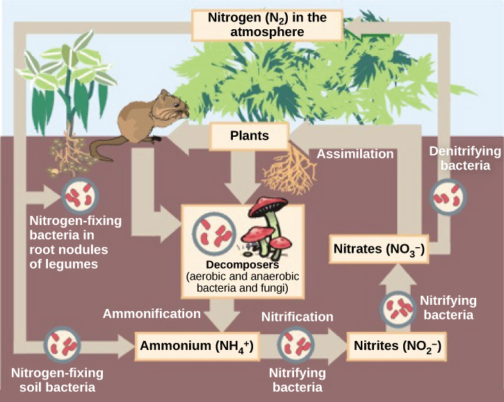
Ammonification is the process by which _____.
- ammonia is released during the decomposition of nitrogen-containing organic compounds
- ammonium is converted to nitrite and nitrate in soils
- nitrate from soil is transformed to gaseous nitrogen compounds such as NO, N2O, and N2
- gaseous nitrogen is fixed to yield ammonia
- Answer
-
A. ammonia is released during the decomposition of nitrogen-containing organic compounds
Footnotes
- 1 Battistuzzi, FU, Feijao, A, and Hedges, SB. A genomic timescale of prokaryote evolution: Insights into the origin of methanogenesis, phototrophy, and the colonization of land. BioMed Central: Evolutionary Biology 4 (2004): 44, doi:10.1186/1471-2148-4-44.
Thumbnail: Scanning electron micrograph of neutrophil ingesting methicillin-resistant Staphylococcus aureus bacteria. (Public Domain; NIAID/NIH).