Biology LibreTexts

Cell Membrane and Transport Coloring

  • Page ID
    27792
  • This page is a draft and is under active development. 

    \( \newcommand{\vecs}[1]{\overset { \scriptstyle \rightharpoonup} {\mathbf{#1}} } \)

    \( \newcommand{\vecd}[1]{\overset{-\!-\!\rightharpoonup}{\vphantom{a}\smash {#1}}} \)

    \( \newcommand{\id}{\mathrm{id}}\) \( \newcommand{\Span}{\mathrm{span}}\)

    ( \newcommand{\kernel}{\mathrm{null}\,}\) \( \newcommand{\range}{\mathrm{range}\,}\)

    \( \newcommand{\RealPart}{\mathrm{Re}}\) \( \newcommand{\ImaginaryPart}{\mathrm{Im}}\)

    \( \newcommand{\Argument}{\mathrm{Arg}}\) \( \newcommand{\norm}[1]{\| #1 \|}\)

    \( \newcommand{\inner}[2]{\langle #1, #2 \rangle}\)

    \( \newcommand{\Span}{\mathrm{span}}\)

    \( \newcommand{\id}{\mathrm{id}}\)

    \( \newcommand{\Span}{\mathrm{span}}\)

    \( \newcommand{\kernel}{\mathrm{null}\,}\)

    \( \newcommand{\range}{\mathrm{range}\,}\)

    \( \newcommand{\RealPart}{\mathrm{Re}}\)

    \( \newcommand{\ImaginaryPart}{\mathrm{Im}}\)

    \( \newcommand{\Argument}{\mathrm{Arg}}\)

    \( \newcommand{\norm}[1]{\| #1 \|}\)

    \( \newcommand{\inner}[2]{\langle #1, #2 \rangle}\)

    \( \newcommand{\Span}{\mathrm{span}}\) \( \newcommand{\AA}{\unicode[.8,0]{x212B}}\)

    \( \newcommand{\vectorA}[1]{\vec{#1}}      % arrow\)

    \( \newcommand{\vectorAt}[1]{\vec{\text{#1}}}      % arrow\)

    \( \newcommand{\vectorB}[1]{\overset { \scriptstyle \rightharpoonup} {\mathbf{#1}} } \)

    \( \newcommand{\vectorC}[1]{\textbf{#1}} \)

    \( \newcommand{\vectorD}[1]{\overrightarrow{#1}} \)

    \( \newcommand{\vectorDt}[1]{\overrightarrow{\text{#1}}} \)

    \( \newcommand{\vectE}[1]{\overset{-\!-\!\rightharpoonup}{\vphantom{a}\smash{\mathbf {#1}}}} \)

    \( \newcommand{\vecs}[1]{\overset { \scriptstyle \rightharpoonup} {\mathbf{#1}} } \)

    \( \newcommand{\vecd}[1]{\overset{-\!-\!\rightharpoonup}{\vphantom{a}\smash {#1}}} \)

    \(\newcommand{\avec}{\mathbf a}\) \(\newcommand{\bvec}{\mathbf b}\) \(\newcommand{\cvec}{\mathbf c}\) \(\newcommand{\dvec}{\mathbf d}\) \(\newcommand{\dtil}{\widetilde{\mathbf d}}\) \(\newcommand{\evec}{\mathbf e}\) \(\newcommand{\fvec}{\mathbf f}\) \(\newcommand{\nvec}{\mathbf n}\) \(\newcommand{\pvec}{\mathbf p}\) \(\newcommand{\qvec}{\mathbf q}\) \(\newcommand{\svec}{\mathbf s}\) \(\newcommand{\tvec}{\mathbf t}\) \(\newcommand{\uvec}{\mathbf u}\) \(\newcommand{\vvec}{\mathbf v}\) \(\newcommand{\wvec}{\mathbf w}\) \(\newcommand{\xvec}{\mathbf x}\) \(\newcommand{\yvec}{\mathbf y}\) \(\newcommand{\zvec}{\mathbf z}\) \(\newcommand{\rvec}{\mathbf r}\) \(\newcommand{\mvec}{\mathbf m}\) \(\newcommand{\zerovec}{\mathbf 0}\) \(\newcommand{\onevec}{\mathbf 1}\) \(\newcommand{\real}{\mathbb R}\) \(\newcommand{\twovec}[2]{\left[\begin{array}{r}#1 \\ #2 \end{array}\right]}\) \(\newcommand{\ctwovec}[2]{\left[\begin{array}{c}#1 \\ #2 \end{array}\right]}\) \(\newcommand{\threevec}[3]{\left[\begin{array}{r}#1 \\ #2 \\ #3 \end{array}\right]}\) \(\newcommand{\cthreevec}[3]{\left[\begin{array}{c}#1 \\ #2 \\ #3 \end{array}\right]}\) \(\newcommand{\fourvec}[4]{\left[\begin{array}{r}#1 \\ #2 \\ #3 \\ #4 \end{array}\right]}\) \(\newcommand{\cfourvec}[4]{\left[\begin{array}{c}#1 \\ #2 \\ #3 \\ #4 \end{array}\right]}\) \(\newcommand{\fivevec}[5]{\left[\begin{array}{r}#1 \\ #2 \\ #3 \\ #4 \\ #5 \\ \end{array}\right]}\) \(\newcommand{\cfivevec}[5]{\left[\begin{array}{c}#1 \\ #2 \\ #3 \\ #4 \\ #5 \\ \end{array}\right]}\) \(\newcommand{\mattwo}[4]{\left[\begin{array}{rr}#1 \amp #2 \\ #3 \amp #4 \\ \end{array}\right]}\) \(\newcommand{\laspan}[1]{\text{Span}\{#1\}}\) \(\newcommand{\bcal}{\cal B}\) \(\newcommand{\ccal}{\cal C}\) \(\newcommand{\scal}{\cal S}\) \(\newcommand{\wcal}{\cal W}\) \(\newcommand{\ecal}{\cal E}\) \(\newcommand{\coords}[2]{\left\{#1\right\}_{#2}}\) \(\newcommand{\gray}[1]{\color{gray}{#1}}\) \(\newcommand{\lgray}[1]{\color{lightgray}{#1}}\) \(\newcommand{\rank}{\operatorname{rank}}\) \(\newcommand{\row}{\text{Row}}\) \(\newcommand{\col}{\text{Col}}\) \(\renewcommand{\row}{\text{Row}}\) \(\newcommand{\nul}{\text{Nul}}\) \(\newcommand{\var}{\text{Var}}\) \(\newcommand{\corr}{\text{corr}}\) \(\newcommand{\len}[1]{\left|#1\right|}\) \(\newcommand{\bbar}{\overline{\bvec}}\) \(\newcommand{\bhat}{\widehat{\bvec}}\) \(\newcommand{\bperp}{\bvec^\perp}\) \(\newcommand{\xhat}{\widehat{\xvec}}\) \(\newcommand{\vhat}{\widehat{\vvec}}\) \(\newcommand{\uhat}{\widehat{\uvec}}\) \(\newcommand{\what}{\widehat{\wvec}}\) \(\newcommand{\Sighat}{\widehat{\Sigma}}\) \(\newcommand{\lt}{<}\) \(\newcommand{\gt}{>}\) \(\newcommand{\amp}{&}\) \(\definecolor{fillinmathshade}{gray}{0.9}\)

    The cell membrane is responsible for moving materials into and out of the cell.  There are two types of transport that can occur across the membrane: passive transport and active transport.  Passive transport does not require energy and includes the processes of osmosis, diffusion and facilitated diffusion.

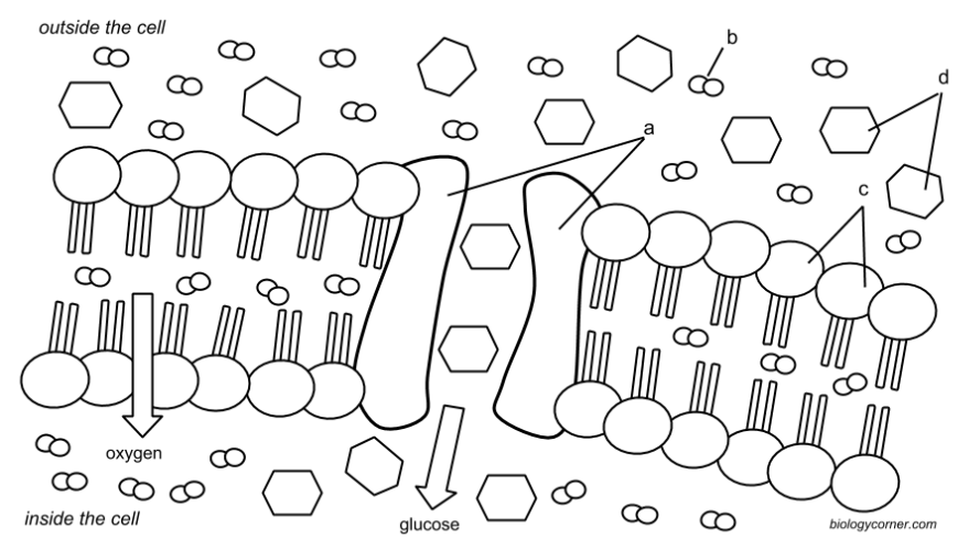
    The graphic shows the phospholipid bilayer and embedded proteins, the two main components of the cell membrane.  Phospholipids are made of hydrophilic (water-loving) heads and hydrophobic (water-hating) tails.  The phospholipids are arranged opposite to each other, with the heads pointing toward the cell’s environment and the tails aligning to each other. Color all the phospholipids (c) of the membrane red.  

    Some small molecules, like oxygen, can move right across the phospholipid bilayer through a process called  diffusion.   Diffusion occurs when molecules move from an area of high concentration to an area of low concentration. Color the oxygen molecules green.  

    Some molecules are too large to pass through the membrane. Molecules like glucose, which is a sugar molecule, must use special proteins embedded in the membrane to move into the cell.  These proteins are called channel proteins, and they provide an opening, like a door, for large molecules to pass.  Glucose will also move from areas of high concentration to low concentration, but because it needs the help of a protein, the process is called facilitated diffusion.  Color all the glucose molecules (d) purple.  Color the channel protein (a) yellow.
    cell membrane and transport.png


    Osmosis is a Special Kind of Diffusion

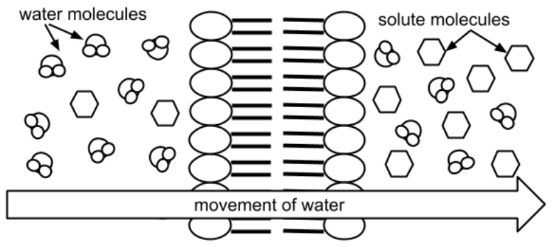
    Osmosis is the process where  water moves across the membrane toward the  area with the highest solute concentration. A solute is a compound, like salt or sugar, that is dissolved in water.  Hypertonic solutions have more solute in them, which cause water to move toward that area.  Cells placed in hypertonic solutions will lose water and shrink. 

    Solutions that have less solute molecules than the cell are called hypotonic.  In this case, water from outside the cell will move into the cell.  The cell will gain water, expand, and could even burst. To summarize, the side with MORE solute is said to be hypertonic, and the side with less is said to be hypotonic.

    Color the water molecules blue, the solute molecules purple and the phospholipids red.
    cell membrane and transport 2.png
    Water will move toward the area with the highest solute concentration.


    1. What type of transport does not require energy?  _________________________________________________
      Provide an example of one process that is a type of passive transport? _____________________________
    2. What are the two main components of the cell membrane?   _______________________________________
    3. How does oxygen cross into the cell?   __________________________________________________________
      How does glucose cross into the cell?  ________________________________________________________
    4. What is the purpose of a channel protein?___________________________________________
    5. What are the two parts of a phospholipid:  ______________________________________________________
    6. Define diffusion.  ___________________________________________________________________________
    7. Define osmosis: ___________________________________________________________________________
    8. What is a solute? ___________________________________________________________________________
    9. Hypertonic solutions have  [ more / less ] solute. Hypotonic solutions have   [ more / less ] solute. (circle)    
      On the image above, which side is HYPERTONIC?   [ left side | right side ]
    10. What will happen to a cell placed in a hypertonic solution?  _______________________________________

    Cell Membrane and Transport Coloring is shared under a not declared license and was authored, remixed, and/or curated by LibreTexts.

    • Was this article helpful?