
18.6: Susceptibility Testing
- Page ID
- 122773

For some microorganisms, susceptibility to chemotherapeutic agents is predictable. However, for many microorganisms (Pseudomonas, Staphylococcus aureus, and Gram-negative enteric bacilli such as Escherichia coli, Serratia, Proteus, etc.) there is no reliable way of predicting which antimicrobial agent will be effective in a given case. This is especially true with the emergence of many antibiotic-resistant strains of bacteria. Because of this, antibiotic susceptibility testing is often essential to determine which antimicrobial agent to use against a specific strain of bacterium.
Several tests may be used to tell a physician which antimicrobial agent is most likely to combat a specific pathogen:
1. Tube dilution tests
In this test, a series of culture tubes are prepared, each containing a liquid medium and a different concentration of a chemotherapeutic agent. The tubes are then inoculated with the test organism and incubated for 16-20 hours at 35C. After incubation, the tubes are examined for turbidity (growth). The lowest concentration of chemotherapeutic agent capable of preventing growth of the test organism is the minimum inhibitory concentration (MIC).
Subculturing of tubes showing no turbidity into tubes containing medium but no chemotherapeutic agent can determine the minimum bactericidal concentration (MBC). MBC is the lowest concentration of the chemotherapeutic agent that results in no growth (turbidity) of the subcultures. These tests, however, are rather time consuming and expensive to perform.
2. The agar diffusion test (Bauer-Kirby test)
A procedure commonly used in clinical labs to determine antimicrobial susceptibility is the Bauer-Kirby disc diffusion method. In this test, the in vitro response of bacteria to a standardized antibiotic-containing disc has been correlated with the clinical response of patients given that drug.
In the development of this method, a single high-potency disc of each chosen chemotherapeutic agent was used. Zones of growth inhibition (see Fig. \(\PageIndex{1}\)) surrounding each type of disc were correlated with the minimum inhibitory concentrations of each antimicrobial agent (as determined by the tube dilution test). The MIC for each agent was then compared to the usually-attained blood level in the patient with adequate dosage. Categories of "Resistant," "Intermediate," and "Susceptible" were then established.

The basic steps for the Bauer-Kirby method of antimicrobial susceptibility testing are given below. This outline of procedure is intended to be used as an adjunct to clinical laboratory instruction. The procedure is highly regulated and controlled by the Clinical and Laboratory Standards Institute (CLSI) and must be accompanied by a rigorous quality assurance proGram including performance by certified and/or licensed personnel when the results are to be reported in clinical settings.
a. Prepare a standard turbidity inoculum of the test bacterium so that a certain density of bacteria will be put on the plate.
- Select 3-5 isolated colonies of the bacterium that is being tested.
- If the organism is a Staphylococcus or is fastidious and grows unpredictably in broth like the streptococci, suspend the colonies is saline, Mueller Hinton broth or trypticase soy broth. If the organism grows rapidly in broth, place the colonies in Mueller Hinton broth or trypticase soy broth and incubate 2-8 hours.
- Match the turbidity of the test suspension or culture with a 0.5 McFarland standard. (McFarland standards are tubes containing either latex particles or barium sulfate and adjusted to a standard turbity.)
- If the bacterial suspension is too turbid, add more saline or broth.
- If the bacterial suspension is too light, pick off more colonies and suspend them in the broth or incubate longer.
b. Inoculate a 150mm Mueller-Hinton agar plate with the standardized inoculum so as to cover the entire agar surface with bacteria.
- Dip a sterile swab into the previously standardized tube of the bacterium being tested.Squeeze the swab against the inner wall of the tube to remove excess liquid.
- Swab the entire plate from top to bottom, edge-to-edge leaving no gaps.
- Rotate the plate approximately 60 degrees and using the same swab, again swab the entire plate from top to bottom.
- Rotate the plate approximately 60 degrees and using the same swab, and swab the entire plate from top to bottom a third time.
c. Place standardized antibiotic-containing discs on the plate.
d. Incubate the plate agar side up. For nonfastidious bacteria, incubate at 35°C for 16-18 hours. For fastidious bacteria, follow CLSI standards.
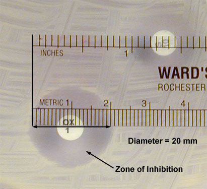
e. Measure the diameter of any resulting zones of inhibition in millimeters (mm) as shown in Fig. \(\PageIndex{2}\) .
f. Determine if the bacterium is susceptible, moderately susceptible, intermediate, or resistant to each antimicrobial agent using a standardized table (see Table 2). (The latest interpretation tables can be found in CLSI document M100 which is updated every January.)
- If there is a double zone of inhibition, measure the diameter of the innermost zone.
- If there is a zone containing colonies, measure the diameter of the colony free zone.
- If there is a feathered zone, measure the diameter of the point where there is an obvious demarcation between growth and no growth.
- When testing swarming Proteus mirabilis, ignore the swarming.
- When testing Staphylococcus aureus, the haze around an oxacillin should not be ignored. Measure the diameter of the zone free of growth or haze.
The term intermediate generally means that the result is inconclusive for that drug-organism combination. The term moderately susceptible is usually applied to those situations where a drug may be used for infections in a particular body site, e.g., cystitis because the drug becomes highly concentrated in the urine.
3. Automated tests
Computerized automated tests have been developed for antimicrobial susceptibility testing. These tests measure the inhibitory effect of the antimicrobial agents in a liquid medium by using light scattering to determine growth of the test organism. Results can be obtained within a few hours. Labs performing very large numbers of susceptibility tests frequently use the automated methods but the equipment is quite expensive.
Contributors and Attributions
Dr. Gary Kaiser (COMMUNITY COLLEGE OF BALTIMORE COUNTY, CATONSVILLE CAMPUS)