
16.2.6: Identification of Microorganisms Using the Direct Fluorescent Antibody Technique
- Page ID
- 122734

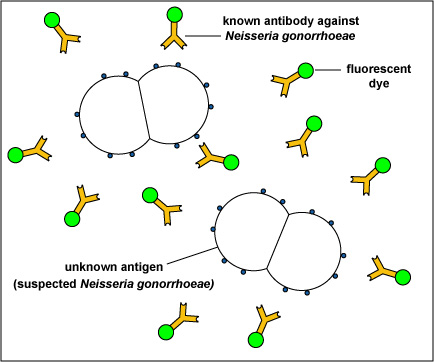
Certain fluorescent dyes can be chemically attached to the known antibody molecules in antiserum. The known fluorescent antibody is then mixed with the unknown antigen,such as a microorganism, fixed to a slide. After washing, to remove any fluorescent antibody not bound to the antigen, the slide is viewed with a fluorescent microscope.
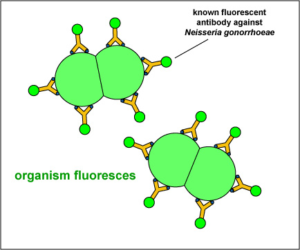
If the fluorescent antibody reacted with the unknown antigen, the antigen will glow or fluoresce under the fluorescent microscope. If the antibody did not react with the antigen, the antibodies will be washed off the slide and the antigen will not fluoresce.
For example, in the direct fluorescent antibody test for Neisseria gonorrhoeae, the unknown antigen, suspected Neisseria gonorrhoeae,is fixed to a microscope slide. Known fluorescent antibodies made against N. gonorrhoeae are then added (see Fig. \(\PageIndex{1}\), step 1) and the slide is then washed to remove any fluorescent antibody not bound to the antigen. The slide is then viewed under a fluorescent microscope.
If the unknown antigen is Neisseria gonorrhoeae, the known antibodies against N. gonorrhoeae with attached fluorescent dye will bind to the bacterium and will not wash off. The bacteria will fluoresce when viewed under a fluorescent microscope (see Fig. \(\PageIndex{1}\), step 2 and Fig. \(\PageIndex{2}\)). If the unknown antigen is not N. gonorrhoeae, the known fluorescent antibodies against will wash off the slide and the bacteria will not fluoresce when viewed under a fluorescent microscope.
|
Fig. \(\PageIndex{1}\): Direct Fluorescent Antibody Test for Neisseria gonorrhoeae, Step-2 |
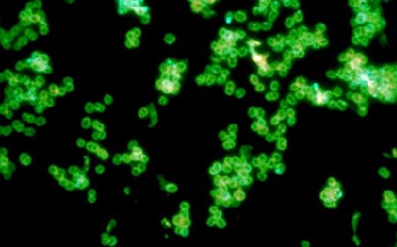
Fig. \(\PageIndex{2}\): A Positive Direct Fluorescent Antibody Test for Neisseria gonorrhoeae |
|---|---|
|
|
| If the unknown antigen is Neisseria gonorrhoeae, the known antibodies against N. gonorrhoeae with attached fluorescent dye will bind to the bacterium and will not wash off. The bacteria will fluoresce when viewed under a fluorescent microscope. | Note green-fluorescent diplococci. |
|
Copyright; Gary E. Kaiser, Ph.D. The Community College of Baltimore County, Catonsville Campus CC-BY-3.0 |
By Content Providers(s): CDC/Public domain. Courtesy of the Centers for Disease Control and Prevention. |
Many bacteria, viruses, and fungi can be identified using this technique.
Contributors and Attributions
Dr. Gary Kaiser (COMMUNITY COLLEGE OF BALTIMORE COUNTY, CATONSVILLE CAMPUS)