16.2.4: Serological Testing to Diagnose Pregnancy
- Page ID
- 122732

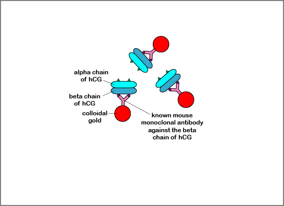
The Alere® hCG Dipstick is an example of a lateral flow immunologic assay. It is a qualitative serologic test for detecting early pregnancy. The hormone human chorionic gonadotropin (hCG), produced by the placenta, appears in the serum and urine of pregnant females. The hCG is composed of two subunits - alpha and beta. The Alere® hCG Dipstick is a one step pregnancy test that detects levels of hCG as low as 25 mlU/ml. Human chorionic gonadotropin (hCG), the unknown antigen for which one is testing, is identified in the urine by using known mouse monoclonal antibodies against the beta subunit of hCG bound to colloidal gold, which is red in color. It also uses known goat polyclonal antibodies against the alpha subunit of hCG which is immobilized on the test result region of the dipstick.
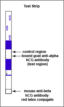
Like the Strep A test mentioned above, this test uses a color immunochromatographic assay to detect the antigen-antibody reaction.The test consists of a membrane strip that is precoated with known mouse anti- beta hCG antibody-colloidal gold conjugate (known antibodies made in mice against the beta chain og hCG with red colloidal gold particles attached) located in a pad at the beginning of the strip. It is also precoated with known goat anti-alpha hCG antibody (known antibodies made in goats against the alpha chain of hCG without attached red colloidal gold) that is immobilized at the test line where the test results are read (see Fig. \(\PageIndex{1A}\)). The red colloidal gold particles attached to the mouse anti-alpha hCG antibody is what ultimately causes the “positive” red band in the test area.
When the test strip is immersed in the urine sample, the hCG begins to move chromatographically up the membrane and binds to the red-colored known anti-beta hCG antibody-gold conjugate in the pad located at the beginning of the strip, forming a hCG antigen-antibody complex (see Fig. \(\PageIndex{1B}\)). This hCG antigen-antibody complex continues to moves up the membrane to the test line region where the immobilized known goat anti-beta hCG antibodies are located.
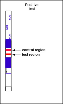
If hCG is present in the urine, a red-colored sandwich of anti-beta antibody/hCG antigen/red gold conjugate anti-alpha antibody forms in the test line region of the strip (see Fig. 7B3). The control region of the strip has immobilized anti-mouse antibodies, that is, antibodies made in a different animal against mouse antibodies. The red color at the control line region appears when molecules of the rabbit anti-hCG antibody-red latex conjugate not trapped at the test line reach the control area and are stopped by binding to the anti-mouse antibodies. The red color at the control line region indicates that the test is finished. As a result, a positive test for hCG antigen appears as a red band in the test result area and a red band in the control area (see Fig. \(\PageIndex{1C}\)).
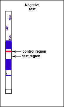
If there is no detectable hCG antigen present in the urine no red band appears in the test result region of the strip and a single red band appears in the control line region, indicating a negative test for hCG antigen (see Fig. \(\PageIndex{1D}\)).