Biology LibreTexts

16.2.3: Serological Typing of Streptococci

  • Page ID
    122731
  • \( \newcommand{\vecs}[1]{\overset { \scriptstyle \rightharpoonup} {\mathbf{#1}} } \)

    \( \newcommand{\vecd}[1]{\overset{-\!-\!\rightharpoonup}{\vphantom{a}\smash {#1}}} \)

    \( \newcommand{\id}{\mathrm{id}}\) \( \newcommand{\Span}{\mathrm{span}}\)

    ( \newcommand{\kernel}{\mathrm{null}\,}\) \( \newcommand{\range}{\mathrm{range}\,}\)

    \( \newcommand{\RealPart}{\mathrm{Re}}\) \( \newcommand{\ImaginaryPart}{\mathrm{Im}}\)

    \( \newcommand{\Argument}{\mathrm{Arg}}\) \( \newcommand{\norm}[1]{\| #1 \|}\)

    \( \newcommand{\inner}[2]{\langle #1, #2 \rangle}\)

    \( \newcommand{\Span}{\mathrm{span}}\)

    \( \newcommand{\id}{\mathrm{id}}\)

    \( \newcommand{\Span}{\mathrm{span}}\)

    \( \newcommand{\kernel}{\mathrm{null}\,}\)

    \( \newcommand{\range}{\mathrm{range}\,}\)

    \( \newcommand{\RealPart}{\mathrm{Re}}\)

    \( \newcommand{\ImaginaryPart}{\mathrm{Im}}\)

    \( \newcommand{\Argument}{\mathrm{Arg}}\)

    \( \newcommand{\norm}[1]{\| #1 \|}\)

    \( \newcommand{\inner}[2]{\langle #1, #2 \rangle}\)

    \( \newcommand{\Span}{\mathrm{span}}\) \( \newcommand{\AA}{\unicode[.8,0]{x212B}}\)

    \( \newcommand{\vectorA}[1]{\vec{#1}}      % arrow\)

    \( \newcommand{\vectorAt}[1]{\vec{\text{#1}}}      % arrow\)

    \( \newcommand{\vectorB}[1]{\overset { \scriptstyle \rightharpoonup} {\mathbf{#1}} } \)

    \( \newcommand{\vectorC}[1]{\textbf{#1}} \)

    \( \newcommand{\vectorD}[1]{\overrightarrow{#1}} \)

    \( \newcommand{\vectorDt}[1]{\overrightarrow{\text{#1}}} \)

    \( \newcommand{\vectE}[1]{\overset{-\!-\!\rightharpoonup}{\vphantom{a}\smash{\mathbf {#1}}}} \)

    \( \newcommand{\vecs}[1]{\overset { \scriptstyle \rightharpoonup} {\mathbf{#1}} } \)

    \( \newcommand{\vecd}[1]{\overset{-\!-\!\rightharpoonup}{\vphantom{a}\smash {#1}}} \)

    \(\newcommand{\avec}{\mathbf a}\) \(\newcommand{\bvec}{\mathbf b}\) \(\newcommand{\cvec}{\mathbf c}\) \(\newcommand{\dvec}{\mathbf d}\) \(\newcommand{\dtil}{\widetilde{\mathbf d}}\) \(\newcommand{\evec}{\mathbf e}\) \(\newcommand{\fvec}{\mathbf f}\) \(\newcommand{\nvec}{\mathbf n}\) \(\newcommand{\pvec}{\mathbf p}\) \(\newcommand{\qvec}{\mathbf q}\) \(\newcommand{\svec}{\mathbf s}\) \(\newcommand{\tvec}{\mathbf t}\) \(\newcommand{\uvec}{\mathbf u}\) \(\newcommand{\vvec}{\mathbf v}\) \(\newcommand{\wvec}{\mathbf w}\) \(\newcommand{\xvec}{\mathbf x}\) \(\newcommand{\yvec}{\mathbf y}\) \(\newcommand{\zvec}{\mathbf z}\) \(\newcommand{\rvec}{\mathbf r}\) \(\newcommand{\mvec}{\mathbf m}\) \(\newcommand{\zerovec}{\mathbf 0}\) \(\newcommand{\onevec}{\mathbf 1}\) \(\newcommand{\real}{\mathbb R}\) \(\newcommand{\twovec}[2]{\left[\begin{array}{r}#1 \\ #2 \end{array}\right]}\) \(\newcommand{\ctwovec}[2]{\left[\begin{array}{c}#1 \\ #2 \end{array}\right]}\) \(\newcommand{\threevec}[3]{\left[\begin{array}{r}#1 \\ #2 \\ #3 \end{array}\right]}\) \(\newcommand{\cthreevec}[3]{\left[\begin{array}{c}#1 \\ #2 \\ #3 \end{array}\right]}\) \(\newcommand{\fourvec}[4]{\left[\begin{array}{r}#1 \\ #2 \\ #3 \\ #4 \end{array}\right]}\) \(\newcommand{\cfourvec}[4]{\left[\begin{array}{c}#1 \\ #2 \\ #3 \\ #4 \end{array}\right]}\) \(\newcommand{\fivevec}[5]{\left[\begin{array}{r}#1 \\ #2 \\ #3 \\ #4 \\ #5 \\ \end{array}\right]}\) \(\newcommand{\cfivevec}[5]{\left[\begin{array}{c}#1 \\ #2 \\ #3 \\ #4 \\ #5 \\ \end{array}\right]}\) \(\newcommand{\mattwo}[4]{\left[\begin{array}{rr}#1 \amp #2 \\ #3 \amp #4 \\ \end{array}\right]}\) \(\newcommand{\laspan}[1]{\text{Span}\{#1\}}\) \(\newcommand{\bcal}{\cal B}\) \(\newcommand{\ccal}{\cal C}\) \(\newcommand{\scal}{\cal S}\) \(\newcommand{\wcal}{\cal W}\) \(\newcommand{\ecal}{\cal E}\) \(\newcommand{\coords}[2]{\left\{#1\right\}_{#2}}\) \(\newcommand{\gray}[1]{\color{gray}{#1}}\) \(\newcommand{\lgray}[1]{\color{lightgray}{#1}}\) \(\newcommand{\rank}{\operatorname{rank}}\) \(\newcommand{\row}{\text{Row}}\) \(\newcommand{\col}{\text{Col}}\) \(\renewcommand{\row}{\text{Row}}\) \(\newcommand{\nul}{\text{Nul}}\) \(\newcommand{\var}{\text{Var}}\) \(\newcommand{\corr}{\text{corr}}\) \(\newcommand{\len}[1]{\left|#1\right|}\) \(\newcommand{\bbar}{\overline{\bvec}}\) \(\newcommand{\bhat}{\widehat{\bvec}}\) \(\newcommand{\bperp}{\bvec^\perp}\) \(\newcommand{\xhat}{\widehat{\xvec}}\) \(\newcommand{\vhat}{\widehat{\vvec}}\) \(\newcommand{\uhat}{\widehat{\uvec}}\) \(\newcommand{\what}{\widehat{\wvec}}\) \(\newcommand{\Sighat}{\widehat{\Sigma}}\) \(\newcommand{\lt}{<}\) \(\newcommand{\gt}{>}\) \(\newcommand{\amp}{&}\) \(\definecolor{fillinmathshade}{gray}{0.9}\)

    The Clearview® Strep A Exact II Dipstick is an example of a lateral flow immunologic assay. It is is a qualitative serologic test for detecting Group A Streptococcal antigen (the unknown antigen) directly from throat swabs and is used as an aid in diagnosing streptococcal pharyngitis caused by Streptococcus pyogenes (Group A Beta Streptococci).

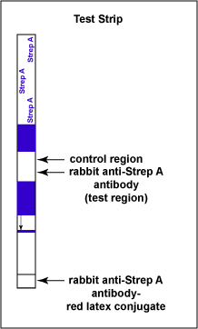
    The test consists of a membrane strip that is precoated with rabbit anti-Strep A monoclonal antibody-red latex conjugate (known antibodies made in rabbits against strep A antigen with red latex particles attached) located in a pad at the beginning of the strip. It is also precoated with rabbit anti-Strep A monoclonal antibody (known antibodies made in rabbits against the strep A antigen but without attached red latex particles) that is immobilized at the test line where the test results are read (see Fig. \(\PageIndex{1A}\)). The red latex particles attached to the rabbit anti-Strep A antibodies are what ultimately causes the “positive” red band.

    Illustration of the test strip for detecting Group A Streptococcus antigen.
    Figure \(\PageIndex{1A}\): Serologic Testing to Identify Group A Streptococcus Antigen: The Test Strip. ( Copyright; Gary E. Kaiser, Ph.D. The Community College of Baltimore County, Catonsville Campus CC-BY-3.0 )

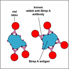
    The throat swab is placed in an extraction solution that lyses the Streptococcus pyogenes, if present, and exposes the strep A antigen in the bacterial cell wall. When the test strip is immersed in the extracted sample, the Group A Streptococcal antigen extracted from the Streptococcus pyogenes on the throat swab of a person with strep throat begins to move chromatographically up the membrane and binds to the known antibody-red latex conjugate in the pad located at the beginning of the strip, forming a Strep A antigen-antibody complex (see Fig. \(\PageIndex{1B}\)). This Strep A antigen-antibody complex continues to moves up the membrane to the test line region where the immobilized rabbit anti-Strep A antibodies are located.

    Illustration of cell wall fragments containing Strep A antigen binding to known anti-Strep A antibodies bound to red latex particles.
    Figure \(\PageIndex{1B}\): Serologic Testing to Identify Group A Streptococcus Antigen: Known Antibodies Against Strep A Antigen binding to Strep A Antigens. Cell wall fragments containing Strep A antigen binds to known anti-Strep A antibodies bound to red latex particles. ( Copyright; Gary E. Kaiser, Ph.D. The Community College of Baltimore County, Catonsville Campus CC-BY-3.0 )

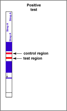
    If Group A Streptococcal antigen is present in the throat swab, a red-colored sandwich of known antibody/Strep A antigen/red latex conjugate forms in the test line region of the strip (see Fig. 6C). The control region of the strip has immobilized anti-rabbit antibodies, that is, antibodies made in a different animal against rabbit antibodies. The red color at the control line region appears when molecules of the rabbit anti-Strep A antibody-red latex conjugate not trapped at the test line reach the control area and are stopped by binding to the anti-rabbit antibodies. This indicates that the test is finished. As a result, a positive test for Group A Strep antigen appears as a red band in the test result area and a red band in the control area (see Fig. \(\PageIndex{1C}\)).

    Illustration of a positive test for Group A Strepyococcus showing twp red bands.
    Figure \(\PageIndex{1C}\): Serologic Testing to Identify Group A Streptococcus Antigen: A Positive Test. Note two red stripes. ( Copyright; Gary E. Kaiser, Ph.D. The Community College of Baltimore County, Catonsville Campus CC-BY-3.0 )

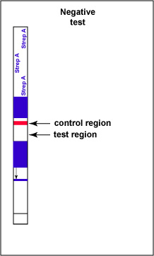
    If there is no Group A Streptococcal antigen present in the throat swab no red band appears in the test result region of the strip and a single red band appears in the control line region, indicating a negative test for Group A Strep antigen (see Fig. \(\PageIndex{1D}\)).

    Illustration of a negative test for Group A Streptoccus antigen showing only one red stripe in the test control area.
    Figure \(\PageIndex{1D}\): Serologic Testing to Identify Group A Streptococcus Antigen: A Negative Test. Note only one red stripe in the test control area. ( Copyright; Gary E. Kaiser, Ph.D. The Community College of Baltimore County, Catonsville Campus CC-BY-3.0 )

    Contributors and Attributions

    • Dr. Gary Kaiser (COMMUNITY COLLEGE OF BALTIMORE COUNTY, CATONSVILLE CAMPUS)


    This page titled 16.2.3: Serological Typing of Streptococci is shared under a not declared license and was authored, remixed, and/or curated by Gary Kaiser.

    • Was this article helpful?