
14.5: Isolation an Identifiation of Pneumococci
- Page ID
- 123470
Videos reviewing techniques used in this lab:
1. Isolation on Blood agar
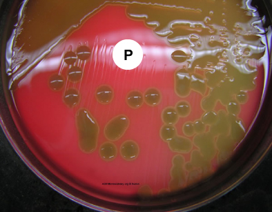
Pneumococci frequently require enriched media and increased CO2 tension for initial isolation. They are usually isolated on Blood agar and incubated in a candle jar (a closed container in which a lit candle is placed to remove O2 and increase CO2 ) at 37C. On Blood agar, colonies appear small, shiny, and translucent. They are surrounded by a zone of alpha hemolysis (see Fig. \(\PageIndex{1}\)). Due to autolysis with age, the colonies may show a depressed center with an elevated rim.
2. Optochin sensitivity
Pneumococci are the only streptococci that are sensitive to the drug optochin (ethylhydrocupreine hydrochloride). This can be detected by a zone of inhibition around a Taxo P® disc (see Fig. \(\PageIndex{1}\)), a paper disc containing the drug optochin, which is placed on the Blood agar plate prior to incubation.
3. Bile solubility test
Most colonies of S. pneumoniae will dissolve within a few minutes when a drop of bile is placed upon them. (This test will not be done in lab today.)
4. Gram stain of sputum
Streptococcus pneumoniae will usually appear as encapsulated, Gram-positive, lancet-shaped diplococci.
The Viridans Streptococci
DISCUSSION
Ten species of streptococci are known as the viridans streptococci. They are the dominant normal flora in the upper respiratory tract. Species include S. mutans, S. sanguis, S. mitis, and S. salivarius. S. mutans is the primary cause of dental caries. Viridans streptococci are responsible for between 50% and 70% of the cases of bacterial endocarditis, especially in people with previously damaged heart valves. They are also frequently associated with bacteremia, deep wound infections, dental abscesses, and abscesses of internal organs. The viridans streptococci show alpha hemolysis or no hemolysis on Blood agar, do not possess Lancefield group antigens, and can be differentiated from other alpha streptococci by biochemical testing.
B. The Genus Enterococcus
Videos reviewing techniques used in this lab:
- How to Interpret the Results of SF Broth and Bile Esculin Agar; Identification of Enterococcus (We are using Bile Esculin Azide Agar only.)
DISCUSSION
Enterococci are Gram-positive streptococci that are normal flora of the intestinal tract. They typically occur singly, in pairs, short chains, and clusters,especially when taken off an agar culture for staining. Â Like the genus Streptococcus, the genus Enterococcus is catalase-negative. Enterococci responsible for a variety of opportunistic infections in humans, and serologically belong to Lancefield group D streptococci.
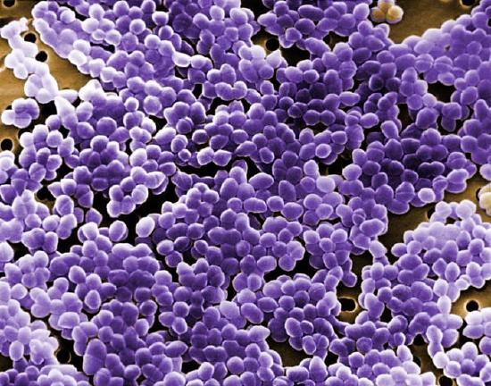
Enterococcus faecalis (see Fig. \(\PageIndex{3}\)) is the most common enterococcus causing human infections, representing 80-90% of human enterococcal clinical isolates. E. faecalis is normal flora of the intestinal tract in humans and is regularly isolated from infections within the peritoneal cavity (especially following penetrating trauma), urinary tract infections, kidney infections, prostate infections, endocarditis, and infections of damaged or compromised skin such as diabetic or decubitus ulcers, burns and surgical wounds. Trauma to the intestines can lead to intra abdominal and pelvic infections. Other opportunistic enterococcal species include E. faecium and E. durans. The enterococci have become the second most common bacterium isolated from nosocomial urinary and wound infections, and the third most common cause of nosocomial bacteremia. Each year in the U.S., in fact, enterococci account for approximately 110,000 urinary tract infections, 40,000 wound infections, 25,000 cases of nosocomial bacteremia, and 1100 cases of endocarditis. Furthermore, the enterococci are among the most antibiotic resistant of all bacteria, with some isolates resistant to all known antibiotics. The ability of enterococci to produce biofilms protects the organism from the body's defenses as well as promotes exchange of genetic material with other pathogens. A scanning electron micrograph of Enterococcus can be seen in Fig. \(\PageIndex{4}\).
|
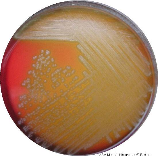
Fig \(\PageIndex{3}\): A Plate of Blood Agar Showing Alpha, Beta, and Gamma Hemolysis (Indirect Lighting) |
Fig. \(\PageIndex{4}\): A Blood Agar Plate of a Throat Culture Showing Possible Streptococcus pyogenes. |
|---|---|
 |
 |
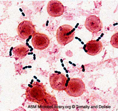
| Enterococcus species are normal glora of the intestinal tract. Enterococcus faecalis frequently causes infections within the peritoneal cavity, especially following penetrating trauma such as gunshot wounds, knife wounds, and surgical wounds, urinary tract infections, kidney infections, prostate infections, and infections of damaged or compromised skin, such as diabetic or decubitus ulcers, burns, and surgical wounds. Other opportunistic fecal streptococci include E. faecium and E. durans. The enterococci have become the second most common bacterium isolated from nosocomial urinary and wound infections, and the third most common cause of nosocomial bacteremia.Furthermore, the enterococci are among the most antibiotic resistant of all bacteria, with some isolates resistant to all known antibiotics. Note Gram-positive streptococci. | Note streptoococcus arrangement (cocci in chains). |
| Image: Enterococcus faecalis in a Blood Culture. © Gloria Delisle and Lewis Tomalty, authors. Licensed for use, ASM MicrobeLibrary. | By Content Providers(s): CDC/ Janice Haney Carr [Public domain] Courtesy of the Centers for Disease Control and Prevention. |
Contributors and Attributions
Dr. Gary Kaiser (COMMUNITY COLLEGE OF BALTIMORE COUNTY, CATONSVILLE CAMPUS)